R E S E A R C H
Open Access
Complex network analysis of thermostable
mutants of
Bacillus subtilis
Lipase A
Nitika Kandhari and Somdatta Sinha
** Correspondence:
Centre for Protein Science Design and Engineering, Department of Biological Sciences, Indian Institute of Science Education and Research, Mohali, Punjab 140306, India
Abstract
Three-dimensional structures of proteins that regulate their functions can be
modelled using complex network based approaches for understanding the structure-function relationship. The six mutants of the protein Lipase A fromBacillus subtilis, harbouring 2 to 12 mutations, retain their function at higher temperatures with negligible variation in their overall three-dimensional crystallographic structures. This enhanced thermostability of the mutants questions the structure-function paradigm. In this paper, a coarse-grained complex network approach is used to elucidate the structural basis of enhanced thermostability in the mutant proteins, by uncovering small but significant local changes distributed throughout the structure, rendering stability to the mutants at higher temperatures. Community structure analysis of the six mutant protein networks uncovers the specific reorganisations among the nodes/ residues that occur, in absence of overall structural variations, which induce enhanced rigidity underlying the increased thermostability. This study offers a novel and significant application of complex network analysis that proposes to be useful in the understanding and designing of thermostable proteins.
Keywords:Protein contact networks, Thermostability, Lipase A, Community structures
Introduction
Proteins are biological macromolecules in the cell that are first synthesized as lin-ear chains of amino acids held together by chemical forces, which then fold into three-dimensional structures through short and long range chemical interactions decided by the chemical nature of the amino acids (Creighton, 1992). Protein function is determined by its three-dimensional structure and significant structural change in the protein due to mutations (i.e., change in the amino acid sequence) often mediates variation in functional properties, known as the structure-function paradigm in protein science. However, there are several cases that are coming to light, where significant changes in function have been ob-served in some proteins due to mutations, which have negligible changes in the overall structures (Srivastava and Sinha, 2013; Srivastava and Sinha, 2014; Kand-hari and Sinha, 2016; Srivastava and Sinha, 2017). Our interest has been to study those proteins and their mutants, having indistinguishable ordered crystallo-graphic structures, but exhibiting large changes in functions. Since structure-based methods cannot easily expose the small changes, we have used the complex network approach to study these protein structures to understand the structural
and mechanistic bases of what leads to the functional improvement, in absence of overall structural variations.
The three-dimensional crystallographic structure of the protein can be represented using a network description – the “Protein Contact Networks” (PCNs) - whose nodes are the amino acids (residues) and the chemical interactions between closely-held residues are the links. Such complex network based approaches have been used for studying the structure and function of proteins (Kannan and Vishveshwara, 1999; Vendruscolo et al. 2002; Bagler and Sinha, 2005; Bagler and Sinha, 2007; Barah and Sinha, 2008; Di Paola et al. 2013). In the present work, we have attempted to utilize this approach for the protein Lipase A, from the bacteria Bacillus subtilis, which has important applications in leather industry, food processing, pulp, and other biotech-nological applications, where it needs to retain its structural integrity and functional activity at higher temperatures (at which normally proteins unfold) (Liszka et al., 2012). Six mutants of the wild-type (WT) Lipase A showing increased thermostabil-ity are chosen for this study because all the proteins have structures very similar to the WT in spite of having 2 to 12 mutations (Srivastava and Sinha, 2014; Kandhari and Sinha, 2016). The PCNs of these seven Lipase A proteins (WT and its six mu-tants) have been studied to uncover those small changes that do not change the structure but confer thermostability to retain functionality.
Earlier studies on thermostability have invoked the role of many structural fac-tors that arise due to mutations and contribute to structural rigidity, such as higher number of hydrogen-bonds and salt-bridges, secondary structure stabilization, disulphide linkages, higher polar surface area, more number of Pro-line residues, shortening and stabilization of loops, etc. (Matsumura et al. 1989; Pjura and Matthews, 1993; Watanabe et al. 1994; Yip et al. 1995; Salminen et al. 1996; Haney et al. 1997; Russell et al. 1997; Vogt and Argos, 1997; Vogt et al. 1997; Nicholson et al. 1998; Watanabe and Suzuki, 1998; Chan et al. 2011; Gro-miha et al. 2013). We have shown, using the complex network approach that ther-mostability in Lipase A, imbibed through several structural factors that arise due to mutations in its mesophilic counter-part, can be elaborated through the study of small changes in the contact patterns in the PCNs. We show the detailed ana-lysis of the small conformational changes, occurring through-out the proteins, by analyzing the contact patterns, their positional information, the local network parameters, and the changes in the community memberships, with respect to their structural stability and functionality at higher temperatures.
Methods and data
Construction of protein contact networks
PCNs were constructed by considering the C-αatom of each residue in the protein as a node and the spatial proximity between any two of them as the link.
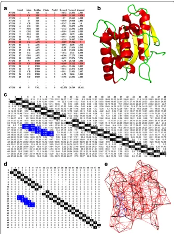
The Protein Data Bank (PDB) is a database for storing solved crystallographic struc-tures of proteins (RCSB Protein Data Bank (PDB) 1971). Figure 1 gives the method of
construction of the coarse-grained PCN for all seven protein structures from the PDB data containing the positions (x, y and z coordinates) of all the atoms of all amino acids in 3-dimensional space. Fig. 1a shows the crystallographic coordinates of all atoms of the first three residues of the protein WT Lipase A (shown in Fig. 1b). We use a coarse-grained approach (Bagler and Sinha 2005; Bagler and Sinha 2007; Srivastava and Sinha 2014) to construct the network by considering the spatial positions of only the protein backbone atoms (i.e. C-αatoms) of each residue (coloured in red in Fig. 1a; the backbone connections are highlighted in black in Fig. 1c and d). A pair-wise Euclidean distance matrix is computed between the C- αatoms of all residues (Fig. 1c). The dis-tance matrix is converted into an Adjacency matrix, A (Fig. 1d), using the following rule–
Aij¼1;if Dij<¼7A
;else;Aij¼0
where, Dijis the Euclidian distance between C-αatoms of the ithand jthresidues. This cut-off has been widely used for constructing C-α based Protein Contact Networks, and provides a reasonable estimate of the interacting residue pairs in the first inter-action shell (Haliloglu et al. 1997). However there are other cut-offs that have been used in all atom network models (Gromiha and Selvaraj 2004). It is obvious that there will be changes in the number of edges if the cut-off is varied. For C-αbased PCNs, the most commonly used values are 0.7 nm to 0.8 nm (Bagler and Sinha 2005; Bagler and Sinha 2007; Srivastava and Sinha 2014; Srivastava and Sinha 2017). The results pre-sented in this work have been checked with 0.75 nm and 0.80 nm cut-offs, and they are comparable to the results presented here.
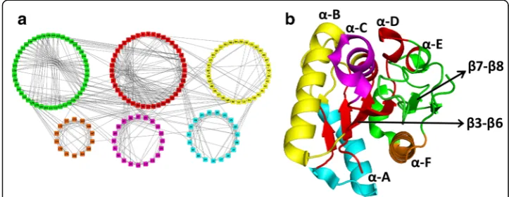
The two-dimensional representation of the coarse-grained PCN, corresponding to the three dimensional structure of Lipase A (Fig. 1b), is shown in Fig. 1e. The black lines in Fig. 1e are the back-bone contacts, the red lines are the contacts be-tween the other nodes present throughout the protein, the and the blue lines (in Fig. 1c and d) are long range contacts between pairs of nodes that are far apart in the linear chain but have come closer in space due to protein folding (e.g., contacts between nodes 5–36, 6–36, 9–39).
The data-set: Lipase A and its mutants fromBacillus subtilis
corresponding to this protein is shown in Fig. 2b. The correspondence between the different secondary structures (α-helices, β-sheets, loop regions, etc.) in Fig. 2a and c are circled to compare with the contacts between the nodes in the PCN (Fig. 2b).
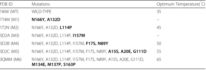
Our dataset from PDB comprising of Lipase A wild-type (WT) protein and its six thermostable mutants are shown in Table 1. These mutants, with 2–12 mutations, were obtained in a manner so that the previous mutations are preserved and new mutations are incorporated (Acharya and Rao 2004; Ahmad et al. 2008; Kamal et al. 2011). All the mutant proteins are stable and functional at increasingly higher temperatures.
Network parameters
Degree of a node is the total number of direct links that it has to other nodes. For PCN all backbone nodes, except the terminal nodes, have a minimum degree of 2 since they connect the protein chain. Larger degree indicates secondary structures and long range contacts due to folding of the chain.
Shortest Path Length (SPL)between a pair of nodes is the smallest number of links that need to be traversed in order to reach from one node to the other. It has been
Fig. 2Schematic representation to show the correspondence between protein secondary structures to the PCN. Theα-helices are denoted byα, and theβ-sheets byβ
Table 1The PDB structure IDs of Lipase A and its six mutants used for analysis, with the mutated sites and their optimum functional temperature
PDB ID Mutations Optimum Temperature(̊C)
1I6W (WT) WILD-TYPE 35
1T4M (M1) N166Y, A132D –
1T2N (M2) N166Y, A132D,L114P 45
3D2A (M3) N166Y, A132D, L114P,I157M –
3D2B (M4) N166Y, A132D, L114P, I157M,F17S, N89Y 50
3D2C (M5) N166Y, A132D, L114P, I157M, F17S, N89Y,A15S, A20E, G111D 55
3QMM (M6) N166Y, A132D, L114P, I157M, F17S, N89Y, A15S, A20E, G111D,
M134E, M137P, S163P
65
shown that fibrous and globular protein and helices and sheets have different path lengths (Bagler and Sinha 2005).
Clustering Coefficient (CC) measures the cliquishness of the neighbourhood of the node, i.e. to what extent the nodes in a network tend to cluster together. It is defined as the ratio of the number of edges among the linked neighbours of the node to the total number of edges possible amongst them. It has been shown that globular proteins and nodes in helices have higher CC compared to fibrous proteins and sheets (Bagler and Sinha 2005).
Betweenness Centrality (BC) of a node is the number of shortest paths passing through that node. More the number of paths passing through a node, more crucial it is for communication between different parts of the network.
Closeness Centrality (CCen) is defined as the inverse of the sum of Shortest Paths from that node to all other nodes in the network. High CCen of a node indicates closeness to other nodes.
For more details see (Newman 2010).
Community structure analysis
Finding communities or modules in a network attempts to divide the network nodes into groups, such that there is a higher density of edges within the groups than between them. Community structure analysis was performed for all the seven PCNs using the fast greedy algorithm (Clauset et al. 2004).
Protein data extraction from PDB was done using in-house PERL scripts. Network analysis and visualization have been done with CYTOSCAPE (Shanon et al. 2003). All statistical analysis was done using R (https://cran.r-project.org/).
Results
Mutant protein networks have larger number of contacts
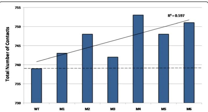
No change in the structures at global scale despite the change in function points towards careful analysis at the local scale. Even though the total number of nodes in all PCNs is the same, the number of contacts (edges) in each PCN, shown in Fig. 4, indicates vari-ation. New contacts are formed and old ones lost due to mutations. All the mutant PCNs have larger number of contacts compared to the WT, and the trend line shows that there is a general increase in the number of edges in mutants with increased thermostability (black solid line). Even though this increase does not directly correlate with increased thermostability (e.g., M4 has the largest number of edges (753) but is not the most thermostable among the mutants), this result indicates that larger number of contacts may introduce higher rigidity in the mutant proteins. This raises the question as to how these contacts are distributed in the PCNs, which can lead to thermostability.
Network parameter analysis of PCNs corroborate negligible structural variations
Increase in the total number of edges in the mutant PCNs can influence the network parameters. The five network parameters studied for the seven PCNs are - Degree,
Fig. 3Network and structural representations of WT and M6: Ring Graph representation of the PCNs of (a) WT and (c) M6. (b) Three-dimensional structural overlap of WT (blue) and M6 (purple) proteins
Betweenness Centrality (BC), Shortest Path Length (SPL), Clustering Coefficient (CC) and Closeness Centrality (CCen). These were first studied at a global-scale i.e. the aver-age network parameters were computed for each PCN. The ranges for averaver-age network parameter values for the seven PCNs are:
Degree: 8:2918:413
Betweenness Centrality:0:0180‐0:0184
Closeness Centrality:0:240‐0:244
Clustering Coefficient:0:527‐0:539
Shortest Path Length:4:173‐4:239
The very low variations of the average network parameters in all PCNs reflect the low cross-structural RMSD of the proteins, thereby corroborating similar three-dimensional structures of Lipase A and its thermostable mutants.
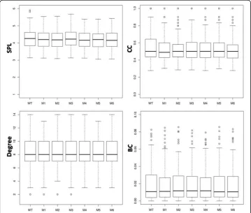
To find any local changes, the network parameters (SPL, CC, Degree, and BC) were computed at individual node level in all seven PCNs (WT, and M1 to M6), and the de-tails of their distributions are shown in the box-plots in Fig. 5. For all parameters, the distributional characteristics of all network parameters seem to be similar for all PCNs in that group (i.e., WT and its mutants) with small variations in the extreme values. If the distributions are skewed, then all PCNs show the same behaviour (see Degree and BC). For Degree, the distributional variation is the least among all PCNs. The box-plot
for CC shows that, even though the median values are similar, all mutant PCNs have smaller box size and more outliers compared to the WT. This indicates that most nodes in the mutant PCNs have CC values close to their average/median value, but they also have more nodes with higher CC. The Kruskal-Wallis test for the null hypoth-esis of equal means show no significant difference (p< 0.01) in the means of all PCNs for all network parameters. These results point towards careful analysis of local contacts in each mutant that may be responsible for increased thermostability.
Contacts analysis reveals their specific roles in imparting stability to the mutant proteins The known factors attributed to increasing stability/rigidity in proteins that are import-ant for thermostability are - loop stabilization (Vogt and Argos 1997), stabilization of secondary structures (Nicholson et al. 1998), stabilization of termini contacts, higher number of hydrogen bonds (Vogt and Argos 1997; Vogt et al. 1997; Pjura and Matthews 1993), shortening of loops (Vogt and Argos 1997; Nicholson et al. 1998), higher polar surface area, higher number of disulphide linkages (Matsumura et al. 1989) and salt bridges (Yip et al. 1995; Chan et al. 2011; Russell et al. 1997; Nicholson et al. 1998), increased buried surface area after oligomerization (Salminen et al. 1996), increase in number of proline residues (Watanabe et al. 1994; Watanabe and Suzuki 1998), surrounding hydrophobicity (Gromiha et al. 2013), etc. A detailed analysis of the new contacts made in the six PCNs of the mutant proteins reveals their contribution into stabilizing some of these factors (listed in Table 2). It is clear that these contacts are mostly stabilizing the loops and the active site region in the mutants.
Community structure analysis of WT PCN
The overall structural similarity of the mutant proteins and few changes in contacts in their PCNs (Fig. 4) that contribute to specific stabilizing factors aiding to thermostabil-ity (Table 2) prompted a detailed study in the changes in the communthermostabil-ity structures in the seven PCNs. It has been shown that modules in PCNs aid in efficient information transfer between different protein domains (Del Sol et al. 2007). Changes in contacts among the nodes can result in reorganisation of the communities thereby influencing information transfer. This comparative analysis allows a better understanding of the small but distributed changes that helps in retaining functionality at higher tempera-tures (thermostability) in the mutant proteins.
The WT Lipase A protein, shown in Fig. 6b with the known secondary structures (helices: α-A toα-F and β-strands:β3 toβ8), consists of six communities (Fig. 6a). It is clear that the communities correspond to the major secondary structures of the protein and clearly shows the structural regions that are closely connected. The communities in
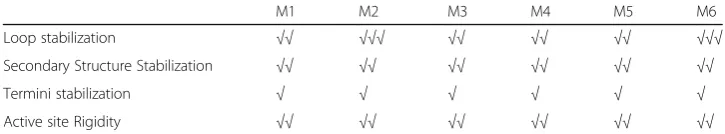
Table 2New contacts in PCNs contribute to the stabilizing factors in mutant proteins
M1 M2 M3 M4 M5 M6
Loop stabilization √√ √√√ √√ √√ √√ √√√
Secondary Structure Stabilization √√ √√ √√ √√ √√ √√
Termini stabilization √ √ √ √ √ √
Active site Rigidity √√ √√ √√ √√ √√ √√
the WT are:Red(having the centralβ-sheet:β3,β4,β5,β6; helixα-D and part of helixα -E); Green(the largest module with portions of helices α-E and α-F, strands β7 andβ8, and major loop region);Yellow(the helixα-B, a small portion of helicesα-C and terminal
α-A);Cyan(a major portion of helixα-A);Magenta(lower portion of the helixα-C), and the smallest communityOrange(portion ofα-F). In the rest of the community structure analysis for the mutant PCNs, these colours of the member nodes have been kept the same for easy identification of their reorganization. The loop regions where many functional sites, including the catalytic triad, exist and mutations were made in the thermostable mutants, are primarily in the Green community in WT PCN.
Community structure analysis unravels increased stability in the mutants
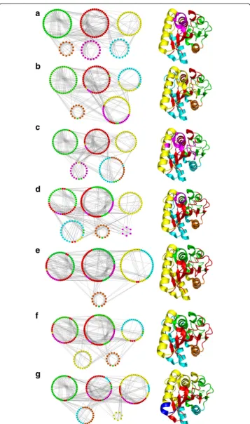
Change in contact patterns in different mutant PCNs can result in changes in their community structures. Figure 7 shows the community structures for all seven Lipase A PCNs. The six mutant PCN communities (Fig. 7(b-g)) reveal changes with respect to the WT PCN communities (Fig. 7a) that arise due to the changed contact patterns occurring due to 2 to 12 mutations in the mutant proteins (M1 to M6). The corre-sponding crystallographic structures for the proteins are also shown along with their communities with same colour code. It is clear from Fig. 7 that the few changes in con-tacts among the mutant PCNs result in quite a few differences in community member-ships among the PCNs. The major secondary structures (α-helices and β-sheets) generally remain in single communities keeping the integrity and retaining the similar-ity of the protein structures, but the nodes that fluctuate among communities in differ-ent PCNs can be iddiffer-entified and correlated with the contacts made/broken - to lead to the thermostability in the mutants. The loop regions are relatively flexible in the protein structure and are expected to change modules. In some cases, the terminal nodes of the regular secondary structures also changed their community if the adjoining loop had changed its community.
The number of communities in WT PCN is the largest (6). The PCN of M3 also shows 6 communities but one of them is very small (only 6 nodes). Rest of the mutants have 4 or 5 communities. This is indicative of the networks becoming more compact, as within the same space the nodes are having larger groupings. For pro-teins this signifies that the larger groups of residues being more connected among
them-selves confer more rigidity to the structures, even when the motions of the atoms increase at higher temperatures.
Below we analyse the communities in the mutant PCNs, vis-à-vis structural regions in the proteins, in Fig. 7 that gives some useful information.
Red module
This module contains nodes primarily involving the central β-sheet, which forms the core of Lipase A structure. Figure 7(b-g) shows that the four β-strandsβ3,β4,β5 and
β6) of the centralβ-sheet forming a single community in WT (Fig. 7a) maintain its in-tegrity in all the mutants. In addition, the Red module is seen to widen as more nodes from adjacent β-strands and the lower loop region come together into one community leading to loop stabilization in mutants. Thus, the community that forms the protein core“expands”thereby strengthening the core of the protein and thus imparting stabil-ity to the protein’s functional region in all thermostable mutants.
Yellow module
The nodes in this community stay constant in most mutant PCNs maintaining the integrity of the secondary structure – the α-B helix. For M2, only one node merges with another module without breaking into smaller modules indicating presence of new contacts. In M6, the two contacts (48–54 and 72–173) tend to pull the helixα-B in opposite directions, thus making the terminal part (nodes 63 to 67) of the helix form a different community altogether (coloured Blue with nodes 63–69 in the structure of M6 in Fig. 7g). In this mutant (M6), the nodes containing the helicesα-B,α-C andα-D form a single community (Yellow) leading tosecondary structure stabilization. Recently several point mutations (N51F, G52 M, V54H, L55F, F58I, V59I in helix α-B) belonging to our Yellow module and I87W in helix α-C were predicted that leads to increase in contacts between helices imparting stability to them and thus causing delay in thermal unfolding (Rathi et al. 2015; Rathi et al. 2016). These results were shown and validated experimentally for few of these mutations (Rathi, et al. 2016). So, stabilization of helices in the Lipase A structure, as shown in our community structure analysis, is crucial for protein stability at higher temperatures.
Cyan module
The nodes in this community also stay constant in most mutant PCNs maintaining the integrity of the secondary structure (helixα-A), except in M4 and M6 where it becomes members of the Yellow community (fully or partially) indicating secondary structure stabilization.
Green module
M3 and other more thermostable mutant PCNs. This leads to loop stabilization and reduced number of modules of the PCN inducing compactness.
Orange module
The orange module consists of nodes 163 to 174. The nodes corresponding to helix α -F (156 to 174) retains its structural integrity in all PCNs but showssecondary structure stabilization with the Cyan moduleα-A in some mutants. The other few nodes, being part of loops, change community memberships.
Magenta module
In WT (Fig. 7a), this module includes nodes containing a portion of helix α-C and nearby loop regions. The nodes of this community change their membership to the other modules in the mutant PCNs extensively often leading to loop stabilization, even though the integrity of the helix is maintained in all PCNs with the nodes stay-ing together always. The contact 85–90 is important for the formation of this mod-ule. Whenever this contact appears, the Magenta module appears. In case of M3, the formation of contacts 48–81 and 52–83 pulls nodes 81, 82 and 83 away from the 86 to 91 stretch. Also, the contacts 88–92 and 91–94 lead to formation of a tiny separ-ate module comprising of only 6 nodes (86 to 91).
Catalytic triad residues
Among these three important functional residues, Asp 133 and His 156 remains in the Green community throughout, but Ser 77 changes its community from Red (in WT, M1, M2) to Green (in M3, M4, M5) to Yellow (in M6). These changes in com-munity membership of Ser 77 (in the loop region) is associated with active site rigidity and more effective functioning at higher temperature, such as, binding of 77 and 156 during hydrolysis, which has been shown experimentally (Kamal et al. 2012).
In addition to the above, our analysis of the modules show that, owing to the other mutations in M6, there are more number of contacts surrounding the α-helices α-D and α-E and the central β-sheet (giving rise to a single module consisting of yellow, cyan, magenta and red nodes in Fig. 7g), which further stabilize M6. This is also sup-ported by results from the structural experiments (Rathi et al. 2015; Kamal et al. 2012).
Information transfer between different modules in the protein
In WT, of the 17 functional nodes taking part in Lipase A activity, 11 nodes connect the Red, Green Yellow and Cyan communities among themselves. On the other hand, in M6 mutant PCN only 7 out of the 17 functional residues are involved in connecting different modules. This clearly suggests that there is less information transfer through the functional residues among the different parts of the protein. This is useful when less transfer of perturbation is needed within the protein for sta-bility at higher temperature.
structure stabilization, and active site rigidity, thereby making the mutants more thermostable.
Discussion
In this study we use the complex network approach to study the protein Lipase A and its six mutants, having 2 to 12 mutations (all at or near the functional sites), from Bacillus subtilis, which do not have any significant difference in their three-dimensional structure, but are functional at very high temperatures (i.e. thermo-stable). There are two important issues that have been addressed in this paper. First, we attempt to understand the small changes arising in these proteins due to muta-tions, which are not easily identifiable using standard structural analysis due to their having similar structures. Secondly, we endeavour to identify those localised changes distributed throughout the proteins that bring about more stability/rigidity in the mutant proteins to be functional at higher temperatures.
The coarse-grained PCN approach is suitable to study this problem as it highlights any change at the node-level contacts in these thermostable mutants that may alter in-formation transfer within the protein structures. Since the over-all crystallographic structures of all seven proteins are similar, the global network parameters are not useful indicators of variations in their thermostabilty. Through a careful and detailed com-parative analysis of the contacts in each of the PCNs, along with their community structure analyses, we have identified what may be the underlying structural correlates for increased thermostability in the mutants of Lipase A. On a careful enumeration of the contacts made and broken in the six mutants PCNs, the total number of contacts show a small but consistent increase in the mutants in comparison to the WT Lipase A. Through a detailed position-specific analyses of these new contacts we could clearly associate them to stabilize the loop regions, secondary structures (forming inter-helical contacts), and in making the active site region more rigid. These features, as mentioned in the introduction, have been attributed to increase structural stability in proteins.
Taking cue from our results, one can study the dynamic consequences, using molecu-lar dynamics simulations (Karplus and McCammon, 2002), of the group of residues that constitute the communities and specifically those which change modules often, to speculate their role in imparting thermostability in Lipase A. Thus, this complex network based approach can be very useful in design and engineering of specific properties in proteins.
Abbreviations
ASPL:Average Shortest Path Length; BC: Betweenness Centrality; CC: Clustering Coefficient; CCen: Closeness Centrality; M1: First Mutant (1T4M); M2: Second Mutant (1T2N); M3: Third Mutant (3D2A); M4: Fourth Mutant (3D2B); M5: Fifth Mutant (3D2C); M6: Sixth Mutant (3QMM); PCN: Protein Contact Networks; PDB: Protein Data Bank; RMSD: Root Mean Square Deviation; SPL: Shortest Path Length; WT: Wild-type protein (1I6W)
Acknowledgements
We thank Shubhakar Reddy for helping with community structure analysis. SS thanks JCBose Fellowship for support.
Funding
The Center for Protein Science Design and Engineering (CPSDE) approved by the Ministry of Human Resource Development (MHRD), Government of India, under its COE-FAST programme at IISER Mohali.
Authors’contributions
SS designed the study; NK performed the analysis. NK and SS wrote the manuscript. Both authors read and approved the final manuscript.
Competing interests
The authors declare that they have no competing interests.
Publisher’s Note
Springer Nature remains neutral with regard to jurisdictional claims in published maps and institutional affiliations.
Received: 17 February 2017 Accepted: 1 June 2017
References
Acharya P, Rajakumara E, Sankarnarayanan R, Rao NM (2004) Structural basis of selection and thermostability of laboratory evolvedBacillus subtilisLipase. J Mol Biol 341(5), 1271-81. doi:10.1016/j.jmb.2004.06.059
Ahmad S, Kamal MZ, Sankaranarayanan R, Rao NM (2008) Thermostable Bacillus Subtilis lipases: in vitro evolution and structural insight. J Mol Biol 381(2):324-340. doi: 10.1016/j.jmb.2008.05.063.
Barah P, Sinha S (2008) Analysis of protein folds using protein contact networks. Pramana journal of physics 71(2):369-378. doi: 10.1007/s12043-008-0170-5
Bagler G, Sinha S (2005) Network properties of protein structures. Physica A 346(1):27-33. doi: 10.1016/j.physa.2004.08. 046
Bagler G, Sinha S (2007) Assortative mixing in protein contact networks and protein folding kinetics. Bioinformatics 23(14):1760-1767. doi: 10.1093/bioinformatics/btm257
Chan CH, Yu TH, Wong KB (2011) Stabilizing salt-bridge enhances protein thermostability by reducing the heat capacity change of unfolding. PLoS One 6(6):e21624. doi: 10.1371/journal.pone.0021624
Clauset A, Newman ME, Moore C (2004) Finding community structure in very large networks. Phys Rev E 70(6 Pt 2): 066111. doi: 10.1103/PhysRevE.70.066111
Creighton TE (1992) Protein Folding. W. H. Freeman, New York.
Del Sol A, Araúzo-Bravo MJ, Amoros D, Nussinov R (2007) Modular architecture of protein structures and allosteric communications: potential implications for signalling proteins and regulatory indices. Genome Biol 8(5), R92. doi:10. 1186/gb-2007-8-5-r92
Di Paola L, De Ruvo M, Paci P, Santoni D, Giuliani A (2013) Protein contact networks: An emerging paradigm in chemistry. Chem Rev 113(3), 1598-1613. doi:10.1021/cr3002356
Gromiha MM, Selvaraj S (2004) Inter-residue interactions in protein folding and stability. Prog Biophys Mol Biol 86(2): 235-277. doi: 10.1016/j.pbiomolbio.2003.09.003
Gromiha MM, Pathak MC, Saraboji K, Ortlund EA, Gaucher EA (2013) Hydrophobic environment is a key factor for the stability of thermophilic proteins. Proteins 81(4):715-721. doi: 10.1002/prot.24232
Haliloglu T, Bahar I, Erman B (1997) Gaussian dynamics of folded proteins. Phys Rev Lett 79(16):3090-3093. doi: 10.1103/ PhysRevLett.79.3090
Haney P, Konisky J, Koretke KK, Luthey-Schulten Z, Wolynes PG (1997) Structural basis for thermostability and identification of potential active site residues for adenylate kinases from the archaeal genus Methanococcus. Proteins 28(1):117-130. doi: 10.1002/(SICI)1097-0134(199705)28:1<117::AID-PROT12>3.0.CO;2-M.
Kamal MZ, Mohammad TA, Krishnamoorthy G, Rao NM (2012) Role of active site rigidity in activity: MD simulation and fluorescence study on a lipase mutant. PLoS One 7(4):e35188. doi: 10.1371/journal.pone.0035188.
Kandhari N, Sinha S (2016) A Complex Network Approach to Understand the Structural Basis of Thermostability in Lipase A. In: Abstracts of the 5th International Workshop on Complex Networks and their Applications, Complex Networks 2016, University of Milan, Italy, 30th Nov-2nd Dec, 2016 ISBN 978-2-9557050-1-8
Kannan N, Vishveshwara S (1999) Identification of side-chain clusters in protein structures by a graph spectral method. J Mol Biol 292(2):441-464. doi: 10.1006/jmbi.1999.3058
Karplus M, McCammon JA (2002) Molecular dynamics simulations of biomolecules. Nat Struct Biol 9(9):646–652. doi: 10. 1038/nsb0902-646
Liszka MJ, Clark ME, Schneider E, Clark DS (2012) Nature versus nurture: developing enzymes that function under extreme conditions. Annu Rev Chem Biomol Eng 3:77–102. doi: 10.1146/annurev-chembioeng-061010-114239 Matsumura M, Signor G, Matthews BW (1989) Substantial increase of protein stability by multiple disulphide bonds.
Nature 342(6247):291-293. doi: 10.1038/342291a0.
Newman M.E.J. (2010) Networks: An Introduction. Oxford University Press; Oxford 978-0199206650
Nicholson H, Becktel WJ, Matthews BW (1988) Enhanced protein thermostability from designed mutations that interact with alpha-helix dipoles. Nature 336(6200):651-656. doi: 10.1038/336651a0.
Pjura P, Matthews BW (1993) Structures of randomly generated mutants of T4 lysozyme show that proteinstability can be enhanced by relaxation of strain and by improved hydrogen bonding via bound solvent. Protein Sci 2(12):2226-2232. doi: 10.1002/pro.5560021222
Rathi PC, Jaeger KE, Gohlke H (2015) Structural rigidity and protein thermostability in variants of lipase A from Bacillus subtilis. PLoS One 10(7):e0130289. doi: 10.1371/journal.pone.0130289
Rathi PC, Fulton A, Jaeger KE, Gohlke H (2016) Application of rigidity theory to the thermostabilization of lipase A from Bacillus subtilis. PLoS Comput Biol 12(3):e1004754 doi:10.1371/journal.pcbi.1004754
RCSB Protein Data Bank (PDB) (1971) University of California, San Diego. http://www.rcsb.org/pdb/home/home.do Russell RJ, Ferguson JM, Hough DW, Danson MJ, Taylor GL (1997) The crystal structure of citrate synthase from the
hyperthermophilic archaeon pyrococcus furiosus at 1.9 A resolution. Biochemistry 36(33):9983-9994. doi: 10.1021/ bi9705321
Salminen T, Teplyakov A, Kankare J, Cooperman BS, Lahti R, Goldman A (1996) An unusual route to thermostability disclosed by the comparison of Thermus thermophilus and Escherichia coli inorganic pyrophosphatases. Protein Sci 5(6):1014-1025. doi: 10.1002/pro.5560050604
Shannon P, Markiel A, Ozier O, Baliga NS, Wang JT, Ramage D, Amin N, Schwikowski B, Ideker T (2003) Cytoscape: a software environment for integrated models of biomolecular interaction networks. Genome Res 13(11):2498-2504. doi: 10.1101/gr.1239303
Srivastava A, Sinha S (2013) Network analysis of inhibition and resistance mechanisms in viral polymerases. J Biomol Struct Dyn 31:130-130. doi: 10.1080/07391102.2013.786441
Srivastava A, Sinha S (2014) Thermostability of in vitro evolved Bacillus subtilis lipase A: a network and dynamics perspective. PLoS One 9(8):e102856. doi: 10.1371/journal.pone.0102856
Srivastava A, Sinha S (2017) Uncoupling of an ammonia channel as a mechanism of allosteric inhibition in anthranilate synthase of Serratia marcescens: dynamic and graph theoretical analysis. Mol Biosyst 13(1):142-155. doi: 10.1039/ C6MB00646A
van Pouderoyan G, Eggert T, Jaeger KE, Dijkstra BW (2001) The crystal structure of Bacillus subtilis lipase: a minimal alpha/beta hydrolase fold enzyme. J Mol Biol 309(1):215-226. doi: 10.1006/jmbi.2001.4659
Vendruscolo M, Dokholyan NV, Paci E, Karplus M (2002) Small-world view of the amino acids that play a key role in protein folding. Phys Rev E 65(6 Pt 1):061910. doi: 10.1103/PhysRevE.65.061910
Vogt G, Argos P (1997) Protein thermal stability: hydrogen bonds or internal packing? Fold Des 2(4):S40-S46. doi: 10. 1016/S1359-0278(97)00062-X
Vogt G, Woell S, Argos P (1997) Protein thermostability, hydrogen bonds, and ion pairs. J Mol Biol 269(4):631-643. doi: 10.1006/jmbi.1997.1042
Watanabe K, Suzuki Y (1998) Protein thermostabilization by proline substitutions. J Mol Catal B: Enzym 4(4):167-180. doi: 10.1016/S1381-1177(97)00031-3
Watanabe K, Masuda T, Ohashi H, Mihara H, Suzuki Y (1994) Multiple proline substitutions cumulatively thermostabilize Bacillus cereus ATCC7064 oligo-1, 6-glucosidase. Biochemistry 226(2):277-283. doi: 10.1111/j.1432-1033.1994.tb20051.x Yip KS, Stillman TJ, Britton KL, Artymiuk PJ, Baker PJ, Sedelnikova SE, Engel PC, Pasquo A, Chiaraluce R, Consalvi, et al