R E S E A R C H A R T I C L E
Open Access
Joint torque variability and repeatability
during cyclic flexion-extension of the elbow
Laurent Ballaz
1,3, Maxime Raison
2,3,5*, Christine Detrembleur
4, Guillaume Gaudet
2,3and Martin Lemay
1,3Abstract
Background:Joint torques are generally of primary importance for clinicians to analyze the effect of a surgery and to obtain an indicator of functional capability to perform a motion. Given the current need to standardize the functional evaluation of the upper limb, the aim of this paper is to assess (1) the variability of the calculated maximal elbow joint torque during cyclic elbow flexion-extension movements and (2) participant test-retest repeatability in healthy young adults. Calculations were based on an existing non-invasive method including kinematic identification and inverse dynamics processes.
Methods:Twelve healthy young adults (malen= 6) performed 10 elbow flexion-extension movement carrying five different dumbbells (0, 1, 2, 3 and 4 kg) with several flexion-extension frequencies (½,1/3, ¼ Hz) to evaluate peak
elbow joint torques.
Results:Whatever the condition, the variability coefficient of trial peak torques remained under 4 %. Bland and Altman plot also showed good test-retest, whatever the frequency conditions for the 0, 1, 2, and 3 kg conditions.
Conclusion:The good repeatability of the flexion-extension peak torques represents a key step to standardize the functional evaluation of the upper limb.
Keywords:Modeling, Inverse dynamics, Kinematic solidification, Elbow joint torques, Variability, Repeatability
Background
In many musculoskeletal diseases muscular weakness leads to functional disability and decreased quality of life. For therapists, it is important to assess and quantify muscle strength in order to choose the most appropriate treatment or to evaluate therapy effects [1, 2]. Joint tor-ques are generally of primary importance for clinicians to analyze the effect of a surgery on symmetry and com-fort, and to obtain an indicator of functional capability to perform a motion. Joint torques are very often ana-lyzed in patients with osteoarthritis (e.g.: [3, 4]) or scoli-osis (e.g.: [5, 6]). Especially at the elbow, the change in elbow torque is an indicator of incremental release of the brachioradialis insertion footprint, for surgeons per-forming open reduction or internal fixation of distal
radius fractures [7]. For physio/ergo-therapists, the elbow torque is an indicator of functional capability to perform a motion, e.g. in stroke patients, and a control variable for assistive devices developed for these patients [8]. In the rehabilitation field, strength is assessed though the measurement of the maximal joint torque [9–11], which represents the resultant action of all mus-cles crossing the joint, but do not provide each muscle force contribution. Studies have shown the potential of musculoskeletal simulation tools to determine the con-tribution of each muscle crossing a joint during move-ment which was otherwise impractical or impossible to obtain experimentally [12]. According to the clinical relevance and accuracy of the used method, such quanti-fication would help clinicians to target the best thera-peutic solution. Indeed, computational model could give the opportunity to predict the effect of the muscle prop-erty modifications on joint torque production [13]. For example, the effect of antagonist muscle release (e.g.: spasticity treatment) on joint torque production could be anticipated.
* Correspondence:[email protected]
2Department of mechanical engineering, École Polytechnique de Montréal,
Montreal, Qc, Canada
3Research & Engineering Chair Applied to Pediatrics (RECAP), Marie Enfant
Rehabilitation Centre (CRME)–Research Center–Sainte-Justine UHC, and École Polytechnique de Montréal, Montreal, Qc, Canada
Full list of author information is available at the end of the article
[18–20]) including exoskeletons and interactive rehabili-tation devices development (e.g. [18, 21]), the under-standing of the mechanisms resulting in joint rigidity (e.g. [22, 23]), or the impact of joint co-contraction on joint constraint (e.g. [17, 24]).
However, it is not always obvious to obtain accurate joint torque results that could be usefully exploited in model [25–27]. Applied to human motion analysis, sev-eral parameters can be a source of error. The major problems are linked to the inverse dynamic solution re-peatability, which is affected by both the data processing and the experimental procedure. More specifically, in a top down approach, inaccuracy in movement coordinate data, joint centre of rotation location, and kinematic data processing can impact on inverse dynamics solution [25]. Indeed, using marker-based optical motion capture systems, marker misallocation and skin movement greatly influence joint centre localisation [28, 29]. The inertia parameters of the body segments can also influ-ence inverse dynamic solution [30]. Lastly, the estimate of internal efforts, i.e. joint torques and muscle forces, is particularly sensitive to accelerations [31–33]. As a re-sult, kinematic data analysis is also of greatest import-ance and mainly impact inverse dynamic results. Riemer et al. found that these various inaccuracies can result in uncertainties of estimated joint torques ranging from 6 % to 232 % of the peak torque during gait. As sug-gested in the literature however, more accurate results can be obtained with corrected kinematics based on a kinematic identification process, named solidification procedure [34], compared to inverse dynamics using either raw kinematic data, smoothing or low-pass filtering [35–37].
Additionally, in order to use inverse dynamics to follow patient progress, the experimental procedure should (1) allow the spontaneous adaptation of the participant to perform the task (e.g.: minimally con-straint movement) and (2) result in within-subject test-retest task repeatability, according to the kine-matic and dynamic movement parameters used in the model.
In light of this information, we have developed a model which quantifies the contribution of muscles
healthy young adults.
Methods
Participants
Twelve healthy young adults (age = 23 ± 2; male n= 6) were included in the present study. Exclusion criteria were known musculoskeletal or orthopaedic pathology, on the basis of a questionnaire in participants. The study was approved by the Research Ethics Board of Ste-Justine Hospital, Montreal, Canada (Ethics case #3362). A written informed consent was obtained from participants. The re-search was in compliance with the Helsinki Declaration.
Procedure
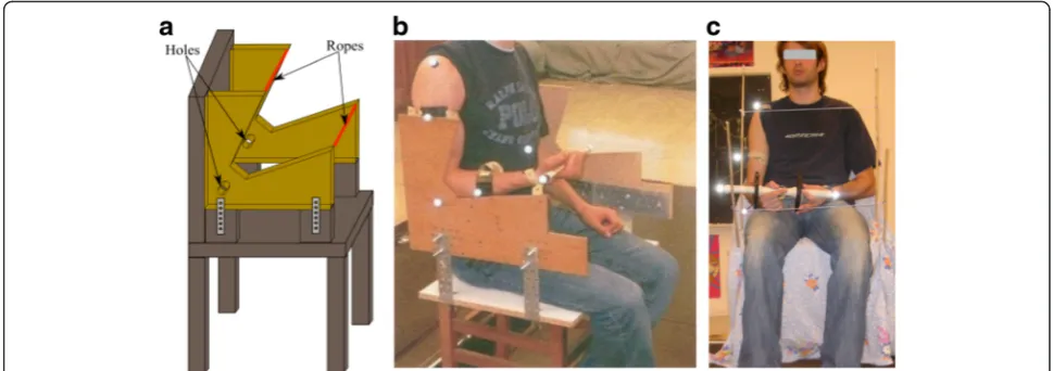
Experimental set-up
dumbbell. The displacement of the markers was filmed by six infrared cameras (Elite-BTS, Milano, Italy) ca-denced at 100 Hz.
Participant instructions
During experimentation, the participant sat on the chair. The participants were asked to perform 10 cycles of flexion-extension, following the rhythm of a given metronome, with and without dumbbells. Participants had to keep the shoulder and elbow joint as motionless as possible and the dumbbell axis horizontal. Partici-pants were involved a few minutes with the dumbbells, before beginning the experiments.
The participants had to perform ten elbow flexion-extension movements with five different masses: 0, 1, 2, 3 and 4 kg, and at three motion frequencies, 0.5 Hz (i.e. a cycle in 2 seconds), 0.33 Hz (1 cycle in 3 seconds) and 0.25 Hz (1 cycle in 4 seconds). The order of the masses and frequencies was drawn ran-domly by the operator. Each male participant per-formed the whole experimental protocol twice in order to assess test and retest reproducibility of the joint torques. The retests were performed approxi-mately 20 min after the tests, without removing the kinematic sensor.
Joint torque quantification process
Using the measurements of kinematic sensors, a 3D multibody model of the human body [17] provides the elbow joint torques via these three consecutive steps:
1. The full model joint kinematics: the system is modeled as a constrained multibody system, using kinematic loops.
2. The joint kinematic identification: the joint coordinatesq, velocities q and accelerations q are numerically determined by an optimization process that estimates the joint coordinates of the multibody model that best fit the
experimental joint positions.
3. The inverse dynamics: using recursive Newton-Euler formalism, a 3D multibody model [17] provides the vectorQinvof joint forces and torques during movement as follows:
Qinv¼f qð ; :q; €q;Fext;Mext;gÞ ð1Þ
wherefis a function of the kinematicsq, q̇, q and
repre-sents the inverse dynamical model of the human body,
on the basis of the external forcesFextand torquesMext
applied to the system, and also gravityg. The inertia
pa-rameters of the body segments have been defined using
the Table from de Leva [39].
These equations were symbolically generated by the ROBOTRAN software [40], UCL, which allows us to straightforwardly interface these equations with any numerical process, such as the optimization process presented above and the time simulation of the trials.
Statistical analysis
ment between tests and retest trial [42]. This method (Bland & Altman, 1986) was extensively used in dif-ferent research fields in test-retest studies [43–46] and is suitable in the case of the present study [41]. A corrected standard deviation of differences for repeated measurements, SDcorrected=√(2●SD
2
), was used based on Bland and Altman (1986) [42]. Statistical analysis was per-formed using SPSS 17.0 (IBM, Chicago, USA).
Results
In each condition, the peak torque values were normally distributed (Kolmogorov–Smirnov test,p> 0.05).
Intra-test variability
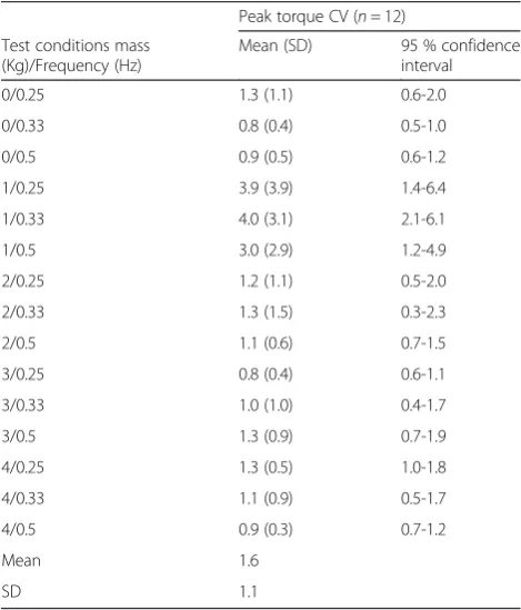
Whatever the test conditions, the variation coefficient of the peak torque ranged between 0.8 and 4 % (see Table 1).
retest difference (See Bland and Altman plots, Fig. 2, right panel).
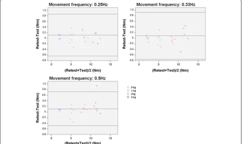
Whatever the mass condition, with a frequency of 0.25, 0.33, and 0.5 Hz, the limits of agreement values were -0.64 Nm to 0.86 Nm, -0.75 Nm to 0.92 Nm, and -0.49 Nm to 0.72 Nm, which represent a variation of 9.1, 9.9, and 7.2 % of the averaged peak torques (8.3 Nm, 8.4 Nm, and 8.3 Nm) around the mean test-retest differ-ences, respectively (see Fig. 3).
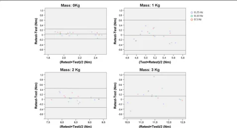
Whatever the frequency condition, with a mass of 0, 1, 2, and 3 kg, the limits of agreement values were -0.16 Nm to 0.24 Nm, -0.64 Nm to 0.60 Nm, -0.34 Nm to 0.44 Nm, -0.74 Nm to 0.99 Nm, which represent a variation of 9.3, 12, 4.6, and 7.6 % of the averaged peak torques torques (2.2 Nm, 5.2 Nm, 8.5 Nm, and 11.4 Nm) around the mean test-retest differences, respectively (see Fig. 4).
Discussion
This study showed that the data processing and the ex-perimental procedure implemented in the present study resulted in a low within-trial variability, i.e. a low vari-ability inside each trial, and a good within-participant test-retest repeatability, i.e. a good repeatability between tests of the same participant, of the elbow peak torque in typically developing young adults. As shown by the limit of agreements, expressed as a percentage of the av-eraged peak torque, the result repeatability was equiva-lent whatever the frequency, amongst 0.25, 0.33, and 0.5 Hz, or the load, amongst 0 1, 2, and 3 kg, imposed during the movement.
This study highlighted that the 4 kg resulted in a more important variability compared to the lower masses. Based on this observation, it can be assumed that in-creasing the mass higher than 4 kg would result in a more important variability that would not be appropri-ated to evaluate the joint torques. On the contrary, using lower masses, such as 0 kg, are recommended for the good repeatability, and certainly do not imply fatigue, es-pecially in female participants.
In summary, to evaluate muscle efforts in the rehabili-tation field, the repeatability of the model at low fre-quencies and with light loads was a key result. In
Table 1Peak torque coefficients of variation within trial
Peak torque CV (n= 12)
Test conditions mass (Kg)/Frequency (Hz)
Mean (SD) 95 % confidence
interval
0/0.25 1.3 (1.1) 0.6-2.0
0/0.33 0.8 (0.4) 0.5-1.0
0/0.5 0.9 (0.5) 0.6-1.2
1/0.25 3.9 (3.9) 1.4-6.4
1/0.33 4.0 (3.1) 2.1-6.1
1/0.5 3.0 (2.9) 1.2-4.9
2/0.25 1.2 (1.1) 0.5-2.0
2/0.33 1.3 (1.5) 0.3-2.3
2/0.5 1.1 (0.6) 0.7-1.5
3/0.25 0.8 (0.4) 0.6-1.1
3/0.33 1.0 (1.0) 0.4-1.7
3/0.5 1.3 (0.9) 0.7-1.9
4/0.25 1.3 (0.5) 1.0-1.8
4/0.33 1.1 (0.9) 0.5-1.7
4/0.5 0.9 (0.3) 0.7-1.2
Mean 1.6
SD 1.1
patients with neurological disorder, muscular strength and movement velocity is potentially very low depending on their functional capacity. As supported by the Bland and Altman analysis (Fig. 3), at low frequency (0.25Hz) the limit of agreement represented 9.1 % of the averaged peak torque, and considering the condition without dumbbells, the limit of agreement represented 9.3 % of the averaged peak torque. Even if the literature still has
no consensus on the clinically important difference in elbow torque for humans, because this torque relates to each joint and each motion, Laitenberger et al. (2015) [47] reported an elbow torque variability up to 24 % in healthy subjects, which confirms that the obtained re-peatability of 8.5 % when all test conditions are viewed together (Fig. 2) is relevant compared to the magnitude of this measurement. As described earlier, kinematic Fig 2Bland and Altman plot for peak torque repeatability. Legend: Bland and Altman plot of the difference between test and retest peak torque values. The left panel illustrates that the homoscedasticity assumption would be violated if the 4 kg condition were included in the analysis (a correlation exists,p< 0.05). The right panel illustrates that the homoscedasticity assumption is met (no correlation exists,p> 0.05) if the 4 kg condition is dropped
data processing, marker misallocation and skin move-ment could greatly influence joint centre localisation [28, 29] and in turn greatly impact inverse dynamic solu-tion repeatability [25]. Riemer et al. found that these various inaccuracies can result in uncertainties of esti-mated joint torques ranging from 6 % to 232 % of the peak torque during gait. The methodology used in the present study in terms of kinematic data processing, based on solidification procedure [34], was adequate to result in a good within-participant test-retest repeatabil-ity. At the same time, these results showed that the de-vice used (Fig. 4) was adequate to obtain a repeatable elbow flexion-extension maximal torque.
Several limits were inherent with this study. First the repeatability of the data processing and the experimental procedure was tested with a limited number of partici-pants. Nevertheless, many conditions were tested (fre-quency*mass), resulting in a test-retest repeatability analysis based on 90 trials. The test-retest repeatability analysis was performed only in male because fatigue could be more present in female compared to male par-ticipants. Secondly, the present study included healthy participants, the repeatability of the data processing and the experimental procedure implemented in the present study should be tested for each targeted disease. Thirdly, the repeatability of the model has been tested without removing the maker. A Further study is required to test the reproducibility with markers replacement because
configurations, is proposed as a satisfying method to es-timate the joint efforts in dynamical context. This prob-lem being deterministic, Qinv becomes a sufficiently
accurate result that can be exploited as a reference for the optimization process that attempts to solve the muscle force redundancy. These results represent the first step leading to the development of an accurate as-sessment of elbow muscle strength in clinical environ-ment. The ability of giving accurate elbow joint net torques during motion, without requiring an important computational cost, is the main benefit of this method. Based on these results, multibody model refinement and clinical analysis will be implemented in further studies.
Conclusion
The aim of this study was to assess the peak torque elbow variability and repeatability. Whatever the flexion-extension movement conditions imposed, within-trial peak torque variability was low and within-participant test-retest repeatability of the elbow joint torques re-sulted in good agreement. This method is promising for potential clinical applications and can be used as a basis for further comparison between efforts quantification methods or refined multibody models in the human body during motion.
Availability of data and materials
The authors’ Research Ethics Board did not allow to publicly share the data and materials of this study. How-ever, these data and materials will be available upon re-quest to the corresponding author and in accordance with the Research Ethics Board.
Competing interests
The authors declare that they have no competing interests.
Authors’contributions
LB has made substantial contributions to the analysis and interpretation of data, and has been involved in drafting the manuscript. MR has made substantial contributions to the study design, the acquisition of data, the analysis and interpretation of data, and has given final approval of the version to be published. CD has made substantial contributions to the study design, and has made substantial contributions to the analysis and interpretation of data. GG has made substantial contributions to the acquisition of data, the analysis and interpretation of data. ML has made substantial contributions to the analysis and interpretation of data, and revising the manuscript critically for important intellectual content. All authors read and approved the final manuscript.
Acknowledgments
The authors wish to thank MÉDITIS program supported by FONCER-CSNG for financial support.
Author details
1
Department of kinanthropology, Université du Québec à Montréal, Montreal, Qc, Canada.2Department of mechanical engineering, École
Polytechnique de Montréal, Montreal, Qc, Canada.3Research & Engineering
Chair Applied to Pediatrics (RECAP), Marie Enfant Rehabilitation Centre (CRME)–Research Center–Sainte-Justine UHC, and École Polytechnique de Montréal, Montreal, Qc, Canada.4Institute of NeuroSciences (IoNS), Université
catholique de Louvain, Bruxelles, Belgium.5CRME–Research Center, Office
GR-123, 5200, East Bélanger Street, H1T 1C9 Montréal, QC, Canada.
Received: 26 April 2015 Accepted: 1 April 2016
References
1. Damiano DL, Abel MF, Pannunzio M, Romano JP. Interrelationships of strength and gait before and after hamstrings lengthening. J Pediatr Orthop. 1999;19:352–8.
2. Hinderer KA, Hinderer SR. Muscle strength development and assessment in children and adolescents, in: H.-R. K. (Ed.), Muscle strength. Edinburgh: Churchill Livingstone 1993;p.93-140
3. Schache AG, Fregly BJ, Crossley KM, Hinman RS, Pandy MG. The effect of gait modification on the external knee adduction moment is reference frame dependent. Clin Biomech (Bristol, Avon). 2008;23(5):601–8.
4. Schwachmeyer V, Damm P, Bender A, Dymke J, Graichen F, Bergmann G. In vivo hip joint loading during post-operative physiotherapeutic exercises. PLoS One. 2013;8(10), e77807.
5. D'Amico M, D'Amico G, Paniccia M, Roncoletta P, Vallasciani M. An integrated procedure for spine and full skeleton multi-sensor biomechanical analysis & averaging in posture gait and cyclic movement tasks. Stud Health Technol Inform. 2010;158:118–26.
6. Raison M, Ballaz L, Detrembleur C, Mahaudens P, Lebleu J, Fisette P, Mousny M. Lombo-sacral joint efforts during gait: comparison between healthy and scoliotic subjects. Stud Health Technol Inform. 2012;176:113–6.
7. Tirrell TF, Franko OI, Bhola S, Hentzen ER, Abrams RA, Lieber RL. Functional consequence of distal brachioradialis tendon release: a biomechanical study. J Hand Surg Am. 2013;38(5):920–6.
8. Cheng HS, Ju MS, Lin CC. Improving elbow torque output of stroke patients with assistive torque controlled by EMG signals. J Biomech Eng. 2003;125(6):881–6.
9. Florence JM, Pandya S, King WM, Robison JD, Baty J, Miller JP, Schierbecker J, Signore LC. Intrarater reliability of manual muscle test (Medical Research Council scale) grades in Duchenne's muscular dystrophy. Phys Ther. 1992;72:115–22.
10. Cuthbert SC, Goodheart Jr GJ. On the reliability and validity of manual muscle testing: a literature review. Chiropr Osteopat. 2007;15:4. 11. Stark T, Walker B, Phillips JK, Fejer R, Beck R. Hand-held dynamometry
correlation with the gold standard isokinetic dynamometry: a systematic review. PM R. 2011;3:472–9.
12. Bessonet G, Sardain P, Chéssé S. Optimal motion synthesis–dynamic modeling and numerical solving aspects. Multibody System Dynamics. 2002;8:257–78.
13. Hoy MG, Zajac FE, Gordon ME. A musculoskeletal model of the human lower extremity: the effect of muscle, tendon, and moment arm on the moment-angle relationship of musculotendon actuators at the hip, knee, and ankle. J Biomech. 1990;23(2):157–69.
14. Massie CL, Fritz S, Malcolm MP. Elbow extension predicts motor impairment and performance after stroke. Rehabil Res Pract. 2011;381978.
15. Amarantini D, Martin L. A method to combine numerical optimization and EMG data for the estimation of joint moments under dynamic conditions. J Biomech. 2004;37:1393–404.
16. De Groote F, Pipeleers G, Jonkers I, Demeulenaere B, Patten C, Swevers J, De Schutter J. A physiology based inverse dynamic analysis of human gait: potential and perspectives. Comput Methods Biomech Biomed Engin. 2009;12:563–74.
17. Raison M, Detrembleur C, Fisette P, Samin JC. Assessment of antagonistic muscle forces during forearm flexion/extension. Comput Methods Appl Sci. 2011;23:215–38.
18. Gilliaux M, Lejeune T, Detrembleur C, Sapin J, Dehez B, Stoquart G. A robotic device as a sensitive quantitative tool to assess upper limb impairments in stroke patients: a preliminary prospective cohort study. J Rehabil Med. 2012;44(3):210–7.
19. Zampagni ML, Casino D, Zaffagnini S, Visani A, Marcacci M. Trend of the carrying angle during flexion-extension of the elbow joint: a pilot study. Orthopedics. 2008;31(1):76.
20. Lin JH, McGorry RW, Banks JJ. Exposures and physiological responses in power tool operations: fastening vs. unfastening threaded hardware. J Occup Environ Hyg. 2010;7(5):290–7.
ed. Hoboken, New-Jersey: John Wiley & Sons, Inc; 2005. p. 720. 28. Kuo YL, Tully EA, Galea MP. Skin movement errors in measurement of
sagittal lumbar and hip angles in young and elderly subjects. Gait Posture. 2008;27:264–70.
29. Stagni R, Leardini A, Cappozzo A, Grazia Benedetti M, Cappello A. Effects of hip joint centre mislocation on gait analysis results. J Biomech. 2000;33:1479–87.
30. Rao G, Amarantini D, Berton E, Favier D. Influence of body segments' parameters estimation models on inverse dynamics solutions during gait. J Biomech. 2006;39:1531–6.
31. Cahouet V, Luc M, David A. Static optimal estimation of joint accelerations for inverse dynamics problem solution. J Biomech. 2002;35:1507–13. 32. Cappozzo A, Leo T, Pedotti A. A general computing method for the analysis
of human locomotion. J Biomech. 1975;8:307–20.
33. Challis JH, Kerwin DG. Quantification of the uncertainties in resultant joint moments computed in a dynamic activity. J Sports Sci. 1996;14:219–31. 34. Cheze L, Fregly BJ, Dimnet J. A solidification procedure to facilitate
kinematic analyses based on video system data. J Biomech. 1995;28:879–84. 35. Cappozzo A, Catani F, Leardini A, Benedetti MG, Croce UD. Position and
orientation in space of bones during movement: experimental artefacts. Clin Biomech (Bristol, Avon). 1996;11:90–100.
36. Chiari L, Della Croce U, Leardini A, Cappozzo A. Human movement analysis using stereophotogrammetry. Part 2: instrumental errors. Gait Posture. 2005;21:197–211.
37. Lu TW, O'Connor JJ. Bone position estimation from skin marker co-ordinates using global optimisation with joint constraints. J Biomech. 1999;32:129–34. 38. Kim K, Song WK, Lee J, Lee HY, Park DS, Ko BW, Kim J. Kinematic analysis of
upper extremity movement during drinking in hemiplegic subjects. Clin Biomech (Bristol, Avon). 2014;29(3):248–56.
39. de Leva P. Adjustments to Zatsiorsky-Seluyanov's segment inertia parameters. J Biomech. 1996;29(9):1223–30.
40. Samin JC, Fisette P. Symbolic modeling of multibody systems. Netherlands: Kluwer Academic Publishers; 2003. p. 479.
41. Atkinson G, Nevill AM. Statistical methods for assessing measurement error (reliability) in variables relevant to sports medicine. Sports Med. 1998;26(4):217–38.
42. Bland JM, Altman DG. Statistical methods for assessing agreement between two methods of clinical measurement. Lancet. 1986;1:307–10.
43. Høyer E, Opheim A, Strand LI, Moe-Nilssen R. Temporal and spatial gait parameters in patients dependent on walking assistance after stroke: reliability and agreement between simple and advanced methods of assessment. Gait Posture. 2014;40(1):101–6.
44. Thompson P, Beath T, Bell J, Jacobson G, Phair T, Salbach NM, Wright FV. Test-retest reliability of the 10-metre fast walk test and 6-minute walk test in ambulatory school-aged children withcerebral palsy. Dev Med Child Neurol. 2008;50(5):370–6.
45. Birmingham TB, Hunt MA, Jones IC, Jenkyn TR, Giffin JR. Test-retest reliability of the peak knee adduction moment during walking in patients with medial compartment knee osteoarthritis. Arthritis Rheum. 2007;57(6):1012–7. 46. de Zwart BC, Frings-Dresen MH, van Duivenbooden JC. Test-retest reliability of the Work Ability Index questionnaire. Occup Med (Lond). 2002;52(4):177–81. 47. Laitenberger M, Raison M, Périé D, Begon M. Refinement of the upper limb joint kinematics and dynamics using a subject-specific closed-loop forearm model. Multibody System Dynamics. 2015;33(4):413–38.
48. Kollmitzer J, Ebenbichler GR, Kopf A. Reliability of surface electromyographic measurements. Clin Neurophysiol. 1999;110(4):725–34.
• We accept pre-submission inquiries
• Our selector tool helps you to find the most relevant journal • We provide round the clock customer support
• Convenient online submission • Thorough peer review
• Inclusion in PubMed and all major indexing services • Maximum visibility for your research
Submit your manuscript at www.biomedcentral.com/submit