T E C H N I C A L A D V A N C E
Open Access
Laparoscopic CBD exploration using a V-shaped
choledochotomy
Eun Young Kim, Soo Ho Lee, Jun Suh Lee and Tae Ho Hong
*Abstract
Background:Laparoscopic common bile duct exploration (LCBDE) is a treatment modality for choledocholithiasis. The advantages of this technique are that it is less invasive than conventional open surgery and it permits single-stage management; however, other technical difficulties limit its use. The aim of this article is to introduce our novel technique for LCBDE, which may overcome some of the limitations of conventional LCBDE. Since December 2013, ten patients have undergone LCBDE using a V-shaped choledochotomy (V-CBD). After the confluence of the cystic duct and the CBD were exposed, a V-shaped incision was made along the medial wall of the cystic duct and the lateral wall of the common hepatic duct, which comprise two sides of Calot’s triangle. The choledochoscope was inserted into the lumen of the CBD through a V-shaped incision, and all CBD stones were retrieved using a basket or a Fogarty balloon catheter or were irrigated with saline. After CBD clearance was confirmed using the choledochoscope, the choledochotomy was closed with the bard absorbable suture material known as V-loc.
Results: The diameter of the CBD ranged from 8 to 30 mm, and the mean size of the stones was 11.6 ± 8.4 mm. The mean operative time was 97.8 ± 30.3 min, and the mean length of the postoperative hospital stay was 6.0 ± 4.6 days. All patients recovered without any postoperative complications, except for one patient who developed postoperative pancreatitis. No conversions to laparotomy were observed, and there were no recurrent stones and no need of T-tube insertion.
Conclusions: This report suggests that our novel technique, known as V-CBD, may represent a feasible and straightforward procedure for treating choledocholithiasis, especially when the CBD is not dilated.
Keywords: Choledocholithiasis, Cholelithiasis, Laparoscopy
Background
Surgical common bile duct (CBD) exploration is one of the treatment modalities for choledocholithiasis, which is the second most common complication of cholelithia-sis, occurring in approximately 10–15 % of cholelithiasis patients [1, 2]. This approach has advantages over endo-scopic retrograde cholangiopancreatography (ERCP) with endoscopic sphincterotomy (EST), which is a widely used treatment for choledocholithiasis but carries a significant risk of complications such as acute pancreatitis, duodenal perforation, bleeding, and, importantly, iatrogenic injury to the muscles of the sphincter of Oddi [3, 4].
With advances in laparoscopic techniques and instru-ments, laparoscopic CBD exploration (LCBDE) has been
performed more frequently, and there have been many reports that laparoscopic choledocholithotomy is less invasive than open surgery [5, 6]. However, in some patients with a narrow CBD, LCBDE is associated with a high risk of postoperative CBD stricture and bile leakage due to technical difficulty. To prevent these complica-tions, surgeons have inserted T-tubes during LCBDE; however, T-tube insertion is nevertheless associated with complications, including infections that ascend through the drain, dislocation of the T-tube (which results in bile leakage), and most importantly, patient inconvenience due to prolonged T-tube placement [7]. Surgeons have proposed a variety of techniques for laparoscopic chole-docholithotomy [1, 6, 8–10], although there remains no consensus as to the best surgical treatment method.
The aim of this article is to describe our novel tech-nique for LCBDE, which we have termed ‘laparoscopic * Correspondence:[email protected]
Department of Hepatobiliary and Pancreatic Surgery, College of Medicine, Seoul St. Mary’s Hospital, The Catholic University of Korea, Seoul, Korea
CBD exploration through a V-shaped choledochotomy (V-CBD).’ This novel approach may help to overcome the limitations of conventional LCBDE for the surgical treatment of choledocholithiasis.
Methods
Since December 2013, a total of 10 patients who were diagnosed with concomitant choledocholithiasis and cholelithiasis have undergone surgery using the novel technique of V-CBD at the Department of Surgery, Seoul St. Mary’s Hospital. In patients with concomitant cholelithiasis and choledocholithiasis, the treatment paradigm at our center is to initially perform ERCP to treat the choledocholithiasis, which is then followed by laparoscopic cholecystectomy (LC). However, V-CBD has been selectively used in patients who are not candi-dates for ERCP (due to conditions such as a history of total gastrectomy, periampullary diverticulum, large and impacted stones, or unavailability of ERCP equipment or endoscopists). Preoperative diagnosis was confirmed according to clinical features, laboratory results and radio-logic tests including magnetic resonance cholangiopan-creatography or computed tomography (CT) scan. In patients with septic shock or who had findings indicating the progression of biliary sepsis (such as delirium or un-controllable fever despite antibiotic treatment), patients were diagnosed as having acute cholangitis and were initially managed with conservative treatment and resusci-tated before any intervention. If patients were felt to be surgical candidates, V-CBD was used regardless of the size or number of stones and the history of previous upper abdominal operations.
All medical data were prospectively collected, includ-ing the followinclud-ing: demographic and clinical features (age, sex, American Society of Anesthesiologists (ASA) grade, body mass index (BMI) and preoperative la-boratory results); disease characteristics (size and number of stones, diameter of the CBD and the pres-ence of gallstone pancreatitis); and surgical outcomes (CBD clearance, operative time, conversion to laparotomy, length of postoperative hospital stay, postoperative morbidity and mortality). This study was approved by the ethics committee at our institution (Institutional Review Board of Seoul St. Mary’s hospital, College of Medicine, the Catholic University of Korea, IRB code: KC14RISI0814) and all the patients provided their informed consent for the publication of this study.
Laparoscopic choledocholithotomy using a V-shaped choledochotomy
All patients were placed in the supine position under general anesthesia, and the surgeon and second assistant (who held the laparoscope) were positioned to the left side of the patient. The first assistant stood on the opposite
choledochotomy closure, the cystic duct was divided, and then standard LC was performed. The gallbladder and the extracted stones were bagged and retrieved through the umbilical trocar site. A closed suction drain was inserted through a lateral 5-mm trocar and placed in Morrison’s pouch. The drain was removed on the 2ndpostoperative day, as long as the drainage was <50 ml/day and free of bile. Patients returned to the outpatient department at the 7thday after discharge, at which time we evaluated their general condition.
Results
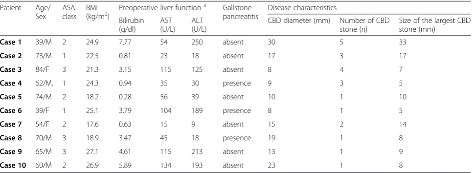
To date, the V-CBD procedure has been performed in a total of 10 patients. These patients’demographic and clin-ical features are shown in Table 1. Seven males and three females were recruited; the mean patient age in the present study was 62.0 ± 14.7 years. Two patients (cases 5 and 7) had a history of open subtotal gastrectomy; the others had
no previous surgical history. Preoperative liver function tests were obtained on the day before surgery. The bilirubin level ranged from 0.28 to 7.77 mg/dl (mean 3.13 ± 2.50 mg/ dl), and gallstone pancreatitis was present in three patients (Cases 4, 6 and 8). These patients were treated preopera-tively in a conservative manner with fluid resuscitation and nutritional support; surgery was performed when the symp-toms were relieved. In terms of disease characteristics, the diameter of the CBD ranged from 8 to 30 mm (mean 15.2 ± 7.2 mm), and the number of CBD stones ranged from 1 to 5. The size of the largest CBD stone in each case ranged from 5 to 33 mm (mean 11.6 ± 8.4 mm). In patients who had large stones (over 10 mm), as in cases 1, 2, 5 and 7, we used stone forceps to fragment and retrieve the stones; this maneuver was successfully completed without any compli-cations or open conversions.
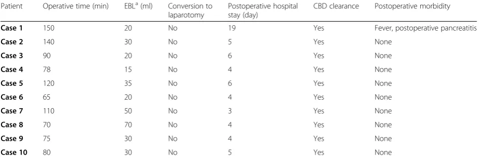
The operative findings and surgical outcomes for each case are shown in Table 2. The mean operative time was
Figure 2Confirmation of CBD clearance using the choledochoscope through a V-shaped incision.(a)Operative view;(b)Illustration
97.8 ± 30.3 min, with a range of 65 to 150 min. Case 1 had the largest and most numerous stones as well as the longest operative time, and it is likely that these charac-teristics affected the operative time. In terms of esti-mated blood loss, minimal blood loss was observed in each case (15 to 70 ml), and no intraoperative transfu-sions were required. In this study, the mean length of the postoperative hospital stay was 6.0 ± 4.6 days (range, 3 to 19 days). The longest hospital stay was 19 days (in case 1), which may have been due to the development of postoperative pancreatitis that required prolonged fast-ing and nutritional support. CBD stones were success-fully cleared in all cases. Postoperative morbidity was observed in only one patient (case 1), who developed fever with postoperative pancreatitis. This patient began an oral diet on postoperative day 12 and improved with-out any additional complications. All other patients recovered normally, and no deaths were observed in our
study. The mean follow up period was 83.0 ± 50.7 days, and no other complications were observed during follow-up.
Discussion
Although LCBDE has a crucial advantage in that it simultaneously treats cholelithiasis and choledocholithia-sis, thereby shortening hospital stays and reducing hos-pital costs, only surgeons with advanced laparoscopic skills can perform LCBDE because the procedure re-quires very specialized laparoscopic techniques and equipment. This study is the first to introduce V-CBD, a novel technique with several characteristics that may overcome the limitations of conventional LCBDE. First, the V-shaped incision more easily provides sufficient space for the introduction of the choledochoscope because the wall of the V-shaped incision includes not only the CBD but also the cystic duct, unlike existing
Figure 3The choledochotomy was closed using the bard V-loc absorbable suture material
Table 1Demographic features and clinical characteristics of patients
Patient Age/ Sex
ASA class
BMI
(kg/m2) Preoperative liver function a
Gallstone pancreatitis
Disease characteristics
Bilirubin (g/dl)
AST (U/L)
ALT (U/L)
CBD diameter (mm) Number of CBD stone (n)
Size of the largest CBD stone (mm)
Case 1 39/M 2 24.9 7.77 54 250 absent 30 5 33
Case 2 73/M 1 22.5 0.81 23 18 absent 17 3 17
Case 3 84/F 3 21.3 3.15 115 125 absent 8 4 7
Case 4 62/M, 1 24.3 0.94 35 30 presence 9 3 5
Case 5 74/M 2 18.2 0.28 56 39 absent 10 1 10
Case 6 39/F 1 25.1 3.79 104 189 presence 8 1 5
Case 7 54/F 2 17.6 0.63 15 9 absent 15 2 14
Case 8 70/M 3 18.9 3.47 45 18 presence 19 1 8
Case 9 65/M 3 27.1 4.61 115 213 absent 13 1 9
Case 10 60/M 2 26.9 5.89 134 193 absent 23 1 8
a
techniques for conventional LCBDE. Therefore, entry of the choledochoscope and stone retrieval can be per-formed without difficulty, even in patients without a dilated CBD. It is difficult to use laparoscopic techniques (especially during primary closure of the CBD) in con-ventional LCBDE for patients whose CBD is less than 1 cm, due to the difficulty of laparoscopic manipulation and concerns for postoperative ductal stricture after suturing. However, V-CBD can be used more easily in patients whose CBD diameter is less than 1 cm. Indeed, V-CBD was used in the present study for patients with small CBDs, as shown in cases 3, 4 and 6. Moreover, V-CBD does not require insertion of a T-tube and there-fore may prevent many problems related to the T-tube, such as infection, dislocation of the tube, prolonged operative times, need for a 2ndprocedure to remove the tube, and patient discomfort, which is particularly important [7, 11]. We believe that V-CBD may offer an option for one-stage management to patients who are not able to undergo surgical treatment due to difficulties in the surgical approach resulting from a narrow cystic duct or CBD.
Suturing using V-loc is one characteristic of V-CBD that may address some limitations of conventional LCBDE, such as the difficulty of laparoscopic manipula-tion. As described above, the V-loc suture has a ring structure on one end with a round needle attached to the other end. Therefore, intracorporeal tying is not needed for the first knot; simply passing the needle through the ring on the end of the thread is sufficient to complete the knot, without complicated manipulation with both hands. The barbed thread, which is another characteristic feature of the V-loc suture, may help to prevent loosening of the suture without the need for keeping continuous traction on the thread (which is usually performed by the assistant). As a result, the sur-geon can suture laparoscopically without the assistant’s
help during the V-CBD procedure, increasing the comfort of handling the laparoscopic instruments, especially given the relatively narrow field of view and small biliary struc-tures. Being able to manipulate the instruments comfort-ably during the V-CBD procedure, without unnecessary motion, may help to reduce the surgeon’s fatigue, thereby increasing the precision of the surgeon’s movements and decreasing the risk of tissue injury.
Despite the advantages of V-CBD, some precautions are needed when performing this procedure. First, although V-CBD has the advantage of being applicable in a wide range of cases, it cannot be used in several situations, including patients with anatomical variations of the cystic duct, severe angulation of the cystic duct to the left side of the CBD, or a very low-lying origin of the cystic duct (near the pancreatic duct). In addition, there may be reasonable doubt about the necessity of V-CBD if the CBD is severely dilated, as conventional LCBDE with primary closure of the choledochotomy site could be suffi-cient for the single-stage treatment of choledocholithiasis.
In the future, a larger number of cases should be stud-ied, including patients undergoing V-CBD and a control group treated with ERCP or conventional LCBDE. A pro-spective comparative study is required for an objective analysis of the results of V-CBD. Additionally, this study does not provide data about other factors, including hos-pital costs; data on cost-effectiveness should therefore also be collected and analyzed in subsequent studies.
Conclusion
In conclusion, this report suggests that our novel technique, V-CBD, may represent a feasible and straightforward pro-cedure for treating choledocholithiasis, especially when the CBD is not dilated. Nevertheless, additional well-designed, randomized, prospective, controlled trials with larger sam-ple sizes should be carried out to confirm the effectiveness of this technique.
Table 2Operative findings and postoperaive outcomes
Patient Operative time (min) EBLa(ml) Conversion to laparotomy
Postoperative hospital stay (day)
CBD clearance Postoperative morbidity
Case 1 150 20 No 19 Yes Fever, postoperative pancreatitis
Case 2 140 30 No 5 Yes None
Case 3 90 20 No 6 Yes None
Case 4 78 15 No 4 Yes None
Case 5 120 35 No 6 Yes None
Case 6 65 20 No 4 Yes None
Case 7 110 50 No 3 Yes None
Case 8 70 70 No 4 Yes None
Case 9 75 30 No 4 Yes None
Case 10 80 30 No 5 Yes None
a
Abbreviations
CBD:Common bile duct; CHD: Common hepatic duct; ERCP: Endoscopic retrograde cholangio pancreatography; EST: Endoscopic sphincterotomy; LCBDE: Laparoscopic CBD exploration.
Competing interests
The authors declare that they have no competing interests.
Authors’contributions
THH conceived and designed the study. EYK, SHL, and JSL were responsible for data collection and writing the article. EYK and THH have made major contributions to analysis and interpretation. All authors have read, edited, and approved the final manuscript.
Acknowledgements
This study did not involve any funding body.
Received: 8 February 2015 Accepted: 4 May 2015
References
1. Huang SM, Wu CW, Chau GY, Jwo SC, Lui WY, P’eng FK. An alternative approach of choledocholithotomy via laparoscopic choledochotomy. Arch Surg. 1996;131:407–11.
2. Cotton PB. Endoscopic management of bile duct stones; (apples and oranges). Gut. 1984;25:587–97.
3. Paik KY, Kim EK. Laparoscopic common bile duct exploration after unsuccessful endoscopic stone removal. J Laparoendosc Adv Surg Tech A. 2013;23:137–40.
4. Lu J, Xiong XZ, Cheng Y, Lin YX, Zhou RX, You Z, et al. One-stage versus two-stage management for concomitant gallbladder stones and common bile duct stones in patients with obstructive jaundice. Am Surg. 2013;79:1142–8.
5. Cuschieri A, Lezoche E, Morino M, Croce E, Lacy A, Toouli J, et al. E.A.E.S. multicenter prospective randomized trial comparing two-stage vs single-stage management of patients with gallstone disease and ductal calculi. Surg Endosc. 1999;13:952–7.
6. Gu AD, Li XN, Guo KX, Ma ZT. Comparative evaluation of two laparoscopic procedures for treating common bile duct stones. Cell Biochem Biophys. 2011;59:159–64.
7. Gurusamy KS, Samraj K. Primary closure versus T-tube drainage after open common bile duct exploration. Cochrane Database Syst Rev.
2007;CD005640.
8. Sinha R. Laparoscopic choledocholithotomy with rigid nephroscope. J Laparoendosc Adv Surg Tech A. 2013;23:211–5.
9. Bandyopadhyay SK, Khanna S, Sen B, Tantia O. Antegrade common bile duct (CBD) stenting after laparoscopic CBD exploration. J Minim Access Surg. 2007;3:19–25.
10. El-Geidie AA. Laparoendoscopic management of concomitant gallbladder stones and common bile duct stones: what is the best technique? Surg Laparosc Endosc Percutan Tech. 2011;21:282–7.
11. Huang SM, Yao CC, Cheng YW, Chen LY, Pan H, Hsiao KM, et al. Laparoscopic primary closure of common bile duct combined with percutaneous cholangiographic drainage for treating choledocholithiasis. Am Surg. 2010;76:517–21.
Submit your next manuscript to BioMed Central and take full advantage of:
• Convenient online submission
• Thorough peer review
• No space constraints or color figure charges
• Immediate publication on acceptance
• Inclusion in PubMed, CAS, Scopus and Google Scholar
• Research which is freely available for redistribution