SPECIAL ARTICLE
Developing Circadian Rhythmicity in Infants
Scott A. Rivkees, MD
ABSTRACT. Circadian rhythms are endogenously gen-erated rhythms with a period length of approximately 24 hours. Evidence gathered during the past decade indi-cates that the circadian timing system develops prena-tally and the suprachiasmatic nuclei, the site of a circa-dian clock, is present by midgestation in primates. Recent evidence also shows that the circadian system of primate infants is responsive to light at very premature stages and that low-intensity lighting can regulate the developing clock. After birth, there is progressive matu-ration of the circadian system outputs, with pronounced rhythms in sleep-wake and hormone secretion generally developing after 2 months of age. Showing the impor-tance of photic regulation of circadian phase in infants, exposure of premature infants to low-intensity cycled lighting results in the early establishment of rest-activity patterns that are in phase with the 24-hour light-dark cycle. With the continued elucidation of circadian system development and influences on human physiology and illness, it is anticipated that consideration of circadian biology will become an increasingly important compo-nent of neonatal care.Pediatrics2003;112:373–381; circa-dian rhythms, infant, human, baboon, suprachiasmatic nuclei.
ABBREVIATIONS. SCN, suprachiasmatic nuclei; RHT, retinohy-pothalamic tract.
THE CIRCADIAN TIMING SYSTEM
C
ircadian rhythms are endogenously drivenrhythms with a period length of approxi-mately 24 hours.1Notable examples of circa-dian rhythms include the sleep-wake cycle and daily rhythms in hormone production. Circadian rhythms are also involved in the pathogenesis of illnesses, such as reactive airway disease and myocardial in-farction.2,3
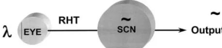
The system responsible for the generation and reg-ulation of circadian rhythms is the circadian timing system. This neural system consists of a biological clock, input pathways, and output pathways1(Fig 1). The paired suprachiasmatic nuclei (SCN) in the an-terior hypothalamus are the site of a biological clock. The SCN are located above the optic chiasm at the base of the third ventricle4(Fig 2). The SCN exhibit
endogenous rhythmicity and have a period of oscil-lation close to 24 hours.
Lesion studies in rodents provided the initial evi-dence that the SCN are the site of a circadian pace-maker.4 In vivo and in vitro studies have since shown day-night rhythms in electrical activity, met-abolic activity, and gene expression. Transplantation of fetal SCN cells into SCN-lesioned animals restores rhythmicity to the recipient, further supporting that the SCN contain a biological clock.4Circadian oscil-lations have been seen in individual rodent SCN cells, and expressed rhythmicity reflects the collec-tive oscillations of many SCN cells.5,6
Evidence gathered during the past several years has yielded new insights into the molecular mecha-nisms resulting in clock oscillations.1,7,8 Twenty-four-hour rhythmicity seems to result from rhythmic expression of transcriptional activating factors, in-cludingper,clock,andtim,which form long feedback loops in SCN cells. Cryptochromes (cry) also play a critical role in regulating clock function by interact-ing withper.1,7,8
Because SCN oscillations are not exactly 24 hours, it is necessary to reset the circadian pacemaker each day to prevent endogenous clock oscillations from drifting (or free-running) out of phase with the ex-ternal light-dark cycle. Input pathways relay photic information from the retina to the SCN to synchro-nize (or entrain) the oscillations of the clock to the 24-hour light-dark cycle.9A direct pathway from the retina to the SCN, the retinohypothalamic tract (RHT; Figs 1 and 3), has been shown to be both necessary and sufficient for photic entrainment.9The raphe nucleus also influences SCN function via se-rotonergic projections.9
Glutamate is the neurotransmitter that mediates RHT action, and the phase shifting actions of gluta-mate involve the nitric oxide system.10Another path-way from the intergeniculate leaflet to the SCN mod-ulates clock entrainment and involves the release of neuropeptide Y.9
Recent evidence suggests that the products of in-termediate-early genes, which include the DNA-binding proteins c-Fos and Jun-B, may mediate en-trainment.11Light exposure at times that reset (phase shift) the clock induces increases in SCN metabolic activity and robust expression of c-Fos and Jun-B in the SCN.11
Output pathways are responsible for the overt ex-pression of circadian rhythms. Several discrete neu-ral pathways projecting from the SCN to seveneu-ral
From the Yale Child Health Research Center, Department of Pediatrics, Yale University School of Medicine, New Haven, Connecticut.
Received for publication Feb 12, 2003; accepted Feb 12, 2003.
Reprint requests to (S.A.R.) Yale Pediatrics, Yale Child Health Research Center, Box 208081, 333 Cedar St, New Haven, CT 06510. E-mail: [email protected]
hypothalamic and nonhypothalamic sites have been defined.11–13 Via these pathways, the circadian sys-tem acts to influence neural physiology broadly. Output pathways of the circadian system also regu-late the rhythmic production of several hormones, including melatonin and cortisol.2,3,11–13
THE PRIMATE CIRCADIAN TIMING SYSTEM Several lines of evidence support that the paired SCN are the site of a biological clock in primates. Similar to rodents, the primate SCN are located above the optic chiasm at the base of the third ven-tricle.14In contrast to rodents, human SCN cells are not densely clustered, making the nuclei less visually apparent.14 –16 However, with the use of probes for melatonin receptors and SCN peptides, the human SCN can be identified.14 –16With the use of 2-deoxy-glucose, day-night oscillations in SCN metabolic ac-tivity have been detected in squirrel monkeys and baboons.17–19
Lesion studies performed in the early 1980s sug-gested the presence of a circadian pacemaker outside the SCN in monkeys.20 However, analysis of these reports revealed that either the completeness of the lesions was not verified or monkeys were not studied
in constant conditions.21Reexamination of this issue challenges the existence of primate circadian pace-makers outside the SCN. Squirrel monkeys with total SCN lesions show a complete absence of circadian rhythmicity when animals are monitored in constant conditions.21Supporting that the SCN are the site of a circadian pacemaker in humans, tumors and con-genital lesions in the SCN region result in the loss of temperature rhythms and organized sleep-wake pat-terns.22,23
The RHT has been anatomically characterized in prosimian (lemurs and shrews) and simian (squirrel monkeys, rhesus macaques, baboons, chimpanzees, and apes) species.14 This tract also has been identi-fied in studies of postmortem human specimens us-ing techniques that label degeneratus-ing retinal ax-ons.24,25 Although it was suggested that cutaneous light exposure can influence circadian function,26 there is little support for the notion that there is extraretinal photoreception in mammals.27–29 Fur-thermore, other investigators have failed to repro-duce phase-shifting effects of cutaneous light expo-sure.17
Outputs of the primate circadian system have been widely characterized in human clinical studies. Many day-night rhythms have been document-ed.2,3,28Several of these rhythms have been shown to persist in constant conditions, indicating that they are true endogenously generated circadian rhythms. Notable examples of circadian rhythms include the sleep-wake cycle, daily rhythms in body temper-ature, and day-night rhythms in cortisol and mela-tonin production.2,3,28 Day-night differences in gonadotropin, testosterone, growth hormone, and thyrotropin secretion are also present.30,31
Fig 1. Schematic representation of the circadian timing system.
DEVELOPMENT OF THE PRIMATE SCN Although rodent studies have led to our under-standing of developmental circadian physiolo-gy,30 –33notable differences between rodents and pri-mates have prevented the extension of rodent data to clinical care. In general, rodents are more immature at birth than humans. Differences in the sensitivity to light and other aspects of circadian physiology be-tween humans and rodents also have been observed. However, on the basis of evidence gathered during the past decade, it seems that the circadian clock in the SCN forms and begins oscillating in utero in primates.
In squirrel monkeys, SCN neurogenesis occurs
early in gestation during days 27 to 48.34 Because monkey and human embryonic development is very similar during the first 100 days of gestation,35 it therefore is likely that the human SCN neurons form early in gestation.
It is not known when the primate SCN are first apparent morphologically, yet with the use of [125I]melatonin and [125I]SKF38393 to label the nuclei, the human SCN have been detected at gestation week 1836,37(Fig 4).
Functional studies suggest that the primate SCN oscillate prenatally. Studies of squirrel monkeys re-veal day-night differences in SCN metabolic activity at the end of gestation.38 It is not known whether
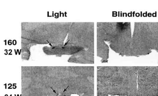
Fig 3. Innervation of the SCN by the RHT in a newborn baboon infant. A, Low-power image showing labeling of retinal fibers in the optic chiasm by horseradish peroxidase. B, Adjacent tis-sue section showing the location of the SCN. C, High-power image showing projections of the RHT into the right SCN. D, Autoradiographic image gener-ated from [14C]2-deoxyglucose uptake
studies showing that light exposure at night induces increases in SCN meta-bolic activity. Areas of increased uptake are dark. Arrows identify the SCN. Scale bar⫽5 mm. Reproduced with permis-sion from Rivkees et al.19
Fig 4. A, Localization of [125I]melatonin binding to
the SCN of an 18-week gestation human fetus. Specific labeling is shown in black. B, The stained section used to generate the autoradiograph in A. Reproduced with permission from Reppert et al.36 C, Localization of
[125I]SKF38393 binding to D1 dopamine receptors in
the SCN of a 20-week postconceptional human infant. Specific labeling is shown in black. D, Nonspecific labeling. Reproduced with permission from Rivkees et al.37OC, optic chiasm; ST, striatum. Arrows identify
SCN oscillations are present at earlier ages. The physiologic processes influenced by the fetal clock have yet to be elucidated in primates.
Similar to rodents, the timing of the onset of labor and birth in humans is influenced by the circadian cycle with peak incidences between midnight and
the early morning.39 However, we do not know
whether the fetal clock plays a role in the circadian gating of birth in humans.
Immunocytochemistry studies show that SCN maturation continues after birth.40The SCN contain distinct populations of neurons that express arginine vasopressin or vasoactive intestinal polypeptide.40In term infants, the number of vasopressinergic neu-rons is 20% of the number present in adults.40 It is not until 1 year of age that infants and adults have
comparable vasopressin neuron numbers.40 The
number of vasoactive intestinal polypeptide-contain-ing SCN cells also increase after birth.40
DEVELOPMENT OF PRIMATE PHOTIC ENTRAINMENT
A critical issue in knowing whether environmental cycles need to be considered in the care of infants is knowing when the primate circadian system be-comes functionally responsive to light. The RHT has been identified in a 36-week-gestation human new-born.41 However, because of human study limita-tions, it has not been possible to determine whether the circadian clock of human infants is functionally responsive to light at birth.
Noninvasive methods used to examine regional changes in brain activity, such as function magnetic resonance imaging or positron emission tomogra-phy, hold promise in being able to examine SCN function directly. In human adults, we have been able to observe acute increases in SCN metabolic activity after light exposure at night using 18 F-2-deoxyglucose in positron emission tomography studies.42However, because of the small size of the SCN, consistent visualization of SCN activity is dif-ficult to achieve and these methods have not been applied to infants.
Because of human study limitations, we have stud-ied baboons, which are excellent models for human infants, to provide insights into the developing hu-man clock. By monitoring changes in SCN metabolic
activity and gene expression (Fig 3), light responsive-ness can be demonstrated at birth in term baboon infants.19The presence of the RHT can also be dem-onstrated (Fig 3).19
By monitoring the effects of different lighting con-ditions on newborn baboon activity patterns, we have been able to show that newborn baboons are entrained by low-intensity (200 lux) lighting.19These findings are similar to those seen in human adults showing that circadian phase can be regulated by low-intensity (ca. 180 lux) lighting.43,44 Thus, it is likely that low-intensity lighting, similar to that found indoors, can regulate the developing primate clock.
To determine when photic responsiveness first oc-curs in primates, we have also studied premature baboon infants.45 To our surprise, we find that the SCN are functionally innervated by the retina at stages equivalent to human infants at 25 weeks postconception45(Fig 5). The primate circadian sys-tem therefore is sensitive to light in very premature infants when postnatal survival with intensive sup-port becomes possible.
DEVELOPMENT OF EXPRESSED RHYTHMICITY IN PRIMATES
The development of expressed rhythmicity has re-ceived attention in both human and nonhuman pri-mates. During pregnancy, day-night rhythms are ob-served for a variety of hormones (esterone and progesterone) and physiologic parameters (uterine contractility) in mothers.46,47 In human fetuses, day-night rhythms in heart rate, respiratory rate, and adrenal steroidogenesis have been detected.46,47 However, these rhythms seem to be driven by the mother.
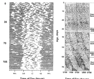
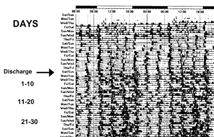
When term human infants are examined, day-night rhythms are difficult to detect in the neonatal period.42,48 –50 Consolidated periods of activity and rest generally are not observed until after the first or second month of life. Activity plots of human new-borns reveal that sleep is generally distributed over the 24-hour day during the first few weeks of life (Fig 6). At 6 weeks of age, infants are awake more during the daytime than at night. By 12 weeks of age, day-time sleep duration decreases further and much more sleep occurs at night. Importantly, although
consolidated periods of rest and activity are not ap-parent until ⬎1 to 2 months after birth, day-night differences in activity can be detected as early as 1 week of age in some infants.
At the age when day-night differences in infant activity become clearly apparent, day-night rhythms in hormone production are observed. Day-night rhythms in melatonin production can be detected at 12 weeks of age.51,52 Circadian variation in cortisol levels appears between 3 and 6 months of age.53–55 With advancing age, circadian rhythms have been detected for a variety of other hormones and circu-lating factors.56
Because infant care influences activity patterns, it is possible that patterns of developing circadian rhythmicity in human infants reflects influences of caregivers rather than endogenous rhythmicity. Thus, to characterize the development of expressed rhythmicity in primates, we have examined the de-velopment of expressed rhythmicity in newborn ba-boons raised in constant conditions (continuous dim lighting, evenly spaced care).19 Similar to human infants, baboon infants do not manifest clear day-night differences in activity patterns in the early neonatal period (Fig 6), yet at 1 month of age, day-night differences in activity patterns are observed. Developing primate rhythmicity thus reflects matu-ration from a state of relative arrhythmicity to rhyth-micity during the first few months of life.
As in rodents, it seems that infant circadian phase is synchronous with that of the mother in baboon and human infants. However, in some humans and baboons,19,57 infant phase may be out of synchrony with that of the mother at birth. Thus, whereas there is mother-infant synchrony of circadian phase in most primates, it may not be universal.
RHYTHMICITY IN PRETERM INFANTS The large number of premature infants who are hospitalized for extended periods has greatly facili-tated studies of rhythmicity in preterm infants. Dur-ing the past decade, studies of patterns of infant activity, heart rate, temperature, and sleep state sur-prisingly have not flourished.58Several of these stud-ies have revealed the presence of ultradian rhythms (rhythms with period lengths of much less than 24 hours). Endogenously driven circadian rhythms, however, are not clearly apparent.
When temperature and heart rate are studied be-ginning at a postconceptional age of 24 to 29 weeks, circadian rhythmicity is generally not apparent even at 17 weeks after birth.59Studies of preterm infants at 32 weeks’ postconceptional age have failed to detect day-night differences in sleep patterns, whereas some differences are noted in term infants.60 Analy-sis of temperature, heart rate, and activity patterns at 35 weeks’ postconceptional age has revealed ultra-dian rhythms but no clear-cut circaultra-dian rhythms.60 – 62 Because feeding and physical contact influence in-fant temperature, heart rates, and activity patterns, it is likely that infant care schedules drive the ultradian rhythms seen in preterm infants. These interventions may also mask the detection of circadian rhythms.
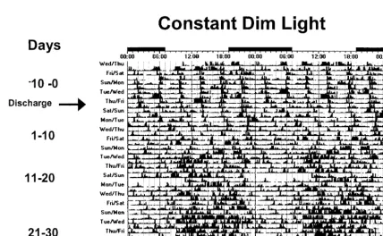
YALE NEONATAL ENTRAINMENT STUDY After the discovery that the primate circadian clock is responsive to light in very premature infants, we next assessed the effects of photic entrainment on premature infants.63 In these studies, the develop-ment of rest-activity patterns was examined in hu-man preterm infants who were exposed to continu-ous dim lighting or low-intensity cycled lighting before discharge from hospital to home.
In general, day-night differences in rest and activ-ity are not apparent in hospitalized control infants (Fig 7), whereas day-night differences in rest and activity are seen in experimental infants (Fig 8). Dur-ing the first 10 days at home, distinct day-night dif-ferences in activity are not seen in control infants, but experimental infants are more active during the day than at night. It was not until 21 to 30 days after discharge that day-night activity ratios in control infants matched those seen in experimental infants shortly after discharge, yet even at this age, experi-mental infants are considerably more active during the day as compared with control infants. Despite the differences in rest-activity patterns among groups, no differences in weight gain or change in head circumference are seen.
These observations show that exposure to low-intensity cycled lighting for 10 days before discharge induces distinct patterns of rest/activity in preterm infants that are in synchrony with the light-dark cycle that they will encounter at home. These effects are even more pronounced as soon as the child is discharged to home. In contrast, the appearance of rest-activity patterns in synchrony with the solar light-dark cycle is delayed in infants who have been exposed to continuous dim lighting in the hospital.
Potential influences of cycled lighting on prema-ture infants have been the subject of a few previous studies. In the Stanford Cycled Lighting Study, dif-ferences in circadian rhythms in temperature were not detected among infants who were exposed either to continuous dim lighting or to cycled lighting be-fore discharge.61,62,64 These infants were studied 1 and 3 months after discharge. Because we observed that infants in both groups manifest similar circadian
phase by 30 days of age, treatment effects on the rhythm of core body temperature may no longer be distinct after 1 month of age.
Other investigators have suggested that exposing infants to light-dark cycles improves infant weight gain. Mann et al65found that exposure to light-dark cycles before discharge resulted in better weight gain and more sleep during the 24-hour day than did chaotic lighting patterns. These effects were seen 6 weeks after discharge and not sooner.65 Because of this lag period, it has been suggested that the ob-served effects were not a direct result of cycled light-ing exposure on the infant.64 More recently, it has been suggested that exposing infants to light-dark cycles improves the in-hospital growth of infants if exposure occurs before 36 weeks of age,66 yet the infants in the near-darkness group in this study seemed more ill than the other groups. Considering that it is difficult to detect circadian activity in pre-mature infants,42,67 the potential mechanisms by which lighting could directly influence the growth of premature infants is not clear. By studying infants that were closely matched at enrollment, we failed to observe influences of lighting on growth either in hospital or at home.
Previous studies have suggested that day-night rhythmicity is not apparent in prematurely born in-fants until nearly 1 month after term-birth age
equiv-alence is reached (⬎42 weeks’ postmenstrual
age).61,62,64,68These conclusions have been based on 24- to 48-hour assessments of rectal temperature and/or sleep patterns. However, using actigraphy to monitor rest-activity patterns continuously, we found that circadian phase can be detected in infants who were exposed to cycled lighting as early as a
postmenstrual age of 34 weeks. In our previous stud-ies of nonhuman primate infants reared in constant conditions, we also found that day-night differences in rest and activity were apparent shortly after term birth.19 Most important, we found that day-night differences in activity could be detected several weeks before it was possible to detect circadian rhythms in core temperature using internal telemetry devices.19Thus, analysis of rest-activity patterns may provide the earliest index of developing circadian rhythmicity in infants.
Although continuous dim lighting is the current practice in most nurseries in the United States, the scientific basis for this practice is not clear.69 It has been suggested that ambient lighting may contribute to eye disease in premature infants,70 yet rigorous clinical studies have failed to show adverse effects of low-intensity lighting on premature infants.71–73 In-vestigators who propose a Neonatal Individualized Developmental Care Assessment Program have sug-gested that because the womb is dark, infants should be reared in the dark.74This approach overlooks that prenatally the infant is exposed to maternal time-of-day cues that synchronize the fetal clock with the external light-dark cycle.32 Rearing premature in-fants in the dark thus deprives inin-fants of the time-of-day information that they would have received during full gestation. Data also show that the Neo-natal Individualized Developmental Care Assess-ment Program approach does not improve develop-mental outcome or sleep of premature infants.75
CONCLUSIONS
Increasing evidence indicates that the circadian timing system is a fundamental homeostatic system
that potently influences human behavior and physi-ology throughout development (Fig 9). After birth, there is progressive maturation of the circadian sys-tem with day-night rhythms in activity and hormone secretion developing between 1 and 3 months of age. Recent evidence shows that the circadian system of primate infants is responsive to light at very pre-mature stages and that low-intensity lighting can regulate the developing clock. With the continued elucidation of circadian system development and in-fluences on human physiology and illness, it is an-ticipated that consideration of circadian biology will become an increasingly important component of neonatal care.
Fig 8. Actograms of rest-activity in a representative infant exposed to cycled lighting. Note the regular patterns of rest and activity in the infant exposed to cycled lighting before and after discharge.
ACKNOWLEDGMENT
This work was supported by National Institutes of Health grant NS32624.
REFERENCES
1. Panda S, Hogenesch JB, Kay SA. Circadian rhythms from flies to hu-man.Nature. 2002;417:329 –335
2. Moore-Ede MC, Czeisler CA, Richardson GS. Circadian timekeeping in health and disease. Part 1. Basic properties of circadian pacemakers.
N Engl J Med. 1983;309:469 – 476
3. Moore-Ede MC, Czeisler CA, Richardson GS. Circadian timekeeping in health and disease. Part 2. Clinical implications of circadian rhythmic-ity.N Engl J Med. 1983;309:530 –536
4. Weaver DR. The suprachiasmatic nucleus: a 25-year retrospective.J Biol Rhythms. 1998;13:100 –112
5. Welsh DK, Logothetis DE, Meister M, Reppert SM. Individual neurons dissociated from rat suprachiasmatic nucleus express independently phased circadian firing rhythms.Neuron. 1995;14:697–706
6. Liu C, Weaver DR, Strogatz SH, Reppert SM. Cellular construction of a circadian clock: period determination in the suprachiasmatic nuclei.
Cell. 1997;91:855– 860
7. Albrecht U. Invited review: regulation of mammalian circadian clock genes.J Appl Physiol. 2002;92:1348 –1355
8. Reppert SM. Cellular and molecular basis of circadian timing in mam-mals.Semin Perinatol. 2000;24:243–246
9. Morin LP. The circadian visual system.Brain Res Brain Res Rev. 1994; 19:102–127
10. Ding JM, Buchanan GF, Tischkau SA, et al. A neuronal ryanodine receptor mediates light-induced phase delays of the circadian clock.
Nature. 1998;394:381–384
11. Kornhauser JM, Mayo KE, Takahashi JS. Light, immediate-early genes, and circadian rhythms.Behav Genet. 1996;26:221–240
12. Watts AG, Swanson LW, Sanchez-Watts G. Efferent projections of the suprachiasmatic nucleus: I. Studies using anterograde transport of Phaseolus vulgaris leucoagglutinin in the rat.J Comp Neurol. 1987;258: 204 –229
13. Watts AG, Swanson LW. Efferent projections of the suprachiasmatic nucleus: II. Studies using retrograde transport of fluorescent dyes and simultaneous peptide immunohistochemistry in the rat.J Comp Neurol. 1987;258:230 –252
14. Moore RY. Organization of the primate circadian system.J Biol Rhythms. 1993;(8 suppl):S3–S9
15. Lydic R, Schoene WC, Czeisler CA, Moore-Ede MC. Suprachiasmatic region of the human hypothalamus: homolog to the primate circadian pacemaker?Sleep. 1980;2:355–361
16. Lydic R, Albers HE, Tepper B, Moore-Ede MC. Three-dimensional structure of the mammalian suprachiasmatic nuclei: a comparative study of five species.J Comp Neurol. 1982;204:225–237
17. Lushington K, Galka R, Sassi LN, Kennaway DJ, Dawson D. Extraocular light exposure does not phase shift saliva melatonin rhythms in sleep-ing subjects.J Biol Rhythms. 2002;17:377–386
18. Schwartz WJ, Reppert SM, Eagan SM, Moore-Ede MC. In vivo metabolic activity of the suprachiasmatic nuclei: a comparative study.Brain Res. 1983;274:184 –187
19. Rivkees SA, Hofman PL, Fortman J. Newborn primate infants are entrained by low intensity lighting.Proc Natl Acad Sci U S A. 1997;94: 292–297
20. Reppert SM, Perlow MJ, Ungerleider LG, et al. Effects of damage to the suprachiasmatic area of the anterior hypothalamus on the daily mela-tonin and cortisol rhythms in the rhesus monkey.J Neurosci. 1981;1: 1414 –1425
21. Edgar DM, Dement WC, Fuller CA. Effect of SCN lesions on sleep in squirrel monkeys: evidence for opponent processes in sleep-wake reg-ulation.J Neurosci. 1993;13:1065–1079
22. Schwartz WJ, Busis NA, Hedley-Whyte ET. A discrete lesion of ventral hypothalamus and optic chiasm that disturbed the daily temperature rhythm.J Neurol. 1986;233:1– 4
23. Rivkees S. Arrhythmicity in a child with septo-optic dysplasia and establishment of sleep-wake cyclicity with melatonin.J Pediatr. 2001; 139:463– 465
24. Friedman DI, Johnson JK, Chorsky RL, Stopa EG. Labeling of human retinohypothalamic tract with the carbocyanine dye, DiI. Brain Res. 1991;560(1–2):297–302
25. Sadun AA, Schaechter JD, Smith LE. A retinohypothalamic pathway in man: light mediation of circadian rhythms.Brain Res. 1984;302:371–377 26. Campbell SS, Murphy PJ. Extraocular circadian phototransduction in
humans.Science. 1998;279:396 –399
27. Hebert M, Martin SK, Eastman CI. Nocturnal melatonin secretion is not suppressed by light exposure behind the knee in humans.Neurosci Lett. 1999;274:127–130
28. Foster RG. Shedding light on the biological clock.Neuron. 1998;20: 829 – 832
29. Jean-Louis G, Kripke DF, Cole RJ, Elliot JA. No melatonin suppression by illumination of popliteal fossae or eyelids.J Biol Rhythms. 2000;15: 265–269
30. Weitzman ED, Czeisler CA, Zimmerman JC, Moore-Ede MC. Biological rhythms in man: relationship of sleep-wake, cortisol, growth hormone, and temperature during temporal isolation.Adv Biochem Psychopharma-col. 1981;28:475– 499
31. Czeisler CA, Klerman EB. Circadian and sleep-dependent regulation of hormone release in humans.Recent Prog Horm Res. 1999;54:97–130 32. Reppert SM, Weaver DR, Rivkees SA. Maternal communication of
circadian phase to the developing mammal.Psychoneuroendocrinology. 1988;13(1–2):63–78
33. Reppert SM. Interaction between the circadian clocks of mother and fetus.Ciba Found Symp. 1995;183:198 –207; discussion 207–211 34. van Eerdenburg FJ, Rakic P. Early neurogenesis in the anterior
hypo-thalamus of the rhesus monkey. Brain Res Dev Brain Res. 1994;79: 290 –296
35. Nowell-Morris L, Faherenbruch CE.Practical and Evolutionary Consider-ations for the Use of Nonhuman Primate Model in Prenatal Research. New York, NY: Alan R. Liss; 1985
36. Reppert SM, Weaver DR, Rivkees SA, Stopa EG. Putative melatonin receptors in a human biological clock.Science. 1988;242:78 – 81 37. Rivkees SA, Lachowicz JE. Functional D1 and D5 dopamine receptors
are expressed in the suprachiasmatic, supraoptic, and paraventricular nuclei of primates.Synapse. 1997;26:1–10
38. Reppert SM, Schwartz WJ. Functional activity of the suprachiasmatic nuclei in the fetal primate.Neurosci Lett. 1984;46:145–149
39. Jolly A. Hour of birth in primates and man.Folia Primatol (Basel). 1972;18:108 –121
40. Swaab DF. Development of the human hypothalamus.Neurochem Res. 1995;20:509 –519
41. Glotzbach SF, Sollars P, Pagano M. Development of the human retino-hypothalamic tract.Soc Neurosci. 1992;18:857
42. Rivkees SA, Hao H. Developing circadian rhythmicity.Semin Perinatol. 2000;24:232–242
43. Boivin DB, Duffy JF, Kronauer RE, Czeisler CA. Dose-response relation-ships for resetting of human circadian clock by light.Nature. 1996;379: 540 –542
44. Shanahan TL, Czeisler CA. Physiological effects of light on the human circadian pacemaker.Semin Perinatol. 2000;24:299 –320
45. Hao H, Rivkees SA. The biological clock of very premature primate infants is responsive to light. Proc Natl Acad Sci U S A. 1999;96: 2426 –2429
46. Seron-Ferre M, Ducsay CA, Valenzuela GJ. Circadian rhythms during pregnancy.Endocr Rev. 1993;14:594 – 609
47. Seron-Ferre M, Torres-Farfan C, Forcelledo ML, Valenzuela GJ. The development of circadian rhythms in the fetus and neonate. Semin Perinatol. 2001;25:363–370
48. Parmelee AH Jr. Sleep cycles in infants.Dev Med Child Neurol. 1969;11: 794 –795
49. Meier-Koll A, Hall U, Hellwig U, Kott G, Meier-Koll V. A biological oscillator system and the development of sleep-waking behavior during early infancy.Chronobiologia. 1978;5:425– 440
50. Kleitman J, Engelman K. Sleep characteristics of infants.J Appl Physiol. 1953;6:127–134
51. Kennaway DJ, Stamp GE, Goble FC. Development of melatonin pro-duction in infants and the impact of prematurity. J Clin Endocrinol Metab. 1992;75:367–369
52. Kennaway DJ, Goble FC, Stamp GE. Factors influencing the develop-ment of melatonin rhythmicity in humans. J Clin Endocrinol Metab. 1996;81:1525–1532
53. Beitins IZ, Kowarski A, Migeon CJ, Graham GG. Adrenal function in normal infants and in marasmus and kwashiorkor. Cortisol secretion, diurnal variation of plasma cortisol, and urinary excretion of 17-hydroxycorticoids, free corticoids, and cortisol. J Pediatr. 1975;86: 302–308
54. Onishi S, Miyazawa G, Nishimura Y, et al. Postnatal development of circadian rhythm in serum cortisol levels in children.Pediatrics. 1983; 72:399 – 404
55. Price DA, Close GC, Fielding BA. Age of appearance of circadian rhythm in salivary cortisol values in infancy.Arch Dis Child. 1983;58: 454 – 456
physio-logical functions in different stages of infancy and childhood.Ann N Y Acad Sci. 1964;117:361–373
57. Parmelee AH Jr, Wenner WH, Akiyama Y, Schultz M, Stern E. Sleep states in premature infants.Dev Med Child Neurol. 1967;9:70 –77 58. Rivkees SA. Developing circadian rhythmicity. Basic and clinical
as-pects.Pediatr Clin North Am. 1997;44:467– 487
59. D’Souza SW, Tenreiro S, Minors D, Chiswick ML, Sims DG, Waterhouse J. Skin temperature and heart rate rhythms in infants of extreme pre-maturity.Arch Dis Child. 1992;67(Spec No):784 –788
60. Anders TF, Keener MA, Kraemer H. Sleep-wake state organization, neonatal assessment and development in premature infants during the first year of life. II.Sleep. 1985;8:193–206
61. Glotzbach SF, Edgar DM, Boeddiker M, Ariagno RL. Biological rhyth-micity in normal infants during the first 3 months of life.Pediatrics. 1994;94:482– 488
62. Glotzbach SF, Edgar DM, Ariagno RL. Biological rhythmicity in preterm infants prior to discharge from neonatal intensive care.Pediatrics. 1995; 95:231–237
63. Rivkees SA, Gross I, Mayes L. Influence of cycled lighting on the development of rest-activity patterns in premature infants [abstract].
Pediatr Res. 2002;51:375
64. Mirmiran M, Ariagno RL. Influence of light in the NICU on the devel-opment of circadian rhythms in preterm infants.Semin Perinatol. 2000; 24:247–257
65. Mann NP, Haddow R, Stokes L, Goodley S, Rutter N. Effect of night and day on preterm infants in a newborn nursery: randomised trial.Br Med J (Clin Res Ed). 1986;293:1265–1267
66. Brandon DH, Holditch-Davis D, Belyea M. Preterm infants born at less
than 31 weeks’ gestation have improved growth in cycled light com-pared with continuous near darkness.J Pediatr. 2002;140:192–199 67. Mirmiran M, Lunshof S. Perinatal development of human circadian
rhythms.Prog Brain Res. 1996;111:217–226
68. McGraw K, Hoffmann R, Harker C, Herman JH. The development of circadian rhythms in a human infant.Sleep. 1999;22:303–310 69. Ariagno RL, Mirmiran M. Shedding light on the very low birth weight
infant.J Pediatr. 2001;139:476 – 477
70. Glass P, Avery GB, Subramanian KN, Keys MP, Sostek AM, Friendly DS. Effect of bright light in the hospital nursery on the incidence of retinopathy of prematurity.N Engl J Med. 1985;313:401– 404
71. Fielder AR, Moseley MJ. Environmental light and the preterm infant.
Semin Perinatol. 2000;24:291–298
72. Reynolds JD, Hardy RJ, Kennedy KA, Spencer R, van Heuven WA, Fielder AR. Lack of efficacy of light reduction in preventing retinopathy of prematurity. Light Reduction in Retinopathy of Prematurity (LIGHT-ROP) Cooperative Group.N Engl J Med. 1998;338:1572–1576 73. Kennedy KA, Fielder AR, Hardy RJ, Tung B, Gordon DC, Reynolds JD.
Reduced lighting does not improve medical outcomes in very low birth weight infants.J Pediatr. 2001;139:527–531
74. Als H, Lawhon G, Duffy FH, McAnulty GB, Gibes-Grossman R, Blick-man JG. Individualized developmental care for the very low-birth-weight preterm infant. Medical and neurofunctional effects. JAMA. 1994;272:853– 858
75. Ariagno RL, Thoman EB, Boeddiker MA, et al. Developmental care does not alter sleep and development of premature infants.Pediatrics. 1997; 100(6). Available at: http://www.pediatrics.org/cgi/content/full/100/ 6/e9
SURPRISE! DIRECT-TO-CONSUMER (DTC) ADVERTISING WORKS!
“. . . Direct-to-consumer (DTC) advertising is perhaps the most visible facet of marketing efforts by drug companies. It is just one portion of an explosion in pharmaceutical promotional advertising. In 2000, the industry spent $15.7 billion on promotion, with $2.5 billion spent of DTC advertising, $4 billion on office-based promotions, and $8 billion on free samples. In 2001, overall promotional spending jumped to $19.1 billion, with $2.7 billion on DTC advertising, $4.8 billion on office-based promotions, and $10.5 billion on free samples, according to IMS, a company based in Plymouth Meeting, Pennsylvania, offering economic informa-tion and analysis to the pharmaceutical industry. . . The Food and Drug Adminis-tration’s numbers imply that DTC advertising is having the effect desired by drug manufacturers. In the survey, 211 of 359 physicians (59%) said a patient had asked them to prescribe a specific brand name drug. And of those 211 physicians, 57% prescribed the brand name drug requested.”
Mitka M. Survey suggesting that prescription drug ads help public is met with skepticism.JAMA.
2003;289:827– 828
DOI: 10.1542/peds.112.2.373
2003;112;373
Pediatrics
Scott A. Rivkees
Developing Circadian Rhythmicity in Infants
Services
Updated Information &
http://pediatrics.aappublications.org/content/112/2/373
including high resolution figures, can be found at:
References
http://pediatrics.aappublications.org/content/112/2/373#BIBL
This article cites 73 articles, 11 of which you can access for free at:
Subspecialty Collections
http://www.aappublications.org/cgi/collection/endocrinology_sub
Endocrinology
following collection(s):
This article, along with others on similar topics, appears in the
Permissions & Licensing
http://www.aappublications.org/site/misc/Permissions.xhtml
in its entirety can be found online at:
Information about reproducing this article in parts (figures, tables) or
Reprints
http://www.aappublications.org/site/misc/reprints.xhtml
DOI: 10.1542/peds.112.2.373
2003;112;373
Pediatrics
Scott A. Rivkees
Developing Circadian Rhythmicity in Infants
http://pediatrics.aappublications.org/content/112/2/373
located on the World Wide Web at:
The online version of this article, along with updated information and services, is
by the American Academy of Pediatrics. All rights reserved. Print ISSN: 1073-0397.