Original Research Article
Morphologic and morphometric analysis of glenoid cavity
of human scapula
Manisha S. Raaj*, Christilda Felicia, Sundarapandian S., Ashma K. A.
INTRODUCTION
Shoulder joint is the most frequently dislocated joint in the body. The lateral angle of scapula has a shallow, pyriform articular surface-the glenoid cavity also known as glenoid fossa of the scapula. The vertical diameter of glenoid cavity is the longest and it is broader below than above.1 Various shapes of glenoid cavity have been
described based on presence of notch on anterior glenoid
rim.2 Glenoid cavity is small and allows only restricted
area for limited fixation devices. Several conditions can cause shoulder pain and disability, and lead patients to consider shoulder joint replacement surgery. They are osteoarthritis, rheumatoid arthritis, post-traumatic arthritis, rotator cuff tear arthropathy, avascular necrosis (osteonecrosis), severe fractures. In shoulder replacement surgery, the damaged parts of the shoulder joint are removed and replaced with artificial components, called a ABSTRACT
Background: To study the morphometry and morphology of glenoid cavity of dried scapula bone and in CT scan of glenoid cavity. The present study aimed at calculating three dimensions i.e. vertical diameter, horizontal diameter of the upper segment and horizontal diameter of the lower segment of the glenoid cavity and describing the incidence of its different shapes.
Methods: This study was done on 100 dry, unpaired adult human scapulae of unknown sex. 50 scapulae belongs to right side and 50 scapulae belongs to left side obtained from Department of Anatomy, SRM Medical College and Research Institute. The above mentioned diameters are measured using Vernier caliper and the mean dimensions are calculated, morphological variations are studied. 3D CT images of scapulae, obtained from Department of Radiology, SRM Medical college and Hospital, Potheri, were also analysed.
Results: In the current study, various dimensions and incidence of various shapes of glenoid cavity were measured. The incidence of both pear and inverted comma shape in right side is greater than in left whereas the incidence of oval shape in left side is greater than right side. The SI diameter of right glenoid is greater than the left, which is statistically significant. The Antero-posterior diameters (AP-1, AP-2, AP-3) of right glenoid is greater than the left, but not statistically significant.
Conclusions: The knowledge of the above said variations of glenoid cavity is important in manufacturing the glenoid component of shoulder prosthesis, repair of Bankart’s lesion, procedures like posterior glenoid osteotomy.
Keywords: 3D CT image of glenoid, Glenoid cavity of scapula morphometry, Glenoid notch, Prosthesis, Shape of glenoid cavity, Shoulder arthroplasty
Department of Anatomy, SRM Medical College Hospital and Research Centre, Potheri, Tamil Nadu, India
Received: 01 February 2018 Revised: 27 April 2018 Accepted: 28 April 2018
*Correspondence: Dr. Manisha S. Raaj,
E-mail: [email protected]
Copyright: © the author(s), publisher and licensee Medip Academy. This is an open-access article distributed under the terms of the Creative Commons Attribution Non-Commercial License, which permits unrestricted non-commercial use, distribution, and reproduction in any medium, provided the original work is properly cited.
prosthesis. The treatment options are either replacement
of just the head of the humerus bone (ball) known as Hemiarthroplasty or replacement of both the ball and the socket (glenoid) known as total shoulder arthroplasty (Figure 1).1
For this reason, knowledge of the morphology of glenoid cavity is important in designing and fitting of glenoid components for total shoulder arthroplasty.1 The
morphology of the glenoid cavity is highly variable and important. Appropriate fixation of glenoid component of scapula is essential to evade loosening which is the common indication of revision surgery.3
Figure 1: Prosthesis used in shoulder joint replacement surgery.
METHODS
This study was done on 100 dry, unpaired adult human scapulae of unknown sex. 50 scapulae belonged to the right side and 50 to the left side from Department of Anatomy, SRM Medical college and Research institute. Only the bones with clear and intact glenoid cavity were used for the study. The age and sex were not known.
Materials used are Vernier calliper, piece of white sheet, lead pencil. All measurements were manually performed with the aid of Vernier callipers and were performed directly by placing the callipers on the glenoid surface. 3D CT Image of scapula obtained from Department of Radiology, SRM Medical college and Hospital, Potheri.
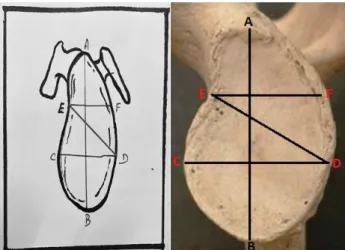
Following diameter of glenoid cavity were measured and analysed (Figure 2).
Superior-inferior diameter (SI): distance from the inferior point on the glenoid margin to the most prominent point of the supraglenoid tubercle, which is also the maximum height of the glenoid cavity,
Anterior-posterior diameter (AP1): measured from maximum breadth of the articular margin of the glenoid cavity.
A-B: Superior-Inferior diameter (SI), C-D: Anterior-Posterior diameter 1 (AP-1), E-F: Anterior-Posterior diameter 2 (AP-2), E-D: Anterior-posterior diameter 3 (AP3)
Figure 2: Various diameters of the glenoid cavity.
Anterior-posterior diameter (AP2): maximum breadth of the top half of the glenoid cavity at the peak of the glenoid notch.
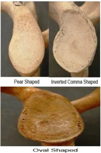
Anterior-posterior diameter (AP3): anterior-posterior diameter between the peak of glenoid notch and the most posterior rim where the maximum width of the glenoid. Following shapes of glenoid cavity were analysed:
• Inverted comma shaped,
• Pear shaped,
• Oval shape.
The shape was made by the slightly raised rim of the glenoid cavity. A piece of white sheet was placed on the glenoid cavity and held firmly in position to trace the shape of the glenoid cavity. The side of a lead pencil was rubbed along the rim of the glenoid cavity to get a tracing of the shape of the glenoid cavity on the paper. variations in shape of the glenoid cavity were classified based on the presence of notch and without notch as inverted comma shaped, pear shaped and oval shape. 3D CT images are obtained, to obtain reproducible images from the 3D reconstructed scapula, true anterior and posterior views were obtained by rotating the 3D image through cranio-caudal axis until glenoid appeared as simple line and rotating this image through the lateral to medial axis until the inferior part of coracoid process reach the upper part of glenoid in the anterior view and until the acromian reach the upper part of glenoid in the posterior view.
Statistical analysis
RESULTS
In the present study, the data were obtained in 50 scapulae of the right and50 scapulae of left. The morphometry of both right and left glenoids were assessed for 100 scapulae and various types of glenoid cavity based on their shape were observed in present study.
Morphology of glenoid cavity
Various shapes of glenoid cavity were observed and classified (Figure 3).
Figure 3: Various shapes of glenoid cavity.
• Type 1 (with notch) : A. Inverted comma shape and pear shape.
• Type 2 (without notch) : Oval shape
Comparison between shape of 50 right and 50 left glenoid cavity were done : x²-0.64, p value-0.72 (Table 1). Hence the incidence of both pear and inverted comma shape in right side is greater than in left whereas the incidence of oval shape in left glenoid is greater than right.
Morphology of glenoid cavity of scapula from 3D CT images were obtained (Figure 4) 5 right and 5 left glenoid
and various shapes were identified. Out of 5 right glenoid, 2 were Inverted comma shape, 2 pear shape and 1 oval shape. Out of 5 left glenoid, 4 were Inverted comma shape, 1 pear shape, no oval shape was found. Hence Inverted comma shape was common in left glenoid.
Figure 4: CT image of a glenoid cavity. A) Pear
shaped glenoid. B) Oval shaped glenoid. C)
Inverted comma shaped glenoid.
Table 1: Comparison between shape of 50 right
and 50 left glenoid cavity.
Shape Right Left
Pear 56% 52%
Inverted comma 36% 32%
Oval 8% 16%
Morphometry of glenoid cavity of scapula
The SI diameter of right is greater than the left, which is statistically significant. The AP-1, AP2, AP3 diameter of right is greater than that of left. But not statistically significant (Table 2).
Comparison of CT images of right and left glenoid
The SI, AP1, AP2, AP3 diameter of CT image of right glenoid is greater than the left glenoid cavity of scapula (Table 3).
A
B
Table 2: Morphometry of glenoid cavity of scapula.
Comparison of measurements of right and left glenoid
Parameter
Range Mean Standard deviation Statistical significance P Value
Right Left Right Left Right Left Right Left
SI diameter 21-40 mm 24-40 mm 33.1 31.6 4.1 3.4 Present Present 0.045 AP-1 diameter 14-28 mm 16-25 mm 21.4 20.5 4.4 2.8 None None 0.191 AP-2 diameter 10-23 mm 11-19 mm 15.6 14.8 3.2 1.7 None None 0.128 AP-3 diameter 15-22 mm 16-20 mm 19.2 18.8 1.9 1.2 None None 0.230
Table 3: Comparison of CT images of right and left glenoid.
Parameter Range Average
Right Left Right Left
SI diameter 30-39mm 27-36mm 35.86mm 30.6mm
AP-1 diameter 19-28mm 19-24mm 24.04mm 21.52mm
AP-2 diameter 15-25mm 13-19mm 19.92mm 15.84mm
AP-3 diameter 22-29mm 21-28mm 25.94mm 23.86mm
DISCUSSION
The present study was compared with several authors who have attempted to determine the glenoid diameters in the course of their research.
Comparison of morphology of glenoid
In this study of 50 right scapulae, 50 left scapulae 36 % of the glenoids had a distinct notch and were inverted comma-shaped, while on the left, 32 % of the glenoids were inverted comma shaped. Mamatha T et al, found right glenoid with distinct notch was 34% and that of left was 33% and Hina B Rajput et al observed inverted comma shaped glenoid in 35% of right and 39% in left glenoids, both the study values were closer to the present study.1,2
In this study 56% of the right glenoids were pear-shaped with an indistinct notch and 52% on the left side were pear-shaped.
Mamatha T et al, observed 46% of right glenoid was pear shaped and that of left it was 43%, Hina B Rajput et al observed 49% of right glenoid and 46% of left glenoid was pear shape and the present study is higher than the above author’s work.1,2
In the present study done on 100 scapulae irrespective of side, 54 (54%) scapulae found to be pear shaped.
Coskun N et al, studied 90 scapulae irrespective of side and found, that in 28% the notch was well expressed and found to be pear shaped, which was lesser than the present study and in the study done by Prescher and Klumpen in 236 scapulae, 55% of scapulae showed
distinct notch and found to be pear shaped which was similar to the present study.5
On the right side 8% were oval and on the left side 16% were oval without any recognizable notch. The comparison between right and left showed that more glenoids on the left did not show any notch and were oval in shape, similar to Mamatha T et al, 20% of glenoid were oval on right whereas 24% on left which is higher than that of present study, Hina B Rajput et al observed 16% of right and 15% of left glenoid were oval, incidence of left glenoid is similar to that of present study which showed 16% of left glenoid were oval.1,2
In the present study of 100 cases 12% were oval glenoid
Prescher and Klumpen observed that 45% of the glenoids irrespective of side did not have a notch and were therefore oval in shape.5 In the study of eighty eight
shoulders, Shortt et al found 85% ovoid glenoids. Coskun N et al, studied 90 scapulae irrespective of side and found that in 72% of the specimens the glenoid notches were absent or oval shaped.4
Comparison of morphometry of glenoid
In the present study the average superior-inferior diameter of the right glenoid was 33.1±4.1mm and the average superior-inferior diameter of the left glenoid was 31.6±3.4mm. The Right glenoid value is slightly more, it is statistically significant.
Hina B Rajput et al, in his study the (SI) diameter of right and left glenoid, the average of right glenoid was 34.76±3mm and of left glenoid was 34.43±3.21mm both the studies are similar to the present study.2 In the present
study the SI diameter of 100 scapulae irrespective of side was 32.7±3.7.
Iannotti et al, of the 140 glenoid irrespective of side to be 39±3.5mm which was more than the value of the present study (32.7±3.7).6 Mallon et al and Von Schroeder et al,
reported the SI diameter of 28, 30 glenoid respectively to be 35±4.1mm and 36±4mm respectively.7,8 Karelse et al,
found the SI diameter to be 35.9±3.6mm.9 The average SI
diameter of the glenoid measured by Churchill et al, irrespective of side was 35.1±2.0mm, and that measured by Ozer et al, was 36.25±2.8mm, Coskun N et al and Karelse et al, found the Superior-Inferior diameter of the glenoid to be 36.33mm and 35.9±3.6 mm respectively, Piyawinijwong S et al found it to be 35.8±2.6mm, all these values are slightly higher than the study, whereas glenoid measured by FrutosLR was 33.62±1.3mm which is closer with the present study (Table 11, Graph 12).10,11,4,9,12,13
In present study the average anterior-posterior diameter (AP-1) of the lower half of the glenoid of the right side was 21.4±4.4mm and that of the left side was 20.5±2.8mm. The combined average on both sides was 20.95±3.6mm. In a study done by Mamatha T et al, the average AP-1 diameter of both right and left glenoid, found to be 23.35mm and 23.05mm, RajputHB, observed the AP-1 of the right glenoid was 23.31±30mm and that of left was 22.92±2.80mm.1,2 These values are similar to
the present study. In a study done by. Churchill et al, irrespective of side recorded the average AP-1 diameter to be 23.6±1.5mm in 344 scapulae and that observed by FrutosLR, in 103 scapulae irrespective of side and found it to be 22.31±1.49mm and Ozer et al in 186 scapulae found to be 22.72±1.9mm, all these values were close with the present study whose diameter was 20.95±3.6mm.10,11,13 Mallon et al, found the AP-1
diameter to be 24±3.3mm, Iannotti et al, recorded 29±3.2mm, Schroeder V et al, observed it to be 28.6±3.3mm and Karelse et al, found the AP-1 diameter to be 27.2±3mm, all these measurements were higher than what was observed in the present study.6-9
The average anterior-posterior diameter (AP-2) of the upper half of the right glenoid was 15.6±3.2mm and that of the left glenoid was 14.8±1.7mm in the current study. This suggested that the right glenoid cavity was slightly broader than the left glenoid cavity. The combined average on both sides was 15.2±2.4mm. This was very close with what was observed by Mamatha T et al, AP-2 of right and left glenoid found to be 16.27±2.0mm and 15.77±1.96mm even in this study the right glenoid is higher than the left.1 Even the present study values are
close with that of Rajput HB et al, whose AP-2 diameter of right glenoid was 16.2±3.23mm and that of left glenoid was 15.24±2.04mm.2
Iannotti et al, done a study in 140 scapulae and found the AP-2 diameter of glenoid irrespective of side which was 23±2.7mm and Piyawinijwong S et al found to be 26±0.26mm. All these values are higher than the present study.6,12
The range for the AP-3 diameter of the right glenoid cavity was 15mm-22mm and the mean for the same was 19.2mm. The AP-3 diameter for the left glenoid varied from 16mm-20mm, while the mean for the left glenoid was 18.8mm.
Hence AP3 diameter of right is greater than that of left. But not statistically significant. Rajput HB et al,observed the average Anterior-Posterior diameter (AP-3) of the right glenoid was 23.08±3.15 mm and that of the left glenoid was 22.64±2.13mm in our study.2
This suggests that the right glenoid cavity was broader than the left glenoid cavity. The combined average of AP-3 diameter of both sides was 19.0±1.5mm.
Piyawinijwong S et al, recorded the average AP-3 diameter of 96 scapulae irrespective of side and found to be 27.8±1.8mm which was higher than the current study whose AP-3 diameter was 19±1.2mm.12
The glenoid morphometric measurements from 3D CT images obtained are compared with those from a previous report by Kwon YW et al.14 In their study the maximum
glenoid length irrespective of side was 37.8mm and the maximum glenoid width was 26.8mm which was higher than the present study in which the maximum glenoid length (SI diameter) irrespective of side was 33.23mm and the maximum glenoid width (AP-1 diameter) irrespective of side was 24.9mm. Information about various 3D CT parameters of glenoid anatomy, would seem to be important information to improve the design of the glenoid component.
Knowledge of the shape and dimensions of the glenoid are important in the design and fitting of glenoid components for total shoulder arthroplasty. An understanding of variations in normal anatomy of the glenoid is essential while evaluating pathological conditions like osseous Bankart lesions and osteochondral defects.
From the result and the discussion it can be implied that the values observed in the present study, though coinciding with that of some of the studies are mostly less than that recorded by many of the observers. The average supero-inferior glenoid diameter in our study is 31-32 mm, however these anatomical specimens were non-arthritic which mean that the average supero-inferior glenoid diameter in an arthritic glenoid would be lesser due to bone loss.
dimensions of the glenoid cavities may have to be taken
into consideration while designing and fitting glenoid components for the total shoulder arthroplasty.
CONCLUSION
By observing the tables in the discussion it can be implied that the values observed in the present study though coinciding with that of some of the studies, are mostly less than that recorded by many of the observers. The average supero-inferior glenoid diameter in our study is 31-32mm, however these anatomical specimens were non-arthritic which mean that the average supero-inferior glenoid diameter in an arthritic glenoid would be lesser due to bone loss. Moreover, the diameter would further decrease following reaming of the glenoid. This implies that the smaller dimensions of the glenoid cavities may have to be taken into consideration while designing and fitting glenoid components for the total shoulder arthroplasty.
ACKNOWLEDGEMENTS
Authors would like to thank Dr. Christilda Felicia (Professor, Department of Anatomy), Dr. S. Sundarapandian (Professor, Department of Anatomy), Dr. A. Ashma for guiding throughout study.
Funding: No funding sources Conflict of interest: None declared
Ethical approval: The study was approved by the Institutional Ethics Committee
REFERENCES
1. Mamatha T, Pai SR, Murlimanju BV, Kalthur SG, Pai MM, Kumar B. Morphometry of glenoid cavity. Online J Health Allied Sci. 2011;10(3):1-4.
2. Rajput HB, Vyas KK, Shroff BD. A study of morphological patterns of glenoid cavity of scapula. Natl J Med Res. 2012 Oct;2(4):504-7.
3. Torrens C, Corrales M, Gonzalez G, Solano A, Cáceres E. Cadaveric and three-dimensional computed tomography study of the morphology of the scapula with reference to reversed shoulder prosthesis. J Orthop Surg Res. 2008 Dec;3(1):49.
4. Coskun N, Karaali K, Cevikol C, Demriel BM, Sindel M. Anatomical basic and variations of the scapula in Turkish adults. Saudi Med J. 2006;27(9):1320-5.
5. Prescher A, Klümpen T. The glenoid notch and its relation to the shape of the glenoid cavity of the scapula. J Anatomy. 1997 Apr;190(3):457-60. 6. Iannotti JP, Gabriel JP, Schneck SL, Evanas BG,
Misra S. The normal glenohumeral relationships. Anatomical study of one hundred and forty shoulders. J Bone Joint Surg Am. 1992;74(4):491-500.
7. Mallon WJ,Brown HR, Vogler JB, Martinez S. Radiograpic and geometric anatomy of the scapula. Clin Orthop Relat Res.1992 Apr;(277):142-54. 8. Von Schroeder HP, Kuiper SD, Botte MJ. Osseous
anatomy of the Scapula Clin Orthop Relat Res. 2001 Feb;(383):131-9.
9. Karelse A, Kegels L, De Wilde L. The pillars of the scapula Clin Anat. 2007 May;20(4):392-9.
10. Churchill RS, Brems JJ, Kotschi H. Glenoid size, inclination, and version: an anatomic study. J Shoulder Elbow Surg. 2001 Jul 1;10(4):327-32. 11. Özer I, Katayama K, Sahgir M, Güleç E. Sex
determination using the scapula in medieval skeletons from East Anatolia. Collegium antropologicum. 2006 Apr 10;30(2):415-9.
12. Piyawinijwong S, Sirisathira N, Chuncharunee A. The Scapula: osseous dimensions and gender dimorphism in Thais. Siriraj Hosp Gaz. 2004;56(7):356-65.
13. Frutos LR. Determination of sex from the clavicle and scapula in Guatemalan contemporary rural indigenous population. Am J Forensic Med Pathol. 2002;23(3):284-8.
14. Kwon YW, Powell KA, Yum JK, Brems JJ, Iannotti JP. Use of three-dimensional computed tomography for the analysis of the glenoid anatomy. J Shoulder Elbow Surg. 2005 Jan 1;14(1):85-90.
Cite this article as: Raaj MS, Felicia C,