i
The Role of Myosin-X in a Putative System of Intrafilopodial Transport
Michael Kerber
A dissertation submitted to the faculty of the University of North Carolina at
Chapel Hill in partial fulfillment of the requirements for the degree of
Doctor of Philosophy in the Department of Cell and Molecular Physiology.
Approved by,
Richard Cheney
Robert Sealock
James Bear
Ken Jacobson
iii
Abstract
Michael Kerber: The Role of Myosin-X in a Putative System of Intrafilopodial Transport (Under the direction of Richard Cheney)
My research focuses on the role of the molecular motor, myosin-X (Myo10), in
the cellular protrusions called filopodia. Chapter one provides an up-to-date review of
Myo10 in a manuscript that is being prepared for submission to the Journal of Cell
Science. Chapter two, my main data chapter, was published in Current Biology and
describes a novel population of fast-moving Myo10 in filopodia that we discovered using
single-molecule imaging techniques. For this paper, I optimized the imaging system used
to detect single Myo10 molecules, performed most of the experiments, and made all of
the figures. I also helped develop a software program, Kymotracker, that exploits a
technique called kymography to track and take measurements of these single molecules
in time-lapse videos. In Chapter three, I describe preliminary experiments investigating
the role of Myo10 in filopodial adhesions. In Chapter four, I summarize conclusions
iv
Table of Contents
List of Figures ... vi
List of Abbreviations ... viii
Chapter 1: Myosin-X: a MyTH4/FERM motor at the filopodial tip ... 1
Summary ... 1
Introduction ... 1
Myosin-X structure and biochemical properties ... 3
Myosin-X localizes to the leading edge of lamellipodia and the tips of filopodia ... 9
Myosin-X potently induces filopodia ... 11
Myosin-X undergoes intrafilopodial motility ... 13
Does Myosin-X transport cargo in filopodia? ... 15
Myosin-X is required for endothelial cell migration and junction formation ... 16
Myosin-X is required for spindle assembly and orientation ... 16
Myosin-X is important for axon outgrowth ... 18
Myosin-X is required during development for neural crest cell migration ... 19
How does Myosin-X select for filopodial actin? ... 20
Discussion ... 21
v
Summary ... 22
Results and Discussion ... 23
Conclusions ... 41
Materials and Methods ... 45
Chapter 3: Investigations of the role of Myo10 in a filopodial tip complex... 52
Summary ... 52
Introduction ... 53
Results ... 56
Discussion ... 73
Materials and Methods ... 75
Chapter 4: Conclusions and Future Directions ... 78
Summary ... 78
Myo10 intrafilopodial motility ... 78
Myo10 as a potential cargo transporter ... 80
The challenges of single-molecule tracking in two colors ... 82
Kymotracker ... 84
Directly visualization of Myo10 dimerization and cargo transport ... 85
Filopodia: an ideal environment for studying stereocilia motors and cargos ... 86
vi
List of Figures
Figure 1.1. Myo10 Structure ... 4 Figure 1.2. Model of possible Myo10 regulation... 6 Figure 1.3. Myo10 localizes to filopodial tips and the leading
edge of lamellipodia ... 10
Figure 1.4. Myo10 overexpression induces filopodia formation ... 12 Figure 1.5. Myo10 undergoes intrafilopodial motility ... 14 Figure 1.6. Knockdown of Myo10 results in the formation of
multipolar spindles ... 17
Figure 2.1. TIRF microscopy reveals fast forward movements
of faint particles of GFP-Myo10 in living cells ... 28
Figure 2.2. Dynamics of GFP-Myo10 in living HeLa cells
imaged with TIRF ... 29
Figure 2.3. Substrate-attached cell extensions in HeLa cells
contain the filopodial markers F-actin and fascin ... 32
Figure 2.4. Faint particles of GFP-Myo10 exhibit characteristics
of single molecules ... 34
Figure 2.5. Fast forward movements require the Myo10 motor
domain and are inhibited by latrunculin B ... 37
Figure 2.6. Ability of different Myo10 constructs to localize to
filopodial tips and TIRF imaging of a motor domain point
mutation and a forced-dimer construct ... 39
Figure 2.7. Particles of GFP-Myo5a also move forward and
rearward in the filopodia of living cells ... 42
Figure 2.8. Faint particles of GFP-VASP move rapidly forward
in filopodia at velocities similar to those of GFP-Myo10... 43
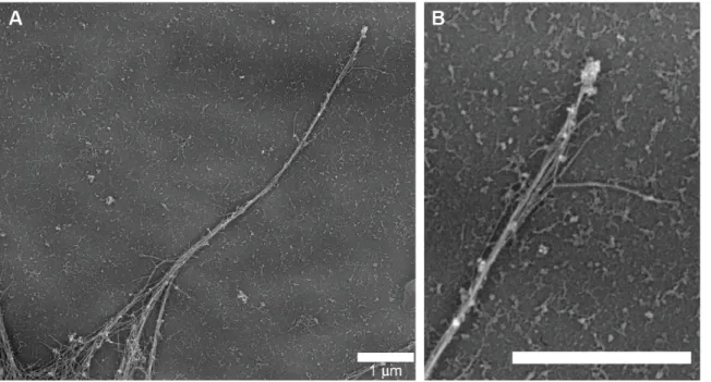
Figure 3.1. Platinum replica electron micrograph of an individual
vii
Figure 3.2. VASP localizes to filopodia in Myo10 knockdown cells ... 58 Figure 3.3. Single-molecule imaging of integrins reveals
occasional forward movements ... 62
Figure 3.4. CALI of GFP-Myo10 tip spots ... 65 Figure 3.5. Filopodial attachments can be pulled away from the
Myo10-labeled tip spot ... 68
Figure 3.6. Myo10 clusters behave like fluids after cytochalasin D
treatement ... 72
Figure 4.1. Single molecules of formin exhibit fast forward
movement in filopodia ... 81
Figure 4.2. Deafness myosins localize to filopodial tips and
viii
List of Abbreviations
ALK6 activin receptor-like kinase 6
ATP adenosine tri-phosphate
AU arbitrary units
BMP6 bone morphogenetic protein 6
CALI chromophore assisted laser inactivation
CytoD cytochalasin D
DCC deleted in colorectal cancer
DIC differential interference contrast
ECM extracellular matrix
ER endoplasmic reticulum
FERM 4.1 ezrin radixin moesin
FRAP fluorescence recovery after photobleaching
HMM heavy meromyosin
GFP green fluorescent protein
LatB latrunculin B
ix MyTH4 myosin tail homology 4
PBS phosphate buffered saline
PCR polymerase chain reaction
PEST proline, glutamic acid, serine, threonine sequence
PH pleckstrin homology
ROS reactive oxygen species
SAH stable α-helix
TIRF total internal reflection fluorescence
1
Chapter 1: Myosin-X: a MyTH4/FERM motor at the filopodial tip
Summary
Myosin-X (Myo10) is a MyTH4-FERM myosin broadly expressed in vertebrate
tissues. Myo10 is best known for its role in the formation of filopodia and its striking
ability to move within filopodia and accumulate at their tips. Its tail region is implicated
in signaling downstream of PI3K, interactions with adhesion-associated proteins, and
binding to microtubules. In addition to its central role in filopodia, Myo10 is also
required for the proper orientation and length of the mitotic spindle. While biophysical
studies of Myo10 have begun to uncover the motor’s single-molecule properties, exciting
progress has also been made in revealing Myo10’s physiological functions. Recent
evidence has demonstrated that Myo10 is required for the migration of cells crucial for
the development of the nervous system.
Introduction
Myosin motors are an ancient group of proteins capable of moving along actin
filaments and binding to cargos via a wide variety of tail domains. Myosin-X (Myo10)
contains a unique combination of domains in its tail and is the founding member of its
own class of myosins. Together with its closest relatives, the class VII and XV myosins,
Myo10 is a member of a larger superclass of myosins defined by the presence of myosin
2
MyTH4-FERM myosins have been implicated in mediating membrane-cytoskeleton
interactions in protrusive structures such as stereocilia and filopodia (Oliver et al., 1999).
MyTH4-FERM myosins are also evolutionarily ancient, appearing very early in
metazoan evolution, although Myo10 is not present in the fly and worm lineages
(Odronitz and Kollmar, 2007).
Myo10 was originally discovered in a screen for novel myosins in bullfrog inner
ear tissue, but has since been detected in most mammalian tissues (Solc et al., 1994;
Yonezawa et al., 2000). It is important to note, however, that its level of expression is
orders of magnitude lower than that of the better-known myosin-II. Myo10 is the only
myosin to boast multiple pleckstrin homology (PH) domains, which confer the ability to
bind directly to the plasma membrane (Berg et al., 2000). Through its unique assortment
of PH, MyTH4, and FERM domains and distinctive motor properties, Myo10 appears to
play several important roles within the cell (Divito and Cheney, 2008; Sousa and Cheney,
2005). In particular, Myo10 has emerged as a central figure in filopodia.
Filopodia provide a point of contact between a cell and its environment and many
cells are thought to rely on these finger-like organelles to probe and interact with their
surroundings (Mattila and Lappalainen, 2008). Filopodia are therefore thought to be
important for developmental processes that rely on directed cell migration, such as axon
guidance and angiogenesis (Eilken and Adams, 2010; Koleske, 2003). Despite clear
implications in human health and disease, the mechanisms underlying filopodial
formation, maintenance, and function remain largely uncertain. Filopodia consist of a
plasma-membrane-encased F-actin bundle, oriented with the actin barbed ends towards
3
polymerization, which contributes to forces that cause the filopodial actin bundle to
continuously slide back towards the cell body, a phenomenon termed “rearward
treadmilling” (Medeiros et al., 2006). One likely master regulator of filopodia is Myo10,
which localizes to filopodial tips, causes an increase in filopodia number, and moves in a
directed manner within filopodia (Berg et al., 2000; Bohil et al., 2006; Kerber et al.,
2009). Here we review key progress in understanding Myo10, from its structural and
single-molecule properties to its role in organismal development.
Myosin-X structure and biochemical properties
Myo10 contains regions referred to as the head, neck, α-helix, and tail (Figure
1.1). The head of Myo10, like all myosins, consists of a conserved motor domain, able to
bind to F-actin and hydrolyze ATP to produce force (Homma et al., 2001). Initial in vitro
experiments performed using an “HMM-like” Myo10 construct, which includes only the
head, neck, and α-helical domains, indicated that Myo10 moved at ~200-300 nm/s with a
processivity somewhere between that of non processive motors like myosin-II and highly
processive motors like myosin-Va (Chen et al., 2001). A monomeric head-neck construct
exhibited an actin activated ATPase of ~4/s at 25°C and a duty ratio of ~16%, again
intermediate between that of nonprocessive and highly processive motors (Kovacs et al.,
2005). On the contrary, a shorter head-neck construct used by a different group yielded a
much higher actin activated ATPase and duty ratio of 13.5/s and ~60%, respectively
(Homma and Ikebe, 2005). These discrepancies illustrate the importance of studying
full-length My10 constructs in the cellular environment to establish the motor properties
4
Figure 1.1. Myo10 Structure
5
Following the motor domain is the Myo10 neck, consisting of three IQ motifs,
each capable of binding to calmodulin or calmodulin-like light chains in a
calcium-dependent manner (Berg et al., 2000; Homma et al., 2001). Recent studies have shown
that calmodulin-like protein actually increases Myo10 expression by interacting
specifically with Myo10’s IQ3 motif (Bennett et al., 2008; Bennett et al., 2007; Bennett
and Strehler, 2008). One explanation for this method of regulation is that during
translation, the growing Myo10 peptide somehow interferes with further translation until
calmodulin-like protein binds to its neck, changing the conformation of the nascent
peptide and allowing translation to proceed (Bennett and Strehler, 2008). Myo10 may
also undergo the kind of self-inhibition exhibited by myosin-VII, wherein part of the tail
region folds into and inhibits the motor (Yang et al., 2009) (Figure 1.2). In the mature
protein, the neck region is thought to function as a rigid lever arm that amplifies the
conformational change in the motor domain, causing the molecule to move a distance
proportional to the length of the lever arm, which is typically dictated by the number of
IQ motifs in the neck. For example, the 3IQ motifs of Myo10’s neck are expected to
span ~10 nm, which would allow a Myo10 dimer to potentially reach across ~20 nm in a
single step.
The precise structure of the Myo10 lever arm is still unclear due to a recent
reanalysis of the ~130 amino acid α-helical region. What was originally predicted to
form a dimer-inducing coiled-coil domain is now accepted to be, at least partially, a
stable single α-helix (SAH). SAH domains function as stiff extensions of the lever arm
and have recently been discovered in several myosins (Baboolal et al., 2009;
6
Figure 1.2. Model of possible Myo10 regulation
Myo10’s predominant intracellular state may resemble a compact, inactive monomer. Binding to actin by the motor domain, or to other cargos by domains in the tail, may cause the monomer to extend. A final, active dimer may be formed either when the local concentration of extended monomers reaches some threshold, or by binding to specific cargos via the Myo10 tail.
7
the first 36 of the α-helical region and possibly much more, extending the lever arm and
leaving a much shorter region that might form a coiled-coil (Berg et al., 2000; Knight et
al., 2005a). The ability of Myo10 to form a dimer has therefore become somewhat
controversial.
Whether through the use of a coiled-coil domain or by some other mechanism,
Myo10 appears to undergo dimerization under certain circumstances. While electron
microscopy revealed that a purified HMM-like Myo10 remained mostly (90%)
monomeric, the 10% of the HMM-Myo10 population that successfully formed dimers
under these in vitro conditions appeared to have lever arms that were much longer than
expected for a neck consisting of only 3IQ motifs. A more recent study by Sun et al.
discovered that HMM-Myo10s readily dimerize when brought into proximity, suggesting
that the distal portion of the predicted coiled-coil domain retains some dimer-forming
ability. To test the necessity of dimerization, many groups have created “forced-dimer”
constructs of Myo10 that include a region that artificially induces dimerization.
Experiments using different forced-dimer versions of HMM-like Myo10 indicate that the
ability to dimerize is necessary for Myo10 to move processively (Kerber et al., 2009;
Nagy et al., 2008; Nagy and Rock, 2010; Ricca and Rock, 2010; Sun et al., 2010).
Consistent with this theory, a construct containing only the head and neck, thus lacking a
coiled-coil domain, is unable to localize to the tips of filopodia (Berg and Cheney, 2002).
It is possible that in the environment of the cell, Myo10 is subject to regulated
dimerization either through cargo binding, as has been reported for Myosin-VI, or
through high local concentrations in the regions of the cell where it accumulates (Iwaki et
8
Immediately following the α-helical region are three proline-rich PEST regions,
which have been implicated as sites of cleavage by calcium-dependent calpain (Berg et
al., 2000; Rechsteiner and Rogers, 1996). While cleavage at the PEST sites can occur in
vitro, generating an HMM-like Myo10 as well as a tail fragment, it is not yet known
whether this is an important process in vivo (Berg et al., 2000). After the PEST regions,
the Myo10 tail contains a cluster of three pleckstrin homology (PH) domains in an
unusual arrangement; the second PH domain (PH2) is inserted into a putative surface
loop of PH1 (Figure 1). The inclusion of PH domains allows Myo10’s tail to bind
directly to the plasma membrane via phosphatidylinositol phosphates (Berg et al., 2000;
Cox et al., 2002; Isakoff et al., 1998; Macias et al., 1994; Musacchio et al., 1993; Tacon
et al., 2004; Yonezawa et al., 2003). This membrane-binding ability is unique among the
many myosins discovered so far and may lead to an increase in the local concentration of
Myo10 to levels that favor dimerization (Sun et al., 2010). Additionally, the presence of
PH domains raise the possibility that Myo10 functions downstream of PI3-kinase
signaling. Indeed, the PH2 domain of Myo10 is known to function downstream of
PI3-kinase in macrophage phagocytosis (Cox et al., 2002) and the inability to bind to
PtdIns(3,4,5)P3 shifts Myo10’s localization from the plasma membrane to Rab7-positive
vesicles (Plantard et al., 2010).
Following the PH domains, the Myo10 tail contains a Myosin Tail Homology 4
(MyTH4) domain, which has been shown to bind to microtubules and may allow Myo10
to act as a link between the actin and microtubule cytoskeletons (Narasimhulu and
Reddy, 1998) (Weber et al., 2004). The tail of Myo10 ends in a FERM domain, named
9
Radixin, and Moesin. Other FERM domain-containing proteins serve to link the actin
cytoskeleton to integral membrane proteins (Chishti et al., 1998), and the FERM domain
of Myo10 has been shown to bind to the NPXY motif of the cytoplasmic domain of β5
integrin (Zhang et al., 2004). In addition to binding to cargos individually, the MyTH4
and FERM domains can also act in conjunction to bind to some cargo proteins (Wei et
al., 2011; Wu et al., 2011). To bind to the netrin receptor, DCC, for example, the
formation of a MyTH4/FERM structural supramodule is apparently required. It is also
likely, according to structure-based sequence analysis, that all MyTH4/FERM tandems
form this supramodule. It will be interesting to see whether this union of the MyTh4 and
FERM domains affects their interactions with other binding partners.
Myosin-X localizes to the leading edge of lamellipodia and the tips of filopodia
One of Myo10’s most defining characteristics is its striking localization to the tips
of the cellular protrusions called filopodia (Figure 1.3). When considering the movement
of motors in filopodia, it is important to note the constant retrograde flow of filopodial
actin. Against this steady rearward flow, Myo10 uses its own motor force to migrate
towards and maintain itself at filopodial tips. As one might expect, the motor domain is
necessary for Myo10’s tip localization while an HMM-like construct is sufficient (Berg
and Cheney, 2002). Although Myo10 is thought to prefer bundled-actin structures, it is
also known to localize to the leading edge of lamellipodia, broadening its range to areas
10
Figure 1.3. Myo10 localizes to filopodial tips and the leading edge of lamellipodia
A bovine aortic endothelial cell fixed and stained with rhodamine-phalloidin (red) and antibodies against My10 (green). Image courtesy of Melinda Divito.
11
Myosin-X potently induces filopodia
While Myo10 exhibits a clear affinity for filopodia, its functions in these
structures have not been completely characterized. There is, however, a large body of
evidence indicating that Myo10 is crucial for the formation of filopodia. Overexpression
of Myo10 is sufficient to induce a massive increase in both substrate-attached and
non-adherent filopodia (Berg and Cheney, 2002) (Figure 1.4). Likewise, knockdown of
Myo10 resulted in a dramatic loss of filopodia in HeLa cells. Myo10 may induce
substrate-attached filopodia via an integrin-dependent mechanism, causing more
filopodia to stick to and become stabilized by the substrate (Zhang et al., 2004). The vast
majority of filopodia, however, are non-adherent, and a Myo10 construct that lacks the
integrin-binding FERM domain is also capable of inducing filopodia (Bohil et al., 2006).
In contrast, an HMM-like construct does not induce filopodia. This evidence indicates
that although the tail of Myo10 is required, there may be an integrin-independent
mechanism of Myo10’s induction of filopodia. One alternate theory proposes that
Myo10 can crosslink actin fibers at the leading edge of the lamellipodium, generating
actin bundles that could form new filopodia (Tokuo et al., 2007). These models do not
exclude the possibility that Myo10 transports some other proteins critical for filopodial
formation or maintenance (Ross et al., 2008). Myo10 may perform a similar function in
the filopodia-like structures, invadopodia, which are protrusions associated with
metastatic cancer cells. Myo10 localizes to the tips of invadopodia and is required for
their elongation, suggesting a provocative link between Myo10 and cancer cell metastasis
12
Figure 1.4. Myo10 overexpression induces filopodia formation
Scanning electron micrographs of Cos-7 cells expressing GFP (A) or GFP-Myo10 (B). Images courtesy of Aparna Bohil.
13
Myosin-X undergoes intrafilopodial motility
Consistent with the hypothesis that Myo10 propels itself along filopodial actin,
GFP-tagged full-length Myo10 constructs have long been known to display striking
motility along the filopodia of living cells (Berg and Cheney, 2002) (Figure 1.5). Bright
puncta of Myo10 not only localize strongly to filopodial tips, but also move rearwards at
rates closely matching those of actin retrograde flow (~15 nm/s). It is thought that these
rearward movements represent Myo10 molecules that have coupled to the rearward
treadmilling filopodial actin bundle, riding it like packages on a conveyor belt.
Occasionally, these Myo10 puncta migrate back towards the tip at ~80 nm/s. More
recently, single-molecule microscopy techniques were employed to detect a previously
uncharacterized, extremely faint population of Myo10 particles that undergo forward
movement at rates that more closely match those exhibited in vitro (~600 nm/s) (Kerber
et al., 2009). These fast-forward events can be quite frequent, with new molecules of
Myo10 moving up the shaft of an individual filopodium as often as every second. The
forward movement of either the large clusters or the single molecules of Myo10 require a
functional motor, and an HMM-like construct was found to be sufficient, indicating that
in both cases Myo10 is propelling itself. Interestingly, an HMM-Myo10 that included an
artificial forced-dimer domain in the α-helical region was also capable of exhibiting fast
forward movement at the single-molecule level, indicating that a head, neck, and the
ability to dimerize is also sufficient. Although it was not clear from initial
“single-molecule” imaging experiments whether the fast, faint particles of Myo10 represented
monomers or dimers, a more recent study reported two-step photobleaching of these
14
Figure 1.5. Myo10 undergoes intrafilopodial motility
(A) Model of Myo10 movement in filopodia. Large clusters of Myo10 (green) are commonly observed both at tips and along the filopodial shaft sliding rearward at ~15 nm/s and migrating forward at ~80 nm/s. These clusters may include as yet undiscovered scaffolding components, actin polymerization machinery, or other cargo (gray).
Meanwhile, individual Myo10 monomers or dimers move much faster towards filopodial tips, ~600 nm/s. This highly motile population of Myo10 may also be transporting either cytoplasmic or membrane-associated cargo proteins (orange). (B) Kymograph of a filopodium from a HeLa cell transfected with GFP-Myo10 and imaged using single-molecule TIRF. Note the complex juxtaposition of the various modes of Myo10 intrafilopodial motility.
15
Does Myosin-X transport cargo in filopodia?
Due to its ability to propel itself along filopodial actin, Myo10 is an excellent
candidate to act as a cargo delivery motor in a putative, filopodia-specific transport
system (Kerber et al., 2009; Nambiar et al., 2010). Consistent with this model, bright
puncta of Myo10 have been reported to undergo cotransport with VASP and VE-cadherin
in filopodia (Almagro et al., 2010; Tokuo and Ikebe, 2004). It should be noted, however,
that it has not yet been shown whether Myo10 is necessary for the transport of its binding
partners. These experiments may require the development of Myo10 knockout cell lines.
It will also be important in the future to show convincing evidence for Myo10 directly
transporting other proteins, a feat that may require more sensitive single-molecule
techniques than those employed so far.
Filopodia are also known to serve as passageways for the transport of cargo
between cells. For example, melanosomes are pigment-producing organelles that appear
to be delivered from melanocytes to keratinocytes along filopodia in a process thought to
be driven by myosin-Va (Scott et al., 2002). A recent study, however, demonstrated the
importance of Myo10 in this transport system. Knockdown of Myo10 in either the
melanocytes or the keratinocytes resulted in inhibition of melanin uptake while
ultraviolet light upregulated both melanosome transfer and Myo10 expression (Singh et
al., 2010). Intriguingly, a similar filopodial transport system may also be exploited by
retroviruses to infect neighboring cells, although the contribution of Myo10 in this
process has not yet been investigated (Lehmann et al., 2005; Sherer et al., 2007; Sherer
16
Myosin-X is required for endothelial cell migration and junction formation
In addition to acting as transport routes for intracellular cargos, filopodia are also
thought to be crucial for signaling and cellular migration. Interestingly,
BMP6-dependent filopodial extension and, consequently, directional migration in endothelial
cells requires Myo10 (Pi et al., 2007). In this process, Myo10 serves as a necessary part
of the signaling pathway that allows the cell to detect BMP6 in the extracellular
environment, possibly by transporting the BMP6-receptor ALK6 to filopodial tips.
Endothelial cell filopodia play an important role in establishing cell-cell junctions, which
are held together by a cell-cell adhesive receptor, VE-Cadherin. VE-Cadherin was
recently reported to immunoprecipitate with Myo10 and undergo coordinate movement
with Myo10 in endothelial cell filopodia. Importantly, expressing a putative dominant
negative Myo10 construct, consisting of the Myo10 FERM domain, inhibited the
localization of VE-Cadherin (Almagro et al., 2010).
Myosin-X is required for spindle assembly and orientation
In addition to its clear association with filopodia-like structures, Myo10 is also
important in a process that has little in common with cellular protrusions: mitotic spindle
orientation. Assembly of the mitotic spindle and anchoring of the nucleus in Xenopus
laevis oocytes are disrupted when Myo10 function is presumably inhibited by either
anti-Myo10 antibodies or a anti-Myo10-tail construct (Weber et al., 2004). More dramatically, the
use of morpholinos to inhibit Myo10 expression in developing embryos results in
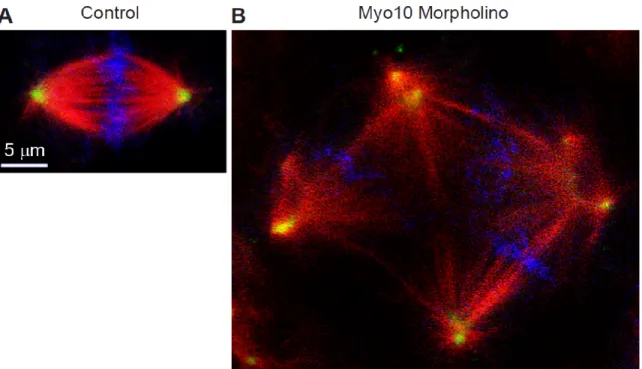
multipolar spindles (Woolner et al., 2008) (Figure 1.6). Proper orientation of the mitotic
17
Figure 1.6. Knockdown of Myo10 results in the formation of multipolar spindles
(A) Control or (B) Myo10 morpholino-treated cells of a Xenopus embryo. Cells were fixed and stained for β-tubulin (red), γ-tubulin (green), and DNA (blue). Image courtesy of Bill Bement.
18
spindles parallel to the substrate in an integrin-mediated, substrate-dependent manner,
lose the ability to correctly orient their spindles when Myo10 expression is knocked
down with siRNA (Toyoshima and Nishida, 2007). Knockdown of Myo10 also increases
the probability of the cancer cell line MDA231 to produce multipolar spindles (Kwon et
al., 2008). It is hypothesized that Myo10 links the spindle-associated F-actin with astral
microtubules and applies a contractile force to the spindle, counteracting the stretching
force applied by cortical F-actin (Wuhr et al., 2008).
Myosin-X is important for axon outgrowth
While most tissues express full-length Myo10, expression in the mouse brain
peaks during the first week after birth, which is also the period of peak synapse
formation. Myo10 expression then plummets in the adult mouse brain, but the brain also
expresses an enigmatic isoform that lacks the motor domain (Sousa et al., 2006). This
“headless” isoform is otherwise intact and may represent a naturally occurring dominant
negative. Indeed, expressing a headless Myo10 construct inhibits the migration of
neuronal cells in vitro (Wang et al., 2009) and impairs chick commissural neuron axon
projection in vivo (Zhu et al., 2007). In neurons, actin and microtubules are both
important for axon guidance and extension, with filopodia being especially important for
axonal path-finding (Dent and Gertler, 2003). Cortical neurons from a mouse line
lacking the three Ena/VASP proteins, Mena, VASP, and EVL, do not form filopodia or
generate neurites. Expressing Myo10 in these triple knockout cells rescues filopodia
formation and neuritogenesis (Dent et al., 2007). Interestingly, Myo10 is upregulated
19
normally active during development. In development, axon path-finding is regulated by a
group of proteins called Netrins, whose receptors include Deleted in Colorectal Cancer
(DCC). DCC localizes to neurite tips in a Myo10-dependent manner and also induces
Myo10-dependent filopodia formation and elongation (Zhu et al., 2007). Together, these
data strongly indicate a developmentally critical role for Myo10 in axon outgrowth.
Myosin-X is required during development for neural crest cell migration
Although there is currently no knockout animal model for Myo10, exciting
studies using Xenopus laevis reveal that Myo10 is required for the proper migration of
cranial neural crest cells (Hwang et al., 2009) (Nie et al., 2009). Neural crest cells are
multipotent, migratory cells that give rise to several cell types including craniofacial
cartilage and bone, melanocytes, and peripheral neurons (Huang and Saint-Jeannet,
2004). In the Xenopus embryo, Myo10 is predominantly expressed in the neural crest
cells in the premigratory and migratory stages. Knocking down Myo10 expression with
morpholinos causes a loss of neural crest cell migration and results in a dramatic decrease
in the size of the cranium. The loss of migration can be rescued in vitro by expressing
exogenous Myo10, confirming that the phenotype is due to the loss of Myo10. This
defect in cell migration appears to be caused by inhibited cellular adhesion and
polarization of the neural crest cells. It will be important to learn whether Myo10 proves
20
How does Myosin-X select for filopodial actin?
Much of Myo10’s filopodia-centric functions rely on its ability to preferentially
localize to bundled actin. Recent advances in the understanding of Myo10’s biophysical
properties are steadily revealing the mechanism underlying Myo10s affinity for bundled
actin in the cell. One hypothesis proposes that Myo10 binds favorably to bundled actin
because its lever arms possess the distinct length and flexibility to endow the myosin with
the unique ability to bind each motor domain to separate, neighboring actin filaments. In
a series of experiments from the Rock group, Myo10 has been shown to initiate longer,
more frequent runs on bundled actin than filamentous actin, and that Fascin, the
filopodial actin bundler, is required for proper Myo10 localization in vivo (Nagy et al.,
2008). Myo10 also appears to prefer parallel actin bundles in ex vivo systems in which
the stabilized cytoskeletons of fixed, detergent-treated cells are used to seed purified
motors (Brawley and Rock, 2009). The specifics of Myo10’s behavior on bundled and
filamentous actin remain unclear, with reported step-sizes ranging from 18 nm to 34 nm
(Ricca and Rock, 2010; Sun et al., 2010). There is also disagreement regarding whether
Myo10 spirals in a right-handed, left-handed, or unbiased fashion on filaments and
bundles. Domain-swapping experiments indicate that Myo10’s bundle selectivity,
however, appears to rely on the properties of its α-helical region and may involve the
amount of flexibility that the region lends to Myo10’s lever arm (Nagy and Rock, 2010).
Much of the discrepancies may be due to differences in the constructs used by different
groups. Some forced-dimer constructs may alter the flexibility of the α-helical region or
21
greatly from a purified, full-length version of Myo10 that could be used to establish the
step size, processivity, and bundle selectivity of the native protein.
Discussion
Although Myo10’s provocative localization to filopodia and similar structures has
long hinted at importance in development, the first confirmation of its role in vivo has
only recently come to light. The requirement for Myo10 in neural crest cell migration
may represent only the first of many potential roles in development, considering its
importance in other in vitro systems (e.g. endothelial cell migration and mitotic spindle
formation). The creation of true knockout animal models may reveal other
developmental processes that require Myo10. Cell lines that truly lack Myo10 would
also provide invaluable new testing grounds to more fully characterize its role in the cell.
Indeed, Myo10 has yet to be fully characterized at the molecular level and many
questions persist. How is Myo10 regulated? Does it dimerize? Does it transport cargo?
Although great progress has been made using various truncated and forced-dimer
versions of Myo10, the field would benefit greatly from a purified full-length Myo10 that
could be used for biophysical studies. Likewise, much has been learned from in vitro and
ex vivo systems, but in vivo approaches may be required to tease apart the actual
dimerization status, step size, and bundled actin behavior that Myo10 exhibits in the cell.
A better understanding of Myo10’s molecular characteristics will undoubtedly lead to a
22
Chapter 2: Imaging Myosin-X at the Single-Molecule Level Reveals a Novel Form of Motility in Filopodia
Michael L. Kerber, Damon T. Jacobs, Luke Campagnola, Brian D. Dunn, Taofei Yin,
Aurea D. Sousa, Omar A. Quintero, and Richard E. Cheney
Summary
Although many proteins, receptors, and viruses are transported rearward along
filopodia by retrograde actin flow (Hu et al., 2007; Lidke et al., 2005; Sherer et al., 2007),
it is less clear how molecules move forward in filopodia. Myosin-X (Myo10) is an
actin-based motor hypothesized to use its motor activity to move forward along actin filaments
to the tips of filopodia (Berg and Cheney, 2002). Here we use a sensitive total internal
reflection fluorescence (TIRF) microscopy system to directly visualize the movements of
GFP-Myo10. This reveals a novel form of motility at or near the single-molecule level in
living cells wherein extremely faint particles of Myo10 move in a rapid and directed
fashion towards the filopodial tip. These fast forward movements occur at ~600 nm/s
over distances of up to ~10 um and require Myo10 motor activity and actin filaments. As
expected for imaging at the single-molecule level, the faint particles of GFP-Myo10 are
23
stepwise bleaching. Faint particles of GFP-Myo5a can also move towards the filopodial
tip, but at a slower characteristic velocity of ~250 nm/s. Similar movements were not
detected with GFP-Myo1a, indicating that not all myosins are capable of intrafilopodial
motility. These data indicate the existence of a novel system of long-range transport
based on the rapid movement of myosin molecules along filopodial actin filaments.
Results and Discussion
Filopodia are slender actin-based extensions thought to function as cellular
sensors in processes such as nerve growth and blood vessel development. Filopodia have
a relatively simple structure consisting of a bundle of parallel actin filaments surrounded
by the plasma membrane (Mattila and Lappalainen, 2008; Wood and Martin, 2002).
Each actin filament has its barbed end oriented towards the tip of the filopodium, and
actin monomers are constantly added to the filament at its barbed end. The actin in
filopodia typically moves rearward at rates of 10-100 nm/s in a process known as
retrograde flow. Although retrograde flow (Albrecht-Buehler and Goldman, 1976) is
now known to be powered by a combination of actin polymerization and myosin-II
mediated contraction (Medeiros et al., 2006), the mechanisms by which molecules move
forward in filopodia are much less clear. Since microtubules and membranous vesicles
are generally absent from filopodia, forward movement in filopodia is likely to depend
either on diffusion or an actin-based mechanism.
Myo10 is an actin-based motor protein that localizes to the tips of filopodia and
24
Myo10 heavy chain consists of a myosin head domain responsible for force production, a
neck domain that provides binding sites for calmodulin or calmodulin-like light chains
(Rogers and Strehler, 2001), and a large tail (Sousa and Cheney, 2005). The tail includes
a segment that was initially predicted to form a coiled coil (Berg et al., 2000), 3 PH
domains that can bind to inositol phospholipids such as PIP3 (Mashanov et al., 2004), a
MyTH4 domain that can bind to microtubules (Weber et al., 2004), and a FERM domain
that can bind to candidate cargoes such as β-integrins (Zhang et al., 2004). Imaging with
conventional epifluorescence revealed that the bright puncta of GFP-Myo10 normally
present at the tips of filopodia sometimes vacate the tip and move slowly rearward at
10-20 nm/s (Berg and Cheney, 10-2002), the rate of retrograde actin flow in HeLa filopodia.
Bright puncta also occasionally moved forward at ~80 nm/s, leading to the hypothesis
that Myo10 moves forward by using its barbed-end motor activity to transport itself along
filopodial actin filaments and that it moves rearward by binding in a rigor-like state to
actin filaments undergoing retrograde flow. Consistent with this, a Myo10 construct
comprised only of the head, neck, and predicted coiled coil (Myo10-HMM) was
sufficient for tip localization (Berg and Cheney, 2002). Although a
baculovirus-expressed Myo10-HMM-like construct appears largely monomeric in vitro (Knight et al.,
2005b), induced dimerization of Myo10 head-neck constructs leads to tip localization in
vivo (Tokuo et al., 2007). Kinetic analyses of Myo10 head-neck constructs indicate they
have duty ratios intermediate between those of highly processive motors such as Myo5a,
and non-processive motors, such as muscle myosin (Homma and Ikebe, 2005; Kovacs et
25
assays show that a HMM-like Myo10 forced-dimer can move rapidly and processively on
artificial actin bundles at 340-780 nm/s (Nagy et al., 2008).
Although in vitro experiments at the single-molecule level have led to many
fundamental insights about motor proteins, tracking the movements of individual motor
molecules in vivo has remained a major challenge. To test whether cells exhibit robust
but previously unsuspected forms of trafficking at the single-molecule level, here we use
TIRF to image the movements of GFP-tagged motor proteins in the filopodia of living
cells. The TIRF system used here provides approximately an order of magnitude increase
in sensitivity and temporal resolution compared to previous conventional fluorescence
microscopy (Berg and Cheney, 2002) while the linear organization and defined polarity
of filopodia greatly facilitates particle tracking and analysis. In addition, the ~100 nm
thickness of a filopodium means that all or most of a filopodium will be within the
100-200 nm penetration distance of the TIRF field. Imaging substrate-attached filopodia with
TIRF thus provides a system that has much of the simplicity of an in vitro motility assay,
but in the context of a living cell.
TIRF reveals a novel form of rapid motility in filopodia
To test the sensitivity of our TIRF system, we adsorbed low concentrations of
pure GFP onto coverslips and imaged with TIRF. As expected for single-molecule
imaging of GFP (Pierce et al., 1997), this resulted in the detection of faint spots that were
diffraction-limited, underwent stepwise bleaching, and exhibited "blinking" (Movie S1).
26
imaged by TIRF under the same conditions, bright labeling was observed at the tips of
filopodia as well as at the ventral surface of the cell (Movie S2). Most importantly, close
inspection of individual filopodia revealed a novel form of movement in which extremely
faint particles of GFP-Myo10 moved rapidly towards the tip (Movies S3-S4).
The movements of the faint particles along a given filopodium are clearly
illustrated in kymographs, which reveal numerous faint tracks corresponding to the rapid
and directed movements of faint particles of GFP-Myo10 towards the tip (Figures 2.1 and
2.2). Approximately a dozen such tracks are visible in the 40 s time-lapse illustrated in
Figure 2.1B. Tracks from these fast forward movements appeared to have relatively
constant intensities and most traveled the entire ~5 µm length of the filopodium.
Although most particles moved in a smooth and apparently processive fashion until they
reached the filopodial tip, particles occasionally paused or transiently reversed,
generating Z-shaped tracks (Figures 2.1D and 2.2). Rapid forward movements of faint
Myo10 particles were detected under a variety of TIRF imaging conditions, including the
use of different camera settings, different magnifications, and a different TIRF
illuminator (Figure 2.2). At 25° C, the faint particles of Myo10 moved forward at an
average velocity of 578 ± 174 nm/s (Figure 2.1E). At 37° C the particles moved faster
(840 ± 210 nm/s), as expected for a motor-driven biological process. The velocities of
GFP-Myo10 particles detected here in living cells are quite similar to the 340-780 nm/s
reported for movements of individual molecules of a Myo10 forced-dimer on artificial
actin bundles (Nagy et al., 2008). The forward movements detected here by TIRF are
28
Figure 7.1. TIRF microscopy reveals fast forward movements of faint particles of GFP-Myo10 in living cells
(A) TIRF image of a single filopodium from a HeLa cell expressing GFP-Myo10 showing a bright punctum of GFP-Myo10 at the tip of the filopodium, several faint particles of GFP-Myo10 along the shaft, and diffuse fluorescence at the base of the filopodium (See Movie S3). (B) Kymograph generated from time-lapse imaging of the same filopodium revealing numerous faint tracks (arrow) sloping gently down to the right that correspond to rapid movements of faint particles towards the tip. The very bright track corresponds to the tip of the filopodium, which was initially extending forward at ~100 nm/s and then stopped. The faint vertical track beyond the tip corresponds to a faint particle of fluorescent debris. (C) Kymograph from a branched retraction fiber in a HeLa cell stably expressing GFP-Myo10. This kymograph shows faint tracks from fast forward movements as well as vertical tracks from stationary particles. One track slopes steeply down to the left and corresponds to GFP-Myo10 that was moving slowly rearward (dashed arrow). (D) Kymograph from a HeLa cell expressing GFP-Myo10 showing numerous faint tracks that terminate midway along a filopodium. One particle moved rapidly forward, transiently reversed, stopped for a few seconds, and then disappeared suddenly (track marked by an asterisk). (E) Velocity histogram for fast forward movements of faint GFP-Myo10 particles (531 measurements from 65 filopodia).
29
Figure 2.2. Dynamics of GFP-Myo10 in living HeLa cells imaged with TIRF
(A) Kymograph of a filopodium imaged at 37° C using a 60x lens, 1.5x tube lens, 2x2
binning, camera set to maximum gain, a pixel size of 142 nm, and a Nikon TIRF II illuminator. Numerous faint tracks slope gradually down to the right, corresponding to fast forward movements of faint particles of GFP-Myo10. Although most particles move at a relatively constant velocity from base to tip, a few particles appeared to slow down as they approached the tip and others appear to transiently reverse direction, creating Z-shaped tracks. Note that the particles move so rapidly that most reach the tip prior to bleaching, even at full laser power. (B) Kymograph of a filopodium imaged using standard imaging conditions (25° C, 60x, 1x tube lens, no binning, zero gain, and a pixel size of 107 nm) except that a Nikon single-molecule TIRF illuminator was used. This kymograph illustrates several different states that Myo10 can exist in, including stationary (vertical lines), moving rapidly forward (tracks that slope gradually down to the right), and moving slowly rearward (tracks that slope steeply down to the left). (C) Kymograph of a filopodium imaged under standard conditions with a Nikon TIRF II illuminator. In this filopodium much of GFP-Myo10 was moving slowly rearward.
30
puncta detected previously by conventional fluorescence in that the particles detected
here are much fainter, move 5-10 fold faster, and move forward much more frequently.
In addition to the fast forward movements of faint particles of Myo10, we also detected
slow rearward movements (Figure 2.1C and 2.2). The average rate of the rearward
movements was 23 ± 8 nm/s (137 measurements from 20 filopodia), a velocity consistent
with the hypothesis that GFP-Myo10 moves rearward by binding to actin filaments
undergoing retrograde flow. The bright puncta of GFP-Myo10 at the tips of filopodia
were generally stationary and thus generated bright vertical tracks that grew gradually
dimmer due to photobleaching. Vertical tracks were also sometimes present at different
points along a filopodium, indicating that some Myo10 within the filopodial shaft is
stationary, perhaps due to association with integrin-based adhesions (Zhang et al., 2004).
It should be noted that we observed obvious movement of Myo10 particles in
slender extensions that extended forward during imaging and that would thus be
functionally defined as filopodia (Figure 2.1A-B). We also observed similar movements
in slender extensions that had the branched morphology of retraction fibers (Figure 2.1C).
Since Myo10 particles exhibited the same kinds of motility in both forms of slender
extension and both forms of extension contained filopodial markers such as F-actin and
the actin bundling protein, fascin (Figure 2.3), we use the convention of Svitkina et al.
32
Figure 2.3. Substrate-attached cell extensions in HeLa cells contain the filopodial markers F-actin and fascin
34
Figure 2.4. Faint particles of GFP-Myo10 exhibit characteristics of single molecules
(A) High magnification TIRF image of a filopodium showing a bright punctum of GFP-Myo10 at the tip and the diffraction limited nature of 3 faint particles within the filopodial shaft. The numbers indicate the background-corrected, integrated intensity for each spot. (B) Images from Kymotracker showing a single faint particle of GFP-Myo10 as it moved rapidly toward the tip of a filopodium and an apparent bleaching event at ~15 s. (C) Images from Kymotracker showing a single molecule of GFP adsorbed to a coverslip and an apparent bleaching event. (D) Intensity histogram of faint particles of GFP-Myo10 moving rapidly forward in filopodia (268 measurements from 8 filopodia). (E) Intensity histogram of single molecules of GFP adsorbed to coverslip surface (1124 measurements). (F, H) Intensity-versus-time plots from Kymotracker for single particles of GFP-Myo10 that underwent apparent bleaching events as they moved rapidly forward in filopodia. Note that each particle disappeared in a single step rather than gradually fading away. (G, I) Intensity-versus-time plots from Kymotracker for single molecules of GFP adsorbed on a coverslip.
35
Faint Myo10 particles have single-molecule characteristics
We next investigated whether the faint particles detected by TIRF exhibited
properties expected of single molecules (Mashanov et al., 2004; Pierce et al., 1997).
High-magnification views show that the faint particles moving within filopodia are
approximately the size expected for a diffraction-limited spot (~0.2 µm at half maximum
intensity), whereas the bright puncta at filopodial tips are generally much larger (Figure
2.4). Manual measurements indicated that the faint particles had integrated intensities of
~100 arbitrary units (AU), which is approximately 1/10th to 1/100th the intensity of a
typical tip punctum. To facilitate particle tracking and quantification, we wrote a
program called Kymotracker, which utilizes the position and time coordinates from a line
on a kymograph to semi-automatically track a particle and measure its intensity through
time. As can be seen from the Kymotracker images of a faint particle of GFP-Myo10 as
it moves along a filopodium, the faint particles appear diffraction-limited and exhibit
relatively constant intensities as they move (Figure 2.4B). In some cases, a particle that
had been tracked through several frames disappeared suddenly, as would be expected for
photobleaching of a single GFP. Using Kymotracker, the average intensity of the faint
particles of GFP-Myo10 in filopodia was found to be 137 ± 53 AU. The intensities of
single GFP molecules adsorbed to coverslips and imaged under the same illumination and
exposure conditions had a similar magnitude and an overlapping distribution (78 ± 35
AU) (Figure 2.4D-E), although it should be noted that the pure GFP was imaged in TBS
rather than cytoplasm. Plots of intensity versus time revealed apparent stepwise
bleaching events both for pure GFP on coverslips and for the faint particles moving
36
system used here can detect single molecules of pure GFP and that the faint particles of
GFP-Myo10 detected in living cells correspond to single molecules or small oligomers.
Particle movements require the Myo10 motor and actin
To investigate the mechanisms responsible for the rapid movement of Myo10, we
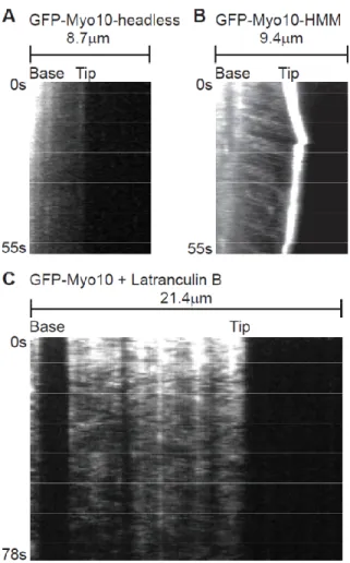
utilized a panel of Myo10 deletion constructs. No rapid particle movements and no tip
localization was detected in HeLa cells transfected with GFP-Myo10-headless, a
naturally occurring form of Myo10 that lacks most of the motor domain and thus lacks
motor activity (Sousa and Cheney, 2005) (Figure 2.5A). In addition, no rapid forward
tracks were detected with a full-length Myo10 construct containing a point mutation in its
motor domain that corresponds to a weak actin-binding mutation in other
myosins(Friedman et al., 1998) (GFP-Myo10-E456K; Figure 2.6). GFP-Myo10-HMM,
which consists of the Myo10 motor, neck, and predicted coiled coil, did undergo rapid
particle movements similar to those of full-length Myo10 (Figure 2.5B). This suggests
that a dimerized Myo10 head-neck domain is sufficient for fast forward movements.
Since systematic analysis of deletion constructs indicated that a forced-dimer construct
consisting of the Myo10 head, neck, and first 34 amino acids of the "coiled coil" fused to
a GCN4 dimerization domain was the minimal construct able to clearly localize to
filopodial tips, we imaged the forced dimer by TIRF and found that it was also capable of
fast forward movements in filopodia (Figure 2.6). Together these experiments indicate
37
Figure 2.5. Fast Forward movements require the Myo10 motor domain and are inhibited by latrunculin B
(A) Kymograph from TIRF imaging of GFP-Myo10-headless in a filopodium. This construct lacks most of the Myo10 motor domain and does not localize to filopodial tips or exhibit obvious fast forward movements. (B) Kymograph from TIRF imaging of GFP-Myo10-HMM. GFP-Myo10-HMM is sufficient for tip localization and faint particles of it undergo fast forward movements. (C) Kymograph showing that fast forward movements of GFP-Myo10 are blocked by latrunculin B. Cells were treated with 1 µM latrunculin B for ~10 minutes to depolymerize actin filaments and the remaining, substrate-attached filopodia were imaged by TIRF.
39
Figure 2.6. Ability of different Myo10 constructs to localize to filopodial tips and TIRF imaging of a motor domain point mutant and a forced dimer construct
(A-I) HeLa cells transfected overnight with the indicated GFP-tagged constructs (green) were replated ~12 hours on glass coverslips, fixed, and then stained with phalloidin (red). (A-D) The full-length, positive control GFP-Myo10 shows clear localization to the filopodial tip, whereas GFP-Myo10 E546K (a motor domain point mutant), as well as the negative controls (GFP-Myo10-headless and GFP alone) show little or no localization to the tip, demonstrating that Myo10 motor activity is necessary for strong tip localization. (E-I) Similar experiment showing that the GFP-Myo10-HMM positive control is clearly sufficient for localizing to filopodial tips, whereas the Myo10-head-neck, GFP-Myo10-head-neck-34, and the GFP-Myo10-head-neck-GCN4 forced dimer show little or no tip localization. The slightly longer GFP-Myo10-head-neck-34-GCN4 forced dimer is able to localize to the filopodial tip, although it also exhibits some diffuse labeling of the cell body. (J) Bar diagram summarizing the domain structure of different Myo10 constructs and their ability to localize to the filopodial tip. (K) Kymograph of a filopodium from a cell transfected with the motor domain point mutant (GFP-Myo10 E456K) and imaged by TIRF. The motor domain point mutant showed little localization to the filopodial tip relative to wild type GFP-Myo10 and did not generate clear tracks corresponding to rapid forward movements. (L) Kymograph of a filopodium from a cell transfected with the GFP-Myo10-head-neck-34-GCN4 forced dimer and imaged by TIRF. This construct generated occasional tracks corresponding to fast forward movements, indicating that a forced dimer is capable of rapid forward movements within filopodia.
40
dimerization of the head-neck region is sufficient for fast forward movement and tip
localization.
To test whether the fast forward movements are dependent on actin, cells were
treated with 1 µM latrunculin B to depolymerize actin filaments. As expected, this
triggered the collapse of filopodia that were not attached to the substrate (not shown).
Although latrunculin did not induce collapse of most substrate-attached filopodia, it did
cause the loss or spreading of the bright puncta of GFP-Myo10 normally present at their
tips (Figure 2.5C). Most importantly, fast forward movements of GFP-Myo10 were not
detected after treatment with latrunculin B, indicating that the fast forward movements
are indeed dependent on F-actin. Treatment of cells with 5 µM nocodazole did not block
fast forward movements of GFP-Myo10 (data not shown).
Faint particles of GFP-Myo5a undergo similar movements
To test whether other myosins were capable of similar movements within
filopodia, we imaged HeLa cells transfected with GFP-Myo1a (brush border myosin I), a
monomeric myosin that is non-processive and localizes to microvilli (Tyska and
Mooseker, 2002). TIRF showed that GFP-Myo1a yielded a diffuse localization along the
filopodia with no obvious enrichment at the filopodial tip (Figure 2.7A). Importantly,
GFP-Myo1a did not undergo detectable fast forward movements in filopodia, indicating
that not all myosins are capable of rapid directed movements in filopodia. We also tested
GFP-Myo5a (Wu et al., 2002), an intensively studied dimeric myosin that is processive
41
1996). Interestingly, GFP-Myo5a was enriched at the tips of filopodia and faint particles
of GFP-Myo5a generated clear tracks corresponding to rapid forward movement (Figure
2.7B). However, the GFP-Myo5a particles moved at only ~251 +/- 121 nm/s (59
measurements from 22 filopodia), significantly (P=0.035) slower than the ~578 nm/s
observed for GFP-Myo10. The velocity of the GFP-Myo5a particles is very similar to
the 270-330 nm/s reported for individual molecules of a dimeric Myo5a construct moving
on actin bundles in vitro (Nagy et al., 2008). It is therefore likely that the rapid and
directed movements of GFP-Myo5a detected here correspond to the visualization of
individual Myo5a molecules moving along the actin filaments of living cells. As with
GFP-Myo10, faint particles of GFP-Myo5a sometimes moved slowly rearward at the
retrograde flow rate of ~10-20 nm/s (Figure 2.7B). This observation provides direct
evidence that Myo5a can indeed undergo retrograde flow, as recently hypothesized (Liu
et al., 2006).
Conclusions
The TIRF experiments reported here reveal a novel form of long-range motility
driven by myosin motors at or near the single-molecule level. The fast movements
require Myo10 motor activity and actin filaments, but not the Myo10 tail. Together,
these results strongly support the hypothesis that Myo10 molecules use their barbed-end
motor activity to move forward along filopodial actin filaments (see model illustrated in
Movie S5). The faint particles detected with TIRF exhibit a size, intensity range, and
bleaching behavior consistent with imaging at the single-molecule level. It is not yet
42
Figure 2.7. Particles of GFP-Myo5a also move forward and rearward in the filopodia of living cells
(A) Kymographs from TIRF imaging of GFP-Myo1a in filopodia. GFP-Myo1a did not localize to the tips of filopodia and no tracks corresponding to rapid forward movement were detected. (B) Kymographs from TIRF imaging of GFP-Myo5a in filopodia. Note that several faint particles of GFP-Myo5a moved rapidly towards the tip while others moved slowly rearward.
43
Figure 2.8. Faint particles of GFP-VASP move rapidly forward in filopodia at velocities similar to those of GFP-Myo10
(A) Kymographs of filopodia from HeLa cells transfected with GFP-VASP and imaged using standard TIRF imaging conditions. Note that GFP-VASP localizes to filopodial tips and generates faint tracks corresponding to rapid forward movement. (B) Velocity histogram for fast forward movements of GFP-VASP particles.
44
oligomers of Myo10. Several factors contribute to this uncertainty, including the
relatively high and variable background fluorescence present in living cells, variable
levels of protein expression, the non-ideal behavior of GFP as a fluorophore, and
variations in the Z-axis position of the filopodium or molecules within it. For example,
even a 0.1 um difference in the Z-axis position (the approximate thickness of a
filopodium) will result in a ~2-fold change in the intensity of the TIRF field. It should
also be noted that although we can clearly detect the movements of some particles, we
cannot guarantee unambiguous detection of every GFP-Myo10 molecule in a filopodium.
Despite these limits, the combination of TIRF and GFP-tagging used here provides a
powerful strategy for imaging the movements of motor proteins and their cargos at or
near the single-molecule level in living cells.
In addition to exhibiting a novel form of rapid, long-range motility in filopodia,
Myo10 also has potent filopodia-promoting activity. Our previous work indicates that
Myo10’s ability to induce numerous filopodia requires (1) elements within the Myo10
tail and (2) the ability to localize to filopodial tips (Bohil et al., 2006). Since we now find
that only Myo10 constructs capable of moving rapidly forward in filopodia are able to
localize to filopodial tips, the novel form of motility reported here is likely to underlie
both tip localization and filopodia promotion. Although the precise mechanism(s) by
which My10 promotes filopodia are not yet clear, it could act by initiating filopodia, by
transporting cargos that facilitate filopodia formation, by functioning as part of a mobile
tip complex, or by some combination of these or other mechanisms (Bohil et al., 2006;
45
As a motor that moves rapidly along filopodia, Myo10 clearly transports itself to
the filopodial tip, but it may also be responsible for the transport of other specific
molecular cargos, such as VASP(Tokuo and Ikebe, 2004). Indeed, preliminary
experiments indicate that faint particles of GFP-VASP exhibit fast, forward movements
in filopodia very similar to those of Myo10 (Figure 2.8). A major goal for the future will
be to test for cotransport at the single-molecule level and to determine whether Myo10 is
required for transport of this and other cargos. It will also be interesting to determine if
Myo3a or Myo15a, myosins that localize to the tips of stereocilia and are necessary for
hearing, undergo similar forms of movement (Belyantseva et al., 2005; Liu et al., 2006;
Schneider et al., 2006). In addition to revealing a novel form of motility, this work also
suggests that motor proteins may power many as yet undetected movements at the
single-molecule level.
Materials and Methods
Constructs. Bovine Myo10 (aa 1-2052), Myo10-HMM (aa 1-947), and
GFP-Myo10-head-neck (aa 1-811) in pEGFP-C2 have been described previously (Berg and
Cheney, 2002). The GFP-Myo10 E456K motor domain point mutant (GAG>AAG, nt
1588-1590) was generated by PCR in pEGFP-C2 and corresponds to a weak
actin-binding mutation in other myosins(Friedman et al., 1998). The
GFP-Myo10-head–neck-GCN4 forced dimer was generated by fusing the 29 aa leucine zipper
(VKQLEDKVEELASKNYHLENEVARLKKLV) from yeast GCN4 to aa 811 of the
bovine Myo10-head-neck construct and cloning this into the BglII-HindIII sites of
46
Myo10 inserted into the BglII-HindIII sites pEGFP-C2. The
GFP-Myo10-head-neck-34-GCN4 construct was identical except that the GFP-Myo10-head-neck-34-GCN4 leucine zipper sequence was added
after aa 845. Myo10 constructs were verified by sequencing and their numbering is
based on GenBank sequence NM_174394. Mouse GFP-Myo1a in pEGFP-C (Tyska and
Mooseker, 2002) was a generous gift from Dr. Matthew Tyska, mouse brain GFP-Myo5a
in pEGFP-C1 ("BR MV")(Wu et al., 2002) was a generous gift of Dr. John Hammer,
human GFP-VASP in pEGFP-N1 was a generous gift from Dr. Frank Gertler, and
pEGFP-C2 was used as a GFP control.
Cells. HeLa cells were transfected with Polyfect (Qiagen) unless indicated otherwise.
To obtain the relatively low levels of expression required to facilitate single-molecule
imaging, HeLa cells were generally transfected for no more than 6-12 hours. Cells were
replated onto #1.5 glass coverslips that had been precoated with fibronectin to facilitate
formation of substrate-attached filopodia. Coverslips were precoated by incubating
acid-washed coverslips for 20 minutes in 10 µg/ml fibronectin in PBS and then acid-washed at least
3x in PBS over 10 minutes prior to plating. To minimize background from cellular
debris, cells were usually plated onto coverslips at <10% confluence, with best results
obtained with less than one cell per camera field. Cells were allowed to attach to the
coverslip for 1-2 hours and the coverslip was mounted in a Rose chamber with a 3 mm
spacer and a #1.5 coverslip for the roof. The chamber was completely filled with
Optimem (Gibco). Tet-off HeLa cells (Clontech) that "stably" express GFP-Myo10 were
generated as per the manufacturer's instructions. These cells were withdrawn from