Cover Page
The handle
http://hdl.handle.net/1887/22208
holds various files of this Leiden University
dissertation
Author: Vaart, Michiel van der
Innate host defense
against intracellular pathogens
Cover: A macrophage (magenta) has phagocytosed Mycobacterium marinum bacteria (black)
Printed and bound by Wöhrmann Print Service, Zutphen, The Netherlands
Innate host defense against intracellular pathogens
Proefschrift
Ter verkrijging van
de graad van Doctor aan de Universiteit Leiden
op gezag van Rector Magnificus prof.mr. C.J.J.M. Stolker,
volgens besluit van het College voor Promoties
te verdedigen op donderdag 14 november 2013
klokke 13.45 uur
door
Michiel van der Vaart
geboren te Maasland
Promotiecommissie:
Promotor: Prof. Dr. H.P. Spaink Co-promotor: Dr. A.H. Meijer Overige leden: Prof. Dr. C.J. ten Cate
Prof. Dr. J. den Hertog
Table of contents
Chapter 1
11
Outline of the thesis
Chapter 2
15
Pathogen recognition and activation of the
innate immune response in zebrafish
Chapter 3
49
Functional analysis of a zebrafish
myd88
mutant
identifies key transcriptional components of the
innate immune system
Chapter 4
79
Mycobacteria counteract a nitrosative host
defense mechanism initiated upon
TLR/IL1R-MyD88 signaling
Chapter
5
101
DRAM1 links pathogen recognition via
TLR-MYD88 to autophagic defense against
tuberculosis
Chapter 6
129
Summary and discussion
Chapter 7
141
Dutch summary
(Nederlandstalige samenvatting)
Chapter 8
145
Curriculum vitae
Chapter 9
149
1
Chapter 1
1
H
umans rely on their immune system for defense against invading microorganisms, such as pathogenic bacteria, fungi, parasites, and viruses. The defense against pathogens can be divided into two general types of host responses: (1) the innate immune response, and (2) the adaptive immune response. The innate immune response consists of cells and proteins that form the first line of defense against invading pathogens. Their function is to quickly arrive at the site of infection and fight off microbes that have breached physical barriers such as the skin or mucosal surfaces. The adaptive immune system is called into action when the innate immune response alone is not sufficient to kill the pathogen. Adaptive immunity involves the production of antibodies that bind and neutralize the pathogen, and the activation of T-cells that directly recognize and kill the pathogen. The innate immune system is the most ancient defense mechanism and many of its components are shared between invertebrate and vertebrate species. The adaptive immune system is unique to vertebrates and arose during the evolution of fishes. Nowadays the zebrafish is one of the most powerful model organisms for the study of the immune system.
This thesis focuses on the recognition of pathogenic bacteria and the defense mechanisms that are activated during the innate immune response. Innate immune detection of pathogens depends on a number of pattern recognition receptors (PRRs) capable of detecting the presence of evolutionary conserved structures on microbes, called pathogen-associated molecular patterns (PAMPs). In chapter 2, we introduce the different classes of PRRs available to the innate immune system and the defense mechanisms which they can activate upon detection of a pathogen. We also review the conservation of these receptors and mechanisms in zebrafish, and discuss recent studies using zebrafish infection models that have advanced our general understanding of the innate immune system.
The most extensively studied class of PRRs is the Toll-like receptor (TLR) family, which signals via adaptor molecules to initiate gene expression and activate the appropriate response upon recognition of a pathogen. In chapter 3,we present a zebrafish mutant line for the TLR signaling adaptor myeloid differentiation factor 88 (MyD88), which is central to the innate immune processes underlying infectious diseases, inflammatory disorders, and cancer. Zebrafish myd88 mutant embryos were more susceptible to infection with the acute bacterial pathogens Salmonella typhimurium and Edwardsiella tarda, as well as the chronic bacterial pathogen Mycobacterium marinum. Further characterization revealed that gene expression of transcription factors and defense proteins important to innate immunity depends on Myd88 during these bacterial infections.
1
immune cells in chapter 4, and found that Mycobacterium marinum could attenuatethis response and thereby avoid the cytostatic and cytotoxic effects of RNS.
Autophagy is a cellular homeostasis process that acts as a quality control system for eukaryotic cells. It has a central role in programmed cell death and survival by providing energy and nutrients and ridding the cytoplasm of unwanted components. Selective autophagy has recently emerged as defense mechanism activated by PRRs against tuberculosis disease, although the responsible molecular signaling pathway remained unclear. In chapter 5, we used zebrafish embryos and human macrophages to demonstrate that anti-mycobacterial autophagy is orchestrated by TLR-MYD88-NFκB signaling via DNA-damage regulated autophagy modulator 1 (DRAM1). Furthermore,
2
Chapter 2
Pathogen recognition and activation of
the innate immune response in zebrafish
Michiel van der Vaart, Herman P. Spaink, and Annemarie H. Meijer
2
Abstract
T
he zebrafish has proven itself as an excellent model to study vertebrate innate immunity. It presents us with possibilities for in vivo imaging of host-pathogen interactions which are unparalleled in mammalian model systems. In addition, its suitability for genetic approaches is providing new insights on the mechanisms underlying the innate immune response. Here, we review the pattern recognition receptors that identify invading microbes, as well as the innate immune effector mechanisms that they activate in zebrafish embryos. We compare the current knowledge about these processes in mammalian models and zebrafish, and discuss recent studies using zebrafish infection models that have advanced our general understanding of the innate immune system. Furthermore, we use transcriptome analysis of zebrafish infected with E. tarda, S. typhimurium, and M. marinum to visualize the gene expression profiles resulting from these infections. Our data illustrate that the two acute disease causing pathogens, E. tarda and S. typhimurium, elicit a highly similar pro-inflammatory gene induction profile, while the chronic disease causing pathogen, M. marinum, induces a weaker and delayed innate immune response.1.
Introduction
The use of adult zebrafish (Danio rerio) and their transparent offspring as hosts to model infectious diseases caused by human pathogens, or closely related animal pathogens, has recently provided novel insights into pathogenesis, which in many cases could not have been achieved using mammalian models1-6. The power of the
zebrafish model lies in its suitability for genetic approaches, high-throughput screening, and live imaging studies. Fluorophore-marked transgenic lines are now available that allow unprecedented visualization of pathogen interactions with macrophages and neutrophils, the major phagocytic innate immune cell types of zebrafish larvae7-11. As
early as one day post fertilization (dpf), zebrafish embryos display phagocytic activity towards microbial infections12 and are able to mount an innate immune response with
a transcriptional signature that resembles responses in mammalian or cell culture systems13. Adaptive immunity becomes active after approximately three weeks of
development14. Therefore innate immunity can be studied during the earlier zebrafish
2
2.
Pattern recognition receptors
The innate immune system is the host’s first line of defense against infection, therefore its main role is to recognize invading pathogens early and trigger an appropriate proinflammatory response15. The innate immune system utilizes a limited number of
germ line-encoded pattern recognition receptors (PRRs) to recognize evolutionary conserved structures on pathogens, named pathogen-associated molecular patterns (PAMPs)15. PRRs are also capable of indirectly sensing the presence of pathogens16, 17. This occurs when infection, inflammation, or other cellular stresses cause host
factors to be present in aberrant locations, or to form abnormal molecular complexes, so called danger-associated molecular patterns (DAMPs)17. PRRs located on the cell
surface are scouting the extracellular environment for the presence of microbes. PRRs located on endosomes identify microbes that have entered the phagolysosomal degradation pathway, and cytoplasmic PRRs recognize intracellular cytosolic pathogens or components of internalized microbes18. Upon PAMP recognition, PRRs signal the
presence of infection and initiate proinflammatory and antimicrobial responses by activating several intracellular signaling pathways19, ultimately leading to activation
of gene expression and synthesis of a broad range of molecules. These include proinflammatory and chemotactic cytokines, and antimicrobial peptides20. The different
families of PRRs present in both humans and zebrafish and their downstream signaling pathways are summarized in Figure 1 and will be discussed below.
2.1
Toll-like receptors
The most extensively studied class of PRRs are the Toll-like receptors (TLRs), a family of 10 proteins in human. TLRs are named after the Drosophila Toll protein, which functions in dorso-ventral patterning and antifungal responses21. TLRs are integral glycoproteins
which possess an extracellular or luminal, ligand-binding domain with leucine-rich repeat (LRR) motifs and a cytoplasmic signaling Toll/Interleukin-1 (IL-1) receptor homology (TIR) domain20, 22. In mammals, the main cell types expressing TLRs are antigen
presenting cells (APCs), including macrophages and dendritic cells, and B lymphocytes18.
However, most cell types are capable of expressing TLRs, for instance in response to a localized infection23. In mammals, TLR4 recognizes Gram-negative bacteria via the lipid
A portion of lipopolysaccharide (LPS), while TLR2 recognizes Gram-positive bacteria via lipoteichoic acid (LTA), lipoproteins, and peptidoglycan, and TLR5 recognizes the motility apparatus protein flagellin, which can be present on both Gram types 18. Other TLRs are
specialized in recognizing nuclear acids in endosomal and phagosomal compartments. TLR3 can detect viral replication by binding to double-stranded RNA (dsRNA), TLR7 and TLR8 specifically recognize single-stranded RNA (ssRNA) of RNA viruses, and unmethylated CpG DNA present in the genomes of viruses and bacteria is detected by TLR918. Ligand binding by a TLR will induce it to form homomeric or heteromeric
oligomers, which triggers intracellular signal transduction via their TIR domains18. The
2
molecules: MYD88, MAL/TIRAP, TRIF/TICAM1, TRAM/TICAM2, and SARM19, 22. Among
these, MYD88 is the most universal adaptor, since it is used for downstream signaling by all TLRs, with the exception of TLR324. Downstream signaling via central intermediate
molecules such as TRAF6, will eventually lead to the activation of transcription factors, mostly members of the ATF, NFĸB, AP-1, IRF, and STAT families, regulating the expression of a battery of antimicrobial and proinflammatory genes24.
Putative orthologs of mammalian TLRs have been identified in zebrafish, in addition to some fish-specific family members25, 26. A genome duplication during the evolution
of teleost fish most likely explains why zebrafish have two counterparts for some of the mammalian TLRs (e.g. tlr4ba/tlr4bb for TLR4 and tlr5a/tlr5b for TLR5), but it is still unknown whether this increase in the number of receptors is associated with diversification in PAMP recognition4. Only some of the zebrafish TLR ligands are currently
known27. The specificity of TLR2, TLR3, and TLR5 is conserved between mammals and
fish, recognizing lipopeptides, dsRNA, and flagellin respectively13, 28, 29. Additionally,
the fish-specific TLR22 has been shown to recognize dsRNA and PolyI:C29. However,
zebrafish TLR4 cannot be stimulated by LPS, illustrating that not all ligand specificities are conserved between mammals and zebrafish30, 31. Signaling intermediates in the
pathway downstream of mammalian TLRs have also been identified in zebrafish, including homologs of four of the adaptor proteins, Myd88, Mal/Tirap, Trif/Ticam1, and Sarm, and the central intermediate Traf632. Among these, Myd88 and Traf6 have
been functionally studied by knockdown analysis in zebrafish embryos, showing their requirement for a proinflammatory innate immune response to microbial presence13, 33-35. Furthermore, triggering of the innate immune response in zebrafish embryos
also leads to induction of members of the ATF, NFĸB, AP-1, IRF, and STAT families of transcription factors1313, 36.
2.2
NOD-like receptors
Pathogens that escape the surveillance of cell surface and endosomal PRRs may end up in the cytosol, where nucleotide-binding oligomerization domain (NOD)-like receptors (NLRs) detect their presence by intracellular PAMPs and DAMPs37. The NLRs
constitute a family of 23 proteins in humans. Their defining features are the presence of a centrally located NOD domain responsible for oligomerization, a C-terminal LRR capable of ligand-binding, and an N-terminal protein-protein interaction domain, such as the caspase recruitment domain (CARD), pyrin (PYD), or baculovirus inhibitor repeat (BIR) domain38. Two of the NLRs, NOD1 and NOD2, can sense bacterial presence by
directly or indirectly detecting molecules produced during synthesis or breakdown of peptidoglycan38. NOD1 recognizes g-D-glutamyl-meso-diaminopimelic acid (iE-DAP), a
2
that these receptors can recognize a broader range of pathogens than was originallyassumed41, 42. Upon ligand-binding, NOD1 and NOD2 recruit the serine/threonine
kinase RIPK2 (also known as RIP2) via CARD-CARD interactions, eventually leading to the activation of NFĸB43, 44. In addition, NOD1/2 stimulation also induces MAP kinase
signaling45. Synergistically, with TLR activation, NOD1/2 signaling cascades induce the
expression of cytokines and chemokines, such as TNF, IL6, IL8, IL10, and IL12, as well as the production of anti-microbial peptides44, 46, 47.
Other NLRs, such as IPAF, NALP1, and NALP3 mainly function to create a multiprotein complex known as the inflammasome, in which they associate with an adaptor called ASC (apoptosis-associated speck-like protein containing a CARD) and with pro-caspase 148. Oligomerization of the proteins in an inflammasome via CARD-CARD interactions
ultimately leads to the cleavage of pro-caspase 1 into its active form, caspase 1, which is then available to catalyze the cleavage of accumulated pro-IL1β and pro-IL18 into their secreted forms, biological active IL1β and IL1838. The NLR family member incorporated
into these complexes determines which PAMPs and DAMPs are recognized by the inflammasome. A role for NALP3 has been established in the recognition of ATP49, uric
acid crystals50, viral RNA51, and bacterial DNA52. Both NALP1 and NALP3 share NOD2’s
ability to respond to MDP53. Furthermore, NALP1 can associate with NOD2 (Hsu 2008),
showing a role for NOD2 in MDP-triggered IL1β activation, separate from its role as an inducer of proinflammatory gene expression.
Although the function of NLR family members in zebrafish is not widely studied, it is known that the canonical members of the mammalian NLR family, NOD1, NOD2, and NOD3 (or Nlrc3), are conserved. Additionally, a subfamily of NLRs resembling the mammalian NALPs and a unique teleost NLR subfamily are present32, 54. Confirmation of
the antibacterial role of NOD1 and NOD2 in zebrafish was achieved by gene knockdown,
resulting in higher bacterial burdens and decreased survival of embryos following
Salmonella enterica infection55. Moreover, nod1/2 depletion significantly decreased
expression of dual oxidase (DUOX), required for production of reactive oxygen species (ROS)55. These findings illustrate that the family of Nod-like receptors and their
down-stream signaling pathways are important for antibacterial innate immunity, both in mammals and in zebrafish.
2.3
RIG-I like receptors
Another family of cytosolic PRRs, the RIG-I like receptors (RLRs), consists of three members: RIG-I (retinoic acid-inducible gene I), MDA5 (melanoma differentiation associated factor 5), and LGP2 (laboratory of genetics and physiology 2). All three members are DExD/H box RNA helicases that can detect the presence of RNA from a broad range of viruses56. While expressed at low levels in most tissues, their expression is
greatly increased upon viral infections or interferon (IFN) exposure57, 58. The RNA helicase
domain of RLRs has the capacity to hydrolyze ATP and bind to RNA59. Furthermore, RIG-I
2
LGP2 lacks the two CARDs and is thought to function as a negative regulator of RIG-I and MDA5 signaling60. Following recognition of viral RNA, the CARDs of RIG-I and MDA5
become available for binding to a common mitochondrial signaling adaptor, IPS-1 or MAVS61. The subsequent signaling cascade culminates in the induction of transcription
factors like interferon regulatory factor 3 (IRF3), IRF7, and NFĸB62. Activation of these
transcription factors leads to the production of type I IFN, which binds to the IFN receptor to initiate expression of interferon-stimulated genes (ISGs)63. Amongst these
ISGs are antiviral proteins, immune-proteasome components, all three RLRs, members of the TLR family, transcription factors like IRF7, and various proinflammatory cytokines and chemokines63. As such, the RLR-induced pathway works cooperatively with TLR
signaling to prepare the cell for elimination of viral infections56.
Zebrafish homologs of RIG-I, MDA5, and DXH58 were identified in a genome search64.
However, in silico analysis of the predicted proteins revealed that the domain distribution differs between humans and zebrafish64. For instance, whilst human RIG-I contains two
CARDs, one DExD/H domain, and a Helicase C domain, zebrafish RIG-I consists of a single CARD and a DExD/H domain64. Whilst functional studies of the RLR pathway are scarce,
it is clear that zebrafish and other teleosts possess a strong antiviral IFN system, which shares a common evolutionary origin with mammals65, 66. The mitochondrial RLR adaptor,
IPS-1/MAVS, was recently cloned from salmon and zebrafish, and overexpression in fish cells led to a constitutive induction of ISGs66. Furthermore, MITA, another adaptor
functioning downstream of IPS-1/MAVS and upstream of Tank-binding kinase 1 (TBK1), was cloned from crucian carp (Carassius auratus) and shown to activate zebrafish IFN promoter gene constructs, dependent on IRF3 or IRF767.
2.4
Scavenger receptors
Scavenger receptors are a large family of transmembrane cell surface receptors, present on macrophages, dendritic cells, mast cells68 and some endothelial and epithelial cell
types69. Although originally defined for their role in uptake of low-density lipoproteins
(LDL), they are now known to act as PRRs for a wide variety of PAMPs, like LPS, LTA, CpG DNA, yeast zymozan, and microbial surface proteins70. Commonly, PAMP binding
to a scavenger receptor will induce the cell to directly phagocytose the pathogen71.
Upregulation of scavenger receptor expression via TLR signaling can be a mechanism to increase phagocytic activity72. Moreover, scavenger receptors can also contribute to
cytokine production as co-receptors for TLRs73, 74. Some of the C-type lectins, discussed
below, also display scavenger receptor activity.
Based upon their multidomain structure, scavenger receptors are divided into eight subclasses (A-H) (Murphy 2005). Subclasses A and B are the most extensively studied, but members from other subclasses have also been shown to recognize bacterial PAMPs70.
2
pneumonia and Escherichia coli75-77. Macrophage receptor with collagenous structure
(MARCO), another subclass A member with established PRR activity78, functions as a
phagocytic receptor for S. pneumonia79 and N. meningitidis80. MARCO was shown to
cooperate with TLR2 to trigger macrophage cytokine responses to the mycobacterial cell wall glycolipid trehalose dimycolate (TDM) and Mycobacterium tuberculosis81. CD36,
the most prominent member of subclass B, is a sensor for LTA and diacylated lipopeptide (MALP-2) and also acts as a co-receptor for TLR273. CD36-mediated phagocytosis of S.
aureus was shown to be required for initiation of TLR2/6 signaling82. SR-BI (or CLA-1),
also in subclass B, can bind to LPS and was implicated in phagocytosis of both Gram-negative and Gram-positive bacteria83. As well as their anti-bacterial roles, CD36 and
SR-BI are also known for increasing the pathogenesis of malaria and hepatitis C virus (HCV). CD36 can function as a receptor for erythrocytes that have been parasitized by
Plasmodium falciparum, adhering these cells to the venular endothelium of various organs70. Furthermore, SR-BI is used by Plasmodium sporozoites and HCV as an entry
site into hepatocytes70.
Many homologs of the mammalian scavenger receptor family can be identified in the zebrafish genome, but a systematic analysis is still awaited. A zebrafish homolog of human MARCO was identified as a specific marker for macrophages and dendritic cells from adult zebrafish84, and this gene is also myeloid-specific in zebrafish embryos85.
Expression of the cd36 gene was up-regulated after exposing zebrafish to haemorrhagic septicemia rhabdovirus86. In contrast, cd36 expression was down-regulated by
Mycobacterium marinum infection in adult zebrafish and larvae87.
2.5
C-type lectins
The C-type lectin receptors (CLRs) are a large family of carbohydrate-binding proteins that are highly conserved amongst mammals88. The diversity of the CLR family is
illustrated by the fact that up to 17 groups are present in vertebrates, with some consisting of soluble serum proteins, whilst others consist of transmembrane proteins. These are mainly expressed in myeloid cells (macrophages and dendritic cells) but also in natural killer cells89, 90. The best known CLR in serum is mannose-binding lectin (MBL),
a member of the collectin class, which binds to a variety of sugar moieties present on viruses, bacteria, fungi, and protozoa, and activates the complement system91. In terms
of their function as PRRs, the transmembrane CLRs that are expressed on myeloid cells are the most interesting. Transmembrane CLRs can be divided into two groups: the mannose receptor family and the asialoglycoprotein receptor family92. CLRs recognize
pathogens mainly via ligand-binding to mannose, fucose, and glucan carbohydrate structures, which means that together they are capable of recognizing most classes of human pathogens92. Like scavenger receptors, CLRs can act as phagocytic receptors
for non-opsonized bacteria, leading to their destruction in acidified phagolysosomes71.
2
induced by CLRs like Dectin-1 is not only important for the lysosomal breakdown of pathogens, but also for antigen presentation94, 95. Besides their role in phagocytosis,
CLRs can directly induce gene expression upon carbohydrate recognition. PAMP recognition by Dectin-1, Dectin-2 and macrophage-inducible C-type lectin (Mincle) ultimately leads to activation of NFĸB96-98. Where Dectin-1 associates with the kinase
Syk to activate NFĸB99, Dectin-2 and Mincle are dependent on Fc receptor Υ-chain as
an adaptor molecule97, 98. Other CLRs, for example DC-specific ICAM3-grabbing
non-integrin (DC-SIGN), induce specific gene expression profiles upon pathogen recognition by modulating TLR signalling92. When DC-SIGN recognizes mannose or fucose moieties
on pathogens such as Mycobacteria, HIV-1, measles virus, and Candida albicans, it activates a Raf-1-dependent signaling pathway that modulates TLR-induced NFĸB activation, increasing the production of IL8 and anti-inflammatory IL10 production100.
Only a few homologs of CLRs have been described in zebrafish. A homolog of the complement activating mannose-binding lectin (MBL) was associated with resistance against Listonella anguillarum101. Expression of another soluble lectin, lgals91l, is
enriched in zebrafish embryonic myeloid cells and is dependent on the Spi1/Pu.1 transcription factor that plays a crucial role in myeloid cell development in vertebrates85.
A membrane type collectin, CL-P1 (collectin placenta 1), was shown to be involved in vasculogenesis during zebrafish embryogenesis102. In humans, CL-P1 is mainly expressed
on vascular endothelial cells and has been shown to act as a scavenger receptor mediating the phagocytosis of bacteria and yeast103. A putative homolog for DC-SIGN
has recently been proposed, and is upregulated in immune-related tissues following infection by Aeromonas anguillarum104. Finally, putative homologs for the mammalian
C-type lectin NK cell receptors have been identified in zebrafish and are differentially expressed on cells from the myeloid and lymphoid lineages105.
3.
Effector mechanisms of the innate immune response in zebrafish
2
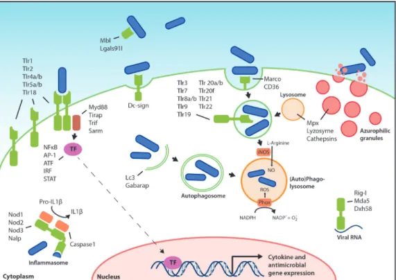
Figure 1: Pattern recognition receptors and effector mechanisms of the innate immune system. The localization of Tlrs on the cell surface or on endosomes is hypothetical and based on the known or proposed functions of their homologs in other fish or mammals. The ability of PRRs (depicted in green) to recognize PAMPs present on various types of microorganisms, like bacteria, viruses, and fungi, has been simplified here by depicting microorganisms as rod-like bacteria (in blue). PAMP recognition by PRRs leads to activation of transcription factors (TF), which translocate to the nucleus and initiate transcription of cytokine genes, antimicrobial genes, and other immune related genes. Defense mechanisms such as autophagy, ROS and NO production, and degranulation can be immediately activated upon microbial recognition, without de novo gene transcription.
3.1
Secreted peptides and lipid mediators of the innate immune response
Cytokines, including interleukins, chemokines, and interferons, are small secreted proteins that steer the host’s immune system into a cytotoxic, humoral, cell-mediated, or allergic response109. Since this review focuses on innate immunity, we will mainly
discuss the cytokines produced by or acting on phagocytic cells. A distinction can be made between cytokines that promote a state of inflammation and cytokines that are anti-inflammatory. The main pro-inflammatory cytokines produced by phagocytes are TNFα, IL1α, IL1β, IL6, and IL8109. TNF-α is processed as a membrane-bound protein and,
when required, the active soluble factor is cleaved off by the TNF-α converting enzyme (TACE)110. Similarly, IL1α and IL1β are synthesized as inactive precursors that are only
secreted as active cytokines after inflammasome-mediated cleavage by caspase 1111.
2
pro-inflammatory cytokine production by macrophages and T-cells112. The IL10/IL12
balance, maintained by cells of the innate immune system, determines whether adaptive immunity polarizes towards a Th1 (promoted by IL12) or Th2 response. A Th1 response, which activates the bactericidal activities of macrophages, is the most important for controlling intracellular pathogens. The single type II IFN, IFNγ, is also required for activating macrophage bactericidal functions, while type I IFNs (IFNα and IFNβ) and type III IFN (IFNλ) function in mounting anti-viral responses. Finally, eicosanoid lipid mediators also promote (e.g. prostaglandins and leukotrienes) or inhibit (e.g. lipoxins) inflammation, thus synergizing with or antagonizing cytokine functions.
Many of the cytokine subfamilies are conserved between zebrafish and mammals32.
However, there has been extensive expansion and diversification of members of the chemokine gene family in zebrafish and their specific functions are yet to be determined113. Several of the main cytokines, like IL1β, IL6, and IL10, have been cloned
and characterized114-116. Furthermore, the zebrafish homolog of Interleukin 10 receptor
1 (IL10R1) has recently been identified and seems to contain all the protein domains that are required for its function in anti-inflammatory signaling117. The pro-inflammatory
chemokine IL8 (CXCL8) and it receptors, CXCR1 and CXCR2, are also conserved between mammals and zebrafish118. In addition, a second IL8/CXCL8 lineage has been
identified in both zebrafish and common carp (Cyprinus carpio), and the chemotactic properties of carp IL8/CXCL8 molecules of both lineages were demonstrated by in vitro chemotaxis assays using carp leukocytes119. Both pro- and anti-inflammatory
cytokines are upregulated upon infection of zebrafish embryos with pathogens such as
S. typhimurium13, P. aeruginosa120, and E. tarda121, 122.
The role of TNF during Mycobacteriummarinum infection of zebrafish embryos was studied by knockdown analysis of the TNF receptor (tnfrsf1a), which revealed that intracellular bacterial burdens, granuloma formation, and necrotic death of macrophages are increased in the absence of TNF signaling123. The importance of TNF signaling during
M. marinum infection was further illustrated when the same model was used to show that a strict balance between pro-inflammatory TNF and anti-inflammatory lipoxins is vital for control of mycobacterial infections, with either too much or too little TNF expression leading to a more severe outcome of the disease1. Another study using the
zebrafish model indicates that TNF-α is a potent activator of endothelial cells, leading to the production of chemokines, whilst it has little effect on the activation status of phagocytes124. This suggests that fish TNF-α mainly functions in the recruitment of
leukocytes to the site of infection, rather than activating them.
The three IFN groups present in humans are not conserved unambiguously in zebrafish and other fish species. The type II group of IFNs in zebrafish consists of IFNγ1 and IFNγ2125. Expression levels of the corresponding genes did not change upon infection of
2
IFNγ1 and IFNγ2 were shown to bind to different receptor complexes and Janus kinase2a (Jak2a), but not Jak2b, was shown to be required for intracellular transmission of the IFNγ signal. Two groups of antiviral IFNs, named IFNφ1 and IFNφ2 exist in zebrafish, and structural analysis showed that these are evolutionarily closer to type I than to type III human IFNs32, 65, 127. IFNφ1 and IFNφ 2 signal via distinct receptor complexes65, 128. All
zebrafish IFNφ genes induce the expression of genes that are predicted to be involved in antiviral activities65.
3.2
Phagocytosis, autophagy, and lysosomal destruction
Internalization of microorganisms is triggered when they are recognized by phagocytic receptors, mainly by scavenger receptors discussed above. This type of direct phagocytosis is termed non-opsonic phagocytosis, while opsonic phagocytosis relies on host-derived proteins that coat the surface of the microbe thereby enhancing phagocytosis efficiency. Opsonins include complement fragments, most notably C3b, which are recognized by complement receptors129. Mannose binding lectin, which can
initiate C3b formation, and antibodies that bind to Fc receptors (IgG) or that activate complement (IgM) are also considered opsonins. Regardless of which receptor initiates the process, phagocytosis requires the activation of kinases and Rab GTPases that control alterations in the phospholipid membrane and remodeling of the actin cytoskeleton130. In
macrophages, fusion of the resulting vesicle with early and late endosomes will decrease the pH of the immature phagosome and alter the proteins present on its membrane. Ultimately, maturing phagosomes turn into phagolysosomes when lysosomes fuse with them, mixing their contents131. Lysosomes are highly acidic endocytic vesicles (pH<5.5),
containing active proteases and lipases, and hydrolytic enzymes such as cathepsin D132.
In addition, phagolysosomes also contain bactericidal peptides (defensins) and have the ability to generate toxic oxidative compounds that help microbial degradation133. Most
of our knowledge about phagosome maturation comes from studies of phagocytosis in macrophages, much less is known about phagosome maturation in neutrophils. While macrophage phagosomes fuse with endosomes and lysosomes, neutrophil phagosomes obtain their bactericidal properties by fusing with secretory vesicles and granules134, 135.
In contrast to phagosome maturation in macrophages, neutrophil phagosomes do not acidify in order to become microbicidal134, 135.
Many intracellular pathogens, like M. tuberculosis, S. typhimurium, and Legionella pneumophila have evolved the ability to prevent phagosome maturation in macrophages and survive inside these vesicles136. To a certain extent such pathogens
can also withstand the hostile environment of the (phago)lysosome. Other pathogens like Listeria monocytogenes, Francisella tularensis, and many viruses can escape the phagosome and enter the cytosol137. Mycobacterium marinum, a pathogen studied
2
M. tuberculosis and is dependent on a virulence factor, the ESX-/RD1 secretion system, shared by all pathogenic mycobacteria140. Together, these data indicate that host cells face
numerous pathogens that have developed multiple strategies to avoid the pathway of phagolysosomal degradation. To counter such threats, cells may use autophagy to clear microbes and microbe containing vesicles from the cytosol. Autophagy is well-known as a metabolic process that recycles nutrients by degrading intracellular organelles and proteins. Only recently it has been recognized that autophagy also plays an important role in the innate immune response against intracellular pathogens141. Autophagy is
initiated when an autophagosomal isolation membrane is formed around its target, enclosing it entirely in a double membrane vesicle. This process relies on Class III phosphatidylinositol 3-kinase (PI3-kinase) and autophagy-related genes (Atgs), such as Atg6 (or Beclin-1)142. The hallmark of autophagosomes is the presence of Atg8 (or LC3) in
their membranes, which is essential for membrane elongation143. Similar to a maturing
phagosome, the autophagosome also fuses with lysosomes to achieve its degradative properties144. In addition, autolysosomes acquire unique antimicrobial properties due
to the function of autophagic adaptor protein p62, which delivers cytosolic components to autolysosomes where they are processed into potent antimicrobial peptides145. As
reviewed elsewhere146, pathogen-targeted autophagy can be induced by several TLRs
and NLRs, TNF-α, NFĸB, and many other immune-related signalling molecules.
The transparency of zebrafish embryos and availability of fluorescent macrophage and neutrophil reporter lines allows for study of the process of phagocytosis in great detail7, 147-149. It was recently shown that zebrafish embryonic macrophages efficiently engulf E.
coli bacteria from blood- and fluid-filled cavities, while neutrophils are hardly capable of phagocytosing bacteria present in fluids149. However, neutrophils did prove to be
highly phagocytic when moving over bacteria present on tissue surfaces. This shows that the type of immune cell that clears an infection not only depends on the PAMPs present on the invading microbe, but also on the characteristics of the infection site. An
in vivo phagocytosis assay was used to show that functions of Wasp1, Wasp2, Abi2 and cofilin regulator 14-3-3ζ (Ywab) in bacterial phagocytosis are conserved in zebrafish150.
The recent generation of a transgenic zebrafish line with GFP-tagged LC3 has enabled
in vivo visualization of the interactions between microbes and this core component of the autophagy machinery151. The importance of autophagy in the innate immune
response of zebrafish remains to be studied, but we have shown that LC3-labeled structures accumulate around M. marinum infection sites in zebrafish embryos (Figure 2). Furthermore, autophagy-related genes were induced in adult zebrafish infected with
Citrobacter freundii and zebrafish embryos infected with S. typhimurium35, 152.
3.3
Oxidative defenses in leukocytes
2
Figure 2: In situ detection of autophagy by Lc3 accumulation. CMV::LC3-GFP transgenic (He et al. 2009) zebrafish embryos (28 hpf) were injected into the caudal vein with 200 colony forming units (CFU) of M. marinum Mma20 expressing a pMST3::mCherry vector. Confocal images were taken of a tail region of the developing larva at 3 days post infection (3 dpi), a point at which the M. marinum infection (A) has been established. Low levels of Lc3-GFP signal (B) can be observed throughout the cells, whilst brighter regions (indicated by arrowheads) are only observed upon Lc3 accumulation and formation of autophagic membranes associated with bacteria (C). Scale bar: 10 µm.
matrix153. Upon recognition of pathogens, neutrophils release their antimicrobial
granules, called azurophils, into phagosomes or the extracellular environment154, 155. Azurophils are packed with acidic hydrolases and antimicrobial proteins, such as
lyzosyme, cathepsins, and myeloperoxidase (MPO)156. The primary function of MPO is to
react with hydrogen peroxide (H2O2), which subsequently oxidates chloride, tyrosine, and nitrite to form hypochloric acid (HOCl), tyrosine radicals, and reactive nitrogen intermediates157. These highly reactive chemicals attack the surface membranes of
microbes. Additionally, microbes can be bound by neutrophil extracellular traps (NETs), which are fibrous networks of granule proteins and chromatin released by neutrophils158.
While MPO is mostly produced in neutrophils, all professional phagocytes produce high levels of reactive oxygen species (ROS), including superoxide, H2O2, and hydroxyl radicals, produced by the enzymes NADPH oxidase (NOX) and dual oxidase (DUOX)159. The NOX
of phagocytes (Phox) is only activated upon exposure to microorganisms or other pro-inflammatory stimuli160. When active, Phox is located in the phagosomal membrane
and catalyzes the respiratory burst, which consists of the large scale production of ROS that helps degrade phagocytosed microbes by non-specifically oxidizing protein, DNA, lipid, and carbohydrate161. H
2O2 produced during the respiratory burst can also function
as a substrate for MPO activity. The oxidative enzyme DUOX may even combine the two functions, by generating H2O2 as a substrate for its own peroxidase domain159.
2
enzymes, neuronal NOS (nNOS or NOS1) and endothelial NOS (eNOS or NOS3), and one inducible NOS (iNOS or NOS2) that is important in innate immunity. Regulation of NOS2 plays an important role in the inflammatory response and many cells of the immune system are capable of producing NO163, 164. NO has cytostatic and cytotoxic antimicrobial
effects when high amounts are excreted by immune cells into mammalian tissues, most likely via reactive nitrogen species (RNS) which are generated when NO interacts with O2165. These RNS subsequently lead to lipid peroxidation, DNA damage, oxidation of
thiols, and nitration of tyrosine residues166. It has recently been shown that Nos2a,
the zebrafish homolog of NOS2, is also required for the expansion of hematopoietic stem cells and progenitor cells during infection, leading to increased numbers of the required immune cells167. This discovery further adds to the importance of NOS2 in the
inflammatory response.
The oxidative defense mechanisms need to be tightly controlled, since high levels of reactive chemicals like ROS and RNS cause tissue damage at sites of infection. Therefore, the resolution phase of inflammation is critical in order to restore the tissue to its normal state and prevent chronic inflammation. The molecules produced during oxidative defenses are often self-limiting and help initiate resolution of inflammation by inducing neutrophil apoptosis159, 168. Furthermore, iNOS-induced NO production can
be countered by activation of arginase (ARG), which depletes the substrate for iNOS by converting L-arginine to the harmless compounds urea and L-ornithine, thus creating conditions more favorable for wound healing 162, 169.
The zebrafish homolog of MPO, officially named MPX, is specifically expressed in neutrophils during embryonic development. Transgenic reporter lines driven by the
mpx promoter have made the zebrafish a highly suitable model organism to study neutrophilic inflammation8, 170. In fact, using one of these lines it was demonstrated
for the first time that H2O2 produced in the context of wounding not only functions as an antiseptic compound, but also forms a gradient that is required for rapid attraction of leukocytes171. However, this H
2O2 gradient is only generated at wounds and does
not occur at infected tissues172. The formation of this H
2O2 gradient was shown to be
dependent on the oxidase activity of Duox. The Src family kinase Lyn has been identified as the redox sensor that mediates neutrophil migration towards the wound173. The
innate immune function of Duox and the importance of ROS in zebrafish was further established by studies showing that knockdown of Duox impaired the ability of zebrafish larvae to control enteric Salmonella infections174. It has also been shown that
zebrafish Phox is important in controlling the in vivo growth of the pathogenic fungus
Candida albicans175. A 5,5-dimethyl-l-pyrroline N-oxide (DMPO) based immuno-spin
trap technique has been adopted for in situ detection of ROS production in zebrafish embryos176. DMPO is a chemical substrate that binds to reactive oxygen, which can
2
to demonstrate that macrophages and neutrophils are the ROS producing cells in zebrafish177. A similar method is available to image the production of NO in zebrafishembryos, using a diamino-fluorescein probe that only becomes fluorescent in the presence of NO178. As mentioned before, nitration of tyrosine residues is a hallmark of
NO production. Forlenza et al. (2008) used an anti-nitro tyrosine antibody on common carp tissue to visualize the tissue nitration that occurs at sites of Trypanoplasma borreli
infection179. We used the same antibody for immunohistochemistry on zebrafish embryos
to visualize the production of RNS in response to M. marinum infection (Figure 3). This technique also visualizes the nitrosative stress that the host tissue suffers upon release of RNS. The resolution of inflammation that should prevent tissue damage following such stresses has also been studied in zebrafish. This has led to new insights on the mechanisms underlying resolution, including apoptosis and retrograde chemotaxis of neutrophils, with the oxygen-sensing transcription factor hypoxia-inducible factor-1α (Hif-1α) playing a role in the control of these mechanisms170, 180.
Figure 3: In situ detection of reactive nitrogen species. Wild type zebrafish embryos (Albino; 28 hpf) were injected into the caudal vein with 200 colony forming units (CFU) of M. marinum Mma20 expressing a pMST3::mCherry vector. Confocal images were taken of a tail region of the developing larva at 3 days post infection (3 dpi), a point at which the M. marinum infection (A) has been established. Embryos were fixed in 4% paraformaldehyde at 3 dpi and immunohistochemistry was performed, using an anti-nitrotyrosine antibody that detects tissue nitration (B) [179]. Co-localization (C) between bacteria and extensive tissue nitration can be observed at this time point. Scale bar: 10 µm.
4.
Gene expression programs reflecting innate immune responses
4.1
Genome-wide expression profiling
The availability of the zebrafish genome sequence facilitates the use of microarray and deep sequencing techniques for genome-wide expression profiling. Zebrafish embryos and larvae are useful for in vivo analysis of gene expression profiles upon infection, since large numbers can be pooled to level out individual variation. However, pooling should be done with caution and it is advisable to verify conclusions by analysis at the single embryo level122. A protocol has been developed for single embryo RNA isolation
that gives sufficient RNA for microarray or RNA sequencing181. Expression profiling can
2
various pathogens has provided insights into the transcriptome during infection, and has provided leads for further functional studies (Table 1). The transcriptional response of both zebrafish embryos and adults showed clear conservation with host responses detected in other vertebrate models and human cells. Genes that were induced upon infection included receptors involved in pathogen recognition, signaling intermediates, their downstream transcription factors (like NFĸB and AP-1), and inflammatory mediators. Furthermore, these studies led to the identification of novel immune responsive genes and infection markers, for example the DNA-damage regulated autophagy modulator 1 gene (dram1), which was identified in a knockdown study of Traf6, a central intermediate in TLR and TNF receptor signaling35.
4.2 Comparison of gene expression profiles induced by different bacterial pathogens
Table 1. Transcriptome profiling studies on infection models in adult and embryonic zebrafish
Bacterial species Strain Infection model Reference
Mycobacterium marinum M; E11 Adult (IP) Meijer et al. (2005)*
Mycobacterium marinum Mma20; E11 28hpf (CV); Adult (IP) Van der Sar et al. (2009)
Mycobacterium marinum M; E11 Adult (IP) Hegedus et al. (2009)*
Salmonella enterica serovar
Typhimurium (Salmonella typhimurium)
SL1027; LPS derivative SF1592 (Ra),
28hpf (CV) Stockhammer et al. (2009)**
Streptococcus suis HA9801 Adult (IP) Wu et al. (2010)
Salmonella enterica serovar
Typhimurium (Salmonella typhimurium)
SL1027; LPS derivative SF1592 (Ra),
28hpf (CV) Ordas et al. (2011)**
Edwardsiella tarda FL6-60 28hpf (CV) Van Soest et al. (2011)
Citrobacter freundii Not specified Adult (IM) Lü et al. (2012)
* and **: these studies used the same samples, but applied microarray analysis and deep sequencing respectively (IP): Intraperitoneal; (CV): Caudal vein; (IM): Immersion.
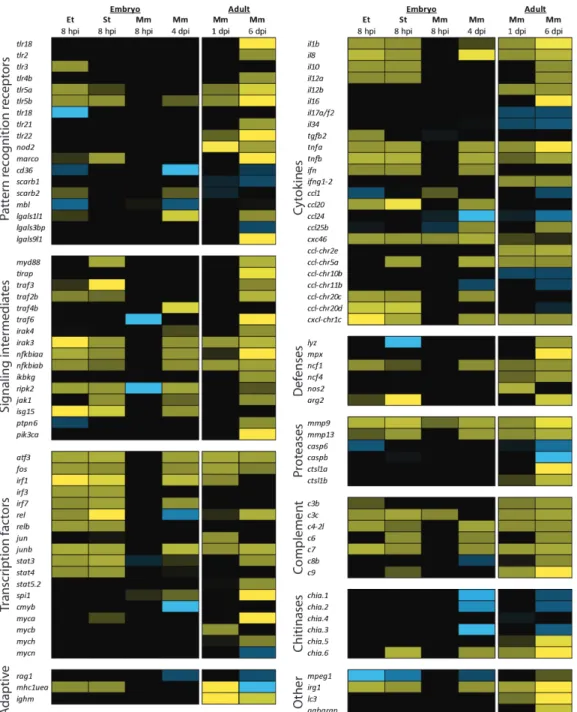
To illustrate the similarities and differences in the innate immune response against different bacterial pathogens, Figure 4 shows a comparison of the gene expression profiles of zebrafish infected with Edwardsiella tarda, S. typhimurium, and M. marinum.
E. tarda is a Gram-negative, naturally occurring fish pathogen that belongs to the
Enterobacteriaceae family. Inside its host, E. tarda is able to resist complement activity and can survive inside macrophages182. It causes a progressive disease when injected into
the caudal vein of 28 hours post fertilization (hpf) embryos, leading to mortality within 2 days post infection (dpi)122. S. typhimurium (short for S. enterica serovar Typhimurium),
also belonging to the Gram-negative Enterobacteriaceae family, causes salmonellosis in a broad range of hosts. S. typhimurium is a facultative intracellular species that can survive within phagocytic and non-phagocytic cells. Following internalization, it survives and replicates in a modified phagosome, known as the Salmonella-containing
2
a progressive disease which leads to mortality of the embryo during the first 30 hourspost infection (hpi)13, 183. In contrast, M. marinum injection at the same stage leads to
a chronic infection that persists during larval development. M. marinum is a natural pathogen of teleost fish and a close relative of M. tuberculosis, the causative agent of tuberculosis in humans. Mycobacteria have a thick, waxy, acid-fast staining cell wall containing characteristic lipids that are important for virulence. Both M. marinum and
M. tuberculosis have the ability to replicate inside macrophages, eventually causing them to undergo apoptosis. Dependent on secreted virulence factors that are conserved between M. marinum and M. tuberculosis, other macrophages are attracted to the initial infection site. These become infected by phagocytosing the apoptotic remains, which ultimately leads to the formation of a granuloma 184. Using the zebrafish embryo
model, Ramakrishan et al. have provided new insights demonstrating the importance of the innate immune system to control M. marinum infection during early stages of pathogenesis1, 2, 123, 185, 186.
Complementary to previously reported transcriptome data (Table 1), here we present new data comparing the gene expression profiles induced by E. tarda, S. typhimurium,
and M. marinum under similar conditions (Figure 4). We injected 200 colony forming units (CFUs) of each pathogen into the caudal vein of 28 hpf zebrafish embryos and analyzed the response at 8 hpi. Since M. marinum develops a chronic infection, we also sampled at 4 dpi, a time point at which granulomas are present. Finally, we compared the transcriptome profile of the embryonic samples with data from a previous study, in which adult zebrafish were infected with the same strain of M. marinum87.
The two progressive Gram-negative pathogens, E. tarda and S. typhimurium, induced a strong early immune response at 8 hpi, while the chronic M. marinum infection hardly induced any response at this time point. At 4 dpi, the transcriptome profile of M. marinum infected embryos did show an immune response, although it was still weaker than the response to E. tarda or S. typhimurium infection at 8 hpi. In adults, the immune response to M. marinum infection has been shown to develop in a similar manner, with hardly any induction of proinflammatory genes at 1 dpi and a stronger response at 6 dpi, when the fish began to show symptoms of disease87. Infections with E. tarda and
S. typhimurium resulted in a remarkably similar transcriptome. Nevertheless, subtle differences were observed, like the up-regulation of Tlr3 that was specific to E. tarda
infection in this data set, and the variation in the panel of cytokines expressed upon these infections.
2
2
receptors. A wide range of transcription factors with well-established functions in immunity (e.g. Atf3, Jun and Fos, Rel, and the IRF and Stat family members) were significantly up-regulated under all conditions tested, except for the 8 hpi time point of
M. marinum infection), whereas we observed up-regulation of transcription factors of the oncogenic Myc family mainly in adult fish. The hematopoietic transcription factor Spi1 (Pu.1) was upregulated in M. marinum infection of embryos and adults. Genes for the key pro-inflammatory cytokines, like TNFα (two genes in zebrafish: tnfa and tnfb), IL1β, and IL8, and for the anti-inflammatory cytokine IL10 were induced by infection with any of the three pathogens. Other cytokines appeared to be more specific for certain pathogens, or might not be expressed at the specific time-point of infection that we sampled.
We also observed increased expression of genes involved in effector mechanisms. However, upregulation of the genes encoding lysozyme, myeloperoxidase, and iNos was detectable only in adult zebrafish infected with M. marinum. Infection with any of the three pathogens led to increased gene expression of ncf1, a subunit of the neutrophil NADPH oxidase complex. Proteases are an important part of the innate immune response, functioning in reorganizing the extracellular matrix to allow leukocyte migration, in degradation of microbes, and in processing of cytokines. In adult zebrafish infected with M. marinum we observed up-regulation of cathepsin-like 1a and 1b (ctsl1a and ctsl1b), members of lysosomal cathepsin family that aids in the destruction of microbes. Expression levels of casp6 and caspb, members of the cysteine-aspartic acid protease (caspase) family involved in apoptosis, were down-regulated at different stages of infection in adults and embryos. The matrix metalloproteinase (mmp) genes 9 and mmp13 proved to be excellent markers for infection, since their gene expression was induced by E. tarda, S. typhimurium and M. marinum.
Our data further suggest that complement activation plays an important role during the early innate immune response, since a large number of complement factor genes show increased expression upon infection. Up-regulated expression of the autophagy marker genes lc3 and gabarap in adults infected with M. marinum hints towards a role
2
for autophagy in the control of this infection. Intriguingly, a macrophage expressed gene with unknown function in immunity, mpeg185, is down-regulated during the
embryonic immune response against all three pathogens. The mouse homolog of this gene encodes a perforin-like protein that is expressed in mature macrophages and prion-infected brain cells187. We have also observed specific up-regulation of
genes with as of yet unknown function in immunity, like immunoresponsive gene 1 (irg1). This gene is highly conserved in vertebrates and has high homology to bacterial methylcitrate dehydrogenase188. We also included some genes involved in adaptive
immunity in our comparison, the lymphocyte marker rag1, the immunoglobulin heavy chain gene ighm, and the antigen presenting major histocompatibility complex class I UEA gene (mhc1uea). Even though no cells of the adaptive immune system are present yet, embryos infected with E. tarda or S. typhimurium increase the expression of the MHC I gene. Finally, upon infection with S. typhimurium and M. marimum we observe up- and down-regulation of chitinases, a family of genes which has been attributed a role during the host-microbial interactions involved in the development of acute and chronic inflammatory conditions189.
5.
Discussion
Zebrafish infectious disease models have started to make an important contribution to the understanding of host-pathogen interaction mechanisms. A good example is the discovery of the mechanism whereby a mycobacterial virulence factor (ESAT6) induces
mmp9 expression in host epithelial cells neighboring infected macrophages, which enhances macrophage recruitment and formation of granuloma-like aggregates that provide a replication niche for mycobacteria2. The combination of genetics and in vivo
imaging in zebrafish embryos is unparalleled in other vertebrate models. Furthermore, zebrafish embryos provide an ideal model for high-throughput in vivo screening of antimicrobial drug candidates or novel vaccine candidates190, 191. Knowledge of the
zebrafish immune system is also important in high throughput screening for cancer in zebrafish embryos192. However, many aspects of zebrafish immunity still require further
characterization and validation.
Currently available transgenic lines clearly distinguish macrophages (marked by csf1r/
fms and mpeg1) from neutrophils (marked by mpx and lyz) in embryos and larvae, but there is insufficient knowledge of surface markers to identify different macrophage and neutrophil subpopulations. Similar to mammals, there is evidence of the existence of subpopulations of classically activated macrophages (M1: high producers of proinflammatory mediators, ROS, and NO) and alternatively activated macrophages (M2: high producers of anti-inflammatory mediators) in fish193. The polarization of
macrophages towards these subtypes plays a critical role in the pathology of both infectious diseases and cancer194. Furthermore, different subpopulations of mammalian
anti-2
functions during infectious disease pathology. Tumor implants in zebrafish embryoswere shown to attract a heterogeneous population of leukocytes, including cells that express arginase, a marker of alternatively activated macrophages176. In addition, the
neutrophil markers mpx, mych and lyz do not show complete overlap176, 196, and markers
such as cxcr3.2 and ptpn6, which are macrophage-specific in one-day old embryos, also label a subset of neutrophils at later stages85. Future development of transgenic lines
that can distinguish these multiple myeloid subsets would further strengthen the use of zebrafish models for innate immunity and infectious disease studies.
As detailed in this review, counterparts of the major vertebrate PRRs and downstream signaling components have been identified in zebrafish, but relatively few have thus far been functionally studied in infectious disease models. Recently, new PRRs have been described in mammals, like the INF-inducible dsRNA-activated protein kinase R (PKR)197,
the cytosolic DNA sensor DNA-dependent activator of IFN-regulatory factors (DAI)198,
and a cytosolic DNA receptor named AIM2 (absent in melanoma 2)199. Thus far only
the zebrafish homolog for PKR has been identified. Furthermore, autophagic adaptors known as sequestosome 1/p62-like receptors (SLRs), conserved between zebrafish and human, have recently been suggested as a new category of PRRs, since they have the ability to recognize and capture targets for immune-related autophagy200.
Various datasets derived from transcriptome analyses have shown the specificity of immune responses to different pathogens. In future studies, the analysis of these responses can be refined by FACS-sorting of immune cell populations from infected embryos, using labeled pathogens in combination with transgenic lines for different immune cell types. For example, it now comes within reach to aim at dissecting the differences in gene expression between M. marinum-infected macrophages inside a granuloma and recently attracted uninfected macrophages. In addition, simultaneous profiling of pathogen and host genes will be a challenging approach to help unravel the complex mechanisms underlying host-pathogen interactions. Transcriptome analysis only reveals altered RNA levels upon infection and therefore the application of proteomic and epigenetic analyses are needed to study the regulation of immune responses on different levels. Transcriptome studies have revealed infection-responsiveness of many genes that have not yet been well studied (for example dram1, mpeg1, irg1, and irg1l, mentioned above), and an emerging immune function for several chitinase-like proteins during infection13, 35, 122. Many zebrafish infection models have been described here and in
other recent reviews4, 201, 202 that can be used to investigate the functions of these genes
2
Acknowledgements
We thank Dan Klionsky (University of Michigan) for the GFP-Lc3 zebrafish line, Maria Forlenza (Wageningen University) for the anti-nitrotyrosine antibody, and Phil Elks for critically reading the manuscript. Infectious disease research in our laboratory is supported by the Smart Mix Program of the Netherlands Ministry of Economic Affairs and the Ministry of Education, Culture and Science, the European Commission 7th framework project ZF-HEALTH (HEALTH-F4-2010-242048), and the European Marie-Curie Initial Training Network FishForPharma (PITN-GA-2011-289209).
References
1. Tobin, D.M. et al. The lta4h locus modulates susceptibility to mycobacterial infection in zebrafish and humans. Cell140, 717-730 (2010).
2. Volkman, H.E. et al. Tuberculous granuloma induction via interaction of a bacterial secreted protein with host epithelium. Science327, 466-469 (2010).
3. Ludwig, M. et al. Whole-body analysis of a viral infection: vascular endothelium is a primary target of infectious hematopoietic necrosis virus in zebrafish larvae. PLoS Pathog7, e1001269 (2011).
4. Meijer, A.H. & Spaink, H.P. Host-pathogen interactions made transparent with the zebrafish model. Curr Drug Targets12, 1000-1017 (2011).
5. Sullivan, C. & Kim, C.H. Zebrafish as a model for infectious disease and immune function. Fish Shellfish Immunol25, 341-350 (2008).
6. Allen, J.P. & Neely, M.N. Trolling for the ideal model host: zebrafish take the bait. Future Microbiol
5, 563-569 (2010).
7. Mathias, J.R., Walters, K.B. & Huttenlocher, A. Neutrophil motility in vivo using zebrafish. Methods Mol Biol571, 151-166 (2009).
8. Renshaw, S.A. et al. A transgenic zebrafish model of neutrophilic inflammation. Blood (2006). 9. Hall, C., Flores, M.V., Storm, T., Crosier, K. & Crosier, P. The zebrafish lysozyme C promoter drives
myeloid-specific expression in transgenic fish. BMC.Dev.Biol.7, 42 (2007).
10. Gray, C. et al. Simultaneous intravital imaging of macrophage and neutrophil behaviour during inflammation using a novel transgenic zebrafish. Thromb Haemost105, 811-819 (2011). 11. Ellett, F., Pase, L., Hayman, J.W., Andrianopoulos, A. & Lieschke, G.J. mpeg1 promoter transgenes
direct macrophage-lineage expression in zebrafish. Blood117, e49-56 (2011).
12. Herbomel, P., Thisse, B. & Thisse, C. Ontogeny and behaviour of early macrophages in the zebrafish embryo. Development126, 3735-3745 (1999).
13. Stockhammer, O.W., Zakrzewska, A., Hegedus, Z., Spaink, H.P. & Meijer, A.H. Transcriptomics and functional analyses of the zebrafish embryonic innate immune response to Salmonella infection.
J.Immunol.in press (2009).
2
16. Beg, A.A. Endogenous ligands of Toll-like receptors: implications for regulating inflammatory and immune responses. Trends Immunol23, 509-512 (2002).
17. Matzinger, P. An innate sense of danger. Ann N Y Acad Sci961, 341-342 (2002).
18. Mogensen, T.H. Pathogen recognition and inflammatory signaling in innate immune defenses.
Clin Microbiol Rev22, 240-273, Table of Contents (2009).
19. Akira, S. & Takeda, K. Toll-like receptor signalling. Nat.Rev.Immunol.4, 499-511 (2004).
20. Akira, S., Uematsu, S. & Takeuchi, O. Pathogen recognition and innate immunity. Cell124, 783-801 (2006).
21. Lemaitre, B., Nicolas, E., Michaut, L., Reichhart, J.M. & Hoffmann, J.A. The dorsoventral regulatory gene cassette spatzle/Toll/cactus controls the potent antifungal response in Drosophila adults.
Cell86, 973-983 (1996).
22. O’Neill, L.A. & Bowie, A.G. The family of five: TIR-domain-containing adaptors in Toll-like receptor signalling. Nat Rev Immunol7, 353-364 (2007).
23. Miettinen, M., Sareneva, T., Julkunen, I. & Matikainen, S. IFNs activate toll-like receptor gene expression in viral infections. Genes Immun2, 349-355 (2001).
24. Takeda, K. & Akira, S. Microbial recognition by Toll-like receptors. J Dermatol Sci34, 73-82 (2004). 25. Jault, C., Pichon, L. & Chluba, J. Toll-like receptor gene family and TIR-domain adapters in Danio
rerio. Mol.Immunol.40, 759-771 (2004).
26. Meijer, A.H. et al. Expression analysis of the Toll-like receptor and TIR domain adaptor families of zebrafish. Mol Immunol.40, 773-783 (2004).
27. Palti, Y. Toll-like receptors in bony fish: from genomics to function. Dev Comp Immunol35, 1263-1272 (2011).
28. Ribeiro, C.M. & Schijns, V.E. Immunology of vaccine adjuvants. Methods Mol Biol 626, 1-14 (2010).
29. Matsuo, A. et al. Teleost TLR22 recognizes RNA duplex to induce IFN and protect cells from birnaviruses. J Immunol181, 3474-3485 (2008).
30. Sepulcre, M.P. et al. Evolution of lipopolysaccharide (LPS) recognition and signaling: fish TLR4 does not recognize LPS and negatively regulates NF-kappaB activation. J Immunol182, 1836-1845 (2009).
31. Sullivan, C. et al. The gene history of zebrafish tlr4a and tlr4b is predictive of their divergent functions. J Immunol183, 5896-5908 (2009).
32. Stein, C., Caccamo, M., Laird, G. & Leptin, M. Conservation and divergence of gene families encoding components of innate immune response systems in zebrafish. Genome Biol8, R251 (2007).
33. Bates, J.M., Akerlund, J., Mittge, E. & Guillemin, K. Intestinal alkaline phosphatase detoxifies lipopolysaccharide and prevents inflammation in zebrafish in response to the gut microbiota.
Cell Host Microbe2, 371-382 (2007).
34. Van der Sar, A.M. et al. MyD88 innate immune function in a zebrafish embryo infection model.
Infect.Immun.74, 2436-2441 (2006).