The Effect of Calcium Binding on Adhesion and Pilus Biogenesis in the PilC Family of Proteins
Michael D. L. Johnson
A dissertation submitted to the faculty of the University of North Carolina at Chapel Hill in partial fulfillment of the requirements for the degree of Doctor of Philosophy in the
Department of Biochemistry and Biophysics.
Chapel Hill 2011
Approved by:
Professor Matthew Redinbo, Ph.D. Professor Edward Collins, Ph.D. Professor Matthew Wolfgang, Ph.D. Professor Jack Griffith, Ph.D.
ii ©2011
iii ABSTRACT
MICHAEL D. L. JOHNSON: The Effect of Calcium Binding on Adhesion and Pilus Biogenesis in the PilC Family of Proteins
(Under the direction of Matthew Redinbo)
Pseudomonas aeruginosa is an opportunistic pathogen prevalent in people on immunosuppressants, recent open wounds, or cystic fibrosis patients. P. aeruginosa attaches to the host cell via polar type IV pili (tfp). These tfp are composed of many proteins including the major pilus subunit PilA, retraction protein PilT, and one focus of this dissertation, 1163 residue protein PilY1. PilY1 shares homology to other bacterial adhesion and pilus biogenesis proteins such as the PilC family of proteins in Neisseria gonorrhoeae, Neisseria meningitidis, and Kingella kingae thus, it is often characterized as a pilus biogenesis protein and the P. aeruginosa adhesin. However, no known mechanism for either situation had been elucidated. In this study, we have identified two calcium binding sites which affect both functional pilus biogenesis and adhesion to host cells. In addition, we have also located an RGD (integrin binding motif) that mediates PilY1 binding to integrin.
iv
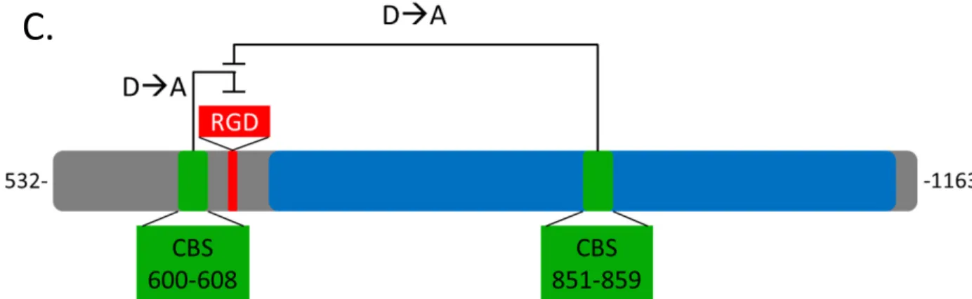
Upon further examination, we also located separate CBS from residues 600-608. This site was in close proximity to an RGD. This CBS was conserved in other strains of P. aeruginosa, but not in other bacteria. We found that purified PilY1 bound integrin in an RGD dependent manner. Furthermore, we found that this interaction was mediated by the calcium bound states of both CBSs.
v Dedication
vi
ACKNOWLEDGEMENTS
First and foremost, I must thank God. On December 14th, 2003, in my then girlfriend, now wife’s church, I was told to pursue science. Since that moment, God has put a great number of people who have been positively essential to in my life as a researcher, a father, and a husband.
Thank you to Dr. Gary Johnson for your wonderful advice, which guided me to giving my CV Paula Harrington, which in turn, led me to first lab mentor, Dr. Jeffery Frelinger. You “grew hair” on my scientific chest; thanks for giving me my first shot in the Show. I’d also like to think Cindy Hensley who was my first lab mom. These relationships led me to Dr. Barbara Vilen who encouraged me to apply to the IBMS program in which I met the program director, Dr. Sharon Milgram. Thanks for the letters of recommendation and scoring me that sweet talk at the NIH. Thanks to my first IBMS family, specifically Kate, Bret, Deb, Rose, Deepak, Erica, Alaina and Pam, you all were my class before I had a class.
I’d like to give a big thanks to my current research mentor, Dr Matthew Redinbo. I still remember our first interview in which we talked about Duke basketball and you told me to a rotation in your lab. I don’t know if I could describe a better interview. Since joining your lab, you have given me a lot of scientific rope and I have been made a better researcher
vii
There are a number of people who are not in the Redinbo lab that have been very helpful. Here I would like to thank Kevin and Michelle for your friendship and Leah for your realism. I have also had the opportunity to mentor people on this journey, Justin, Stephani, Angela, and Brian, you all have taught me so much about myself and who I want to be.
Thank you to my collaborators; Dr. Matthew Wolfgang, Kimberly, Dr, Joe St. Geme, Eric, Dr. Christopher Thomas, Jeff, and Christine. I would like to thank a number of people in the Redinbo lab. Joe, you were always like a big brother/mentor to me and I will always appreciate that, Rebekah, for showing how to think on the other side of the road, and Monica, the other half of the original happy corner, my lab wife, and just a good overall friend, thanks for not making lab seem like work all the time; and yes, the plans are still set. I’d also like to thank Keith, the walking UNC encyclopedia, Yuan, and all the past and present members of the Redinbo Lab for great scientific and mindless conversations.
So much of what I accomplished in graduate school would not have been possible without Pat Phelps. Your Midas touch on my graduate school life has been much
appreciated. Thanks for the outreach opportunities, giving me the tools to succeed, and for being extremely helpful in me getting a postdoc at St. Jude. Meeting you made me a better person.
viii
Table of Contents
List of Tables ... xiii
List of Figures... xiv
List of Abbreviations and Symbols ... xviii
Chapter 1: Introduction to Pseudomonas aeruginosa Type IV Pili ... 1
1.1 Pseudomonas aeruginosa Overview ... 1
1.2 P. aeruginosa Infection ... 2
1.3 Type IV Pili ... 3
1.4 Proteins in the Type IV Pilus ... 3
1.5 The fimUpilEVWXY1Y2E Operon ... 5
1.6 PilY1 ... 5
1.7 References ... 7
Chapter 2: Crystal Structure Analysis Reveals Pseudomonas PilY1 as an Essential Calcium-Dependent Regulator of Bacterial Surface Motility ... 12
2.1 Summary ... 13
2.2 Introduction ... 14
2.3 Results ... 15
2.3.1 PilY1 Exhibits a Modified β-Propeller Fold ... 15
ix
2.3.3 Calcium Binding and Release by PilY1 are Essential for Functional Pili . 18
2.4 Discussion ... 20
2.5 Methods ... 22
2.6 Credits ... 24
2.7 Figure Legends ... 25
2.8 Supplemental Figure Legends ... 26
2.9 Additional Figure Legends ... 28
2.10 References Additional ... 45
Chapter 3: Pseudomonas aeruginosa PilY1 Binds Integrin in an RGD- and Calcium-Dependent Manner ... 48
3.1 Summary ... 49
3.2 Introduction ... 50
3.3 Results ... 51
3.3.1 PilY1 Contains a Conserved Integrin Binding Motif ... 51
3.3.2 PilY1 Binds Integrin in an RGD-Dependent Manner ... 52
3.3.3 PilY1 has a Second Calcium Binding Site ... 53
3.3.4 PilY1 Binds Integrin in a Calcium Dependent Manner ... 54
3.4 Discussion ... 56
3.5 Methods ... 59
3.6 Credits ... 62
3.7 Figure Legends ... 63
x
3.9 Additional Figure Legends ... 66
3.10 References ... 84
Chapter 4: Kingella kingae PilC1 and PilC2 ... 89
4.1 Introduction ... 89
4.2 Methods ... 89
4.3 Results ... 92
4.3.1 Kingella kingae PilC1 and PilC2 Protein Homology ... 92
4.3.2 PilC1 and PilC2 Calcium Binding ... 92
4.3.3 PilC2 Crystal Trials ... 93
4.4 Figure Legends ... 94
4.5 References ... 105
Chapter 5: Effects of Calcium Binding on the Neisseria gonorrhoeae adhesin, PilC1 ... 106
5.1 Summary ... 106
5.2 Introduction ... 107
5.3 Methods ... 108
5.4 Results ... 111
5.4.1 Obtaining Soluble Recombinant Protein ... 111
5.4.2 Recombinant FL-PilC1 blocks N. gonorrhoeae Binding ... 112
5.4.3 Calcium Binding Domain Homology ... 112
5.4.4 Expression of Calcium Binding Mutants in N. gonorrhoeae ... 113
xi
5.4.6 PilC1 Calcium Binding Site Affects Aggregation and
Transformation Frequency ... 114
5.5 Discussion ... 115
5.6 Figure Legends ... 117
5.7 References ... 128
Chapter 6: Past and Future Studies of Pseudomonas aeruginosa PilY1, PilY2, and Neisseria gonorrhea PilC1 ... 130
6.1 Introduction ... 130
6.2 Pseudomonas aeruginosa PilY1 ... 130
6.2.1 PilY1 Purification ... 130
6.2.2 PilY1 Crystallization ... 131
6.2.3 PilY1 Metal Binding ... 132
6.2.4 RGD mutations in PAK PilY1 in vivo assays ... 132
6.2.5 Potential PilY1 Small Molecule Inhibitor Assay ... 134
6.2.6 PilY1 H797 Mutation ... 135
6.2.7 P. aeruginosa Biofilm Formation in the Presence of Host Cells ... 135
6.2.8 Exotoxin Secretion ... 136
6.2.9 Host Cell Viability ... 136
6.3 Pseudomonas aeruginosa PilY2 ... 137
6.3.1 PilY2 Expression ... 137
6.3.2 PilY2 Binding Experiments ... 137
xii
6.4 Neisseria gonorrhoeae PilC1 ... 138
6.4.1 Putative Neisseria gonorrhoeae PilC1 calcium binding domain ... 138
6.5 Figure Legends ... 139
6.6 References ... 155
Chapter 7: Conclusion ... 157
7.1 Introduction ... 157
7.2 Calcium Binding Site Homology ... 157
7.3 Calcium Regulating Pilus Extension and Retraction ... 158
7.4 Calcium Affecting Integrin Binding ... 159
7.5 Other Putative Integrin Binding Mechanisms ... 161
7.6 Figure Legends ... 163
xiii
List of Tables
Table 3.1 Calcium effects on RGD binding proteins ... 72 Supplemental Table 3.1 P. aeruginosa PilY1 is homologous to PilC
family of bacterial adhesin and pilus biogenesis proteins ... 73 Supplemental Table 3.2 Melting temperatures (TM) for mutations of PilY1 ... 74 Table 4.1 List of K. kingae PilC1 and PilC2 constructs made ... 96 Table 5.1 Partial sequence alignment of calcium binding for
various tfp containing species ... 121 Table 5.2 Impact of calcium binding on the thermostability of PilC1 ... 123 Table 6.1 List of PilY1 protein constructs created ... 142 Table 7.1 Bacteria that have homologous calcium binding sites
xiv
List of Figures
Figure 2.1 Crystal structure of the P. aeruginosa PilY1 CTD ... 29 Figure 2.2 PilY1 calcium-binding site ... 30 Figure 2.3 Calcium effects on RGD binding proteins ... 32 Figure 2.4 The PilY1 calcium-binding site is essential for T4P
production and function ... 33 Supplemental Figure 2.1 Topology diagram of the P. aeruginosa PilY1
CTD fold showing the positions of secondary structural elements ... 34 Supplemental Figure 2.2 Sequence alignment of the PilY1 CTDs from
three Pseudomonas strains ... 35 Supplemental Figure 2.3 PilY1 superpositions ... 36 Supplemental Figure 2.4 Comparison of the ligand-to-calcium atom
distances in PilY1and human calmodulin ... 37 Supplemental Figure 2.5 Calcium binding curve for purified
P. aeruginosa PilY1 D859K C-terminal domain ... 38 Supplemental Figure 2.6 Circular dichroism spectropolarimetry
wavelength scans for purified P. aeruginosa PilY1 ... 39 Supplemental Figure 2.7 Melting temperature for purified P.
aeruginosa PilY1 C-terminal domain ... 40 Supplemental figure 2.8 A-D. Modulation of calcium binding
influences T4P production without altering pilin availability ... 41 Supplemental Figure 2.9 Model of the Asp-859-Lys (D859K)
mutation in the PilY1 C-terminal domain calcium binding site ... 42 Additional Figure 2.1 Calcium binding curves for purified P. aeruginosa ... 43 Additional Figure 2.2 Circular dichroism spectropolarimetry
xv
Figure 3.1 P. aeruginosa PilY1 strains contain conserved integrin
binding residues RGD and conserved putative calcium binding site ... 67
Figure 3.2 Purified PilY1 binds to integrin in an RGD dependent manner ... 68
Figure 3.3 PilY1 has two functional calcium binding sites ... 69
Figure 3.4 Calcium binding sites play a role in PilY1’s ability binds to integrin ... 70
Supplemental Figure 3.1 P. aeruginosa PilY1 is homologous to PilC family of bacterial adhesin and pilus biogenesis proteins ... 75
Supplemental Figure 3.2 Circular dichroism for calcium binding mutants ... 76
Supplemental Figure 3.3 Mutational effects on PilY1:Integrin binding ... 77
Additional Figure 3.1 Isothermal titration calorimetry PilY1 ... 78
Additional Figure 3.2 Mutational effects on PilY1:Integrin binding ... 80
Additional Figure 3.3 The structure of αVβ3 integrin bound to RGD peptide ... 81
Figure 4.1 SDS PAGE gel of PilC1 739-1047 K. kingae in LIC-HIS-MBP expression ... 97
Figure 4.2 Homology of PilC1 ... 98
Figure 4.3 Homology of PilC2 ... 99
Figure 4.4 K. kingae PilC1 binds calcium ... 100
Figure 4.5 K. kingae PilC2 has one functional calcium binding site ... 101
Figure 4.6 Stability of PilC2 D1444A mutants ... 102
Figure 4.7 Comparison of C-terminal PilC2 and 532-1163 P. aeruginosa PilY1 by circular dichroism wavelength scan ... 103
Figure 4.8 Dynamic light scattering of C-terminal PilC2 ... 104 Figure 5.1 Purified full-length PilC1 protein inhibits in a dose-
xvi
human epithelial cells ... 120
Figure 5.2 Binding of Tb3+ to different PilC1 constructs ... 122
Figure 5.3 Expression of PilC1 calcium binding mutants in N. gonorrhoeae ... 124
Figure 5.4 Impact of calcium binding on the surface exposure of PilC1 in N. gonorrhoeae ... 125
Figure 5.5 Impact of calcium binding on the adherence of N. gonorrhoeae to human ME180 cells ... 126
Figure 5.6 Impact of calcium binding on aggregation and transformation frequency in N. gonorrhoeae ... 127
Figure 6.1 Dynamic light scattering of 532-1163 PAK PilY1 ... 143
Figure 6.2 Crystal hit of 532-1163 PilY1 ... 144
Figure 6.3 PilY1 protein degradation in crystal ... 145
Figure 6.4 PilY1 binds manganese ... 146
Figure 6.5 Infrared scan of a western blot showing IPTG induced PilY1 in whole cell PAK ... 147
Figure 6.6 Twitching and surface pilin expression in PAK ... 148
Figure 6.7 Adhesion assay with PAK PilY1 RGD mutations ... 149
Figure 6.8 Small molecule inhibition of PilY1 ... 150
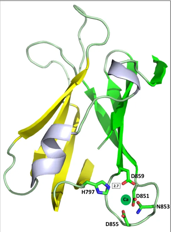
Figure 6.9 PyMOL image of H797 interacting with the CBS of C-terminal PilY1 ... 151
Figure 6.10 Chromatograms and SDS Page gels of PAK PilY2 ... 152
Figure 6.11 Circular dichroism wavelength scan of PilY2 ... 153
Figure 6.12 Putative N. gonorrhoeae PilC1 calcium binding site ... 154
xvii
xviii
List of Abbreviations and Symbols Å – angstrom
α – alpha Ala, A – alanine Amp – ampicillin
ANOVA –
analysis of variance
Asn, N – asparagine Asp, D – aspartic acid
ATP – adenosine triphosphate
β – beta
BSA – bovine serum albumin C – Celsius
Ca – calcium CaM – calmodulin
CBD – calcium binding domain CBS – calcium binding site CD – circular dichroism
CD – cluster of differentiation
CFU – colony forming unit Cl – chloride
CTD- C-terminal domain
C-terminus, C-term, C-terminal – carboxy terminus
∆ – deletion
xix
DxDxDG – aspartic acid-x-aspartic acid-x-aspartic acid-glycine calcium binding motif EC50 – half maximal effective concentration
EDTA – ethylenediaminetetraacetic acid EGTA – ethylene glycol tetraacetic acid EF – helix-loop-helix
F – fluorine FL – full length g – gravity
Glu, E – glutamic acid
GM – sialotetrahexosylganglioside
GRADSP – glycine- arginine-alanine-aspartic acid-serine-proline control peptide H – hydrogen
h – hour
HEPES - 4-(2-hydroxyethyl)-1-piperazineethanesulfonic acid His, H – histidine
HRP – horseradish peroxidase I - isoleucine
IC50 - half maximal inhibitory concentration IPTG – isopropyl β-D-1-thiogalactopyranoside ITC – isothermal titration calorimetry
K, Lys – lysine K – potassium
Kd – dissociation constant kD – kilodalton
xx L – leucine
LB – lurea broth
LDV – leucine-aspartic acid-valine integrin binding motif LIC – ligation independent cloning
MBP – maltose binding protein µ – micro
µg – microgram µL – microliter µm – micron µM - micromolar
MBP- maltose binding protein mdeg – millidegrees
mg – milligram min – minutes mL – milliliters mm – millimeter Mn – manganese mol – mole M - methionine
MW – molecular weight Na – sodium
NHS PEO4 Biotin – 12-biotinylamino-4,7,10-trioxadodecanoic acid N-hydroxysuccinimidyl ester
xxi nN – nano Newton
N-terminus, N-term N-terminal – amino terminus O – oxygen
OD – optical density P – phosphate p – p-value
PAGE - polyacrylamide gel electrophoresis PAK – Pseudomonas aeruginosa strain K PBS – phosphate buffered saline
PBST – phosphate buffered saline .1% Tween PDB – Protein Data Bank
PEG – polyethyleneglycol
pH – negative log (base 10) of the molar concentration of hydronium ions Phe, F – phenylalanine
PMSF - phenylmethanesulfonylfluoride pN – pico Newton
RGD – arginine-glycine-aspartic acid integrin binding motif
RGDS – arginine-glycine-aspartic acid integrin binding inhibitor peptide
RIPL – BL21-CodonPlus (DE3)-RIPL cells
rpm – revolutions per minute s – seconds
SAD – single wavelength anomalous dispersion SDS – sodium dodecyl sulfate
xxii Tb – terbium
Tet – tetracyclin
TEV – tobacco etch virus T4P – Type IV Pili
TFP – Type IV Pili
Tm – melting temperature TPR – tetratricopeptide
TRIS – 2-Amino-2-hydroxymethyl-propane-1,3-diol Tyr, Y – tyrosine
UNC-CH – University of North Carolina at Chapel Hill U.S. – United States
WT – wild type
xxiii % – percent
Chapter 1
Introduction to Pseudomonas aeruginosa Type IV Pili
1.1 Pseudomonas aeruginosa Overview
Pseudomonas aeruginosa is a rod-shaped Gram-negative opportunistic pathogen that is highly prevalent in people with cystic fibrosis, on immunosuppressants, or with recent open wounds or burns. Needing only minimal nutrients to live, P. aeruginosa has been found living on or in a variety of environments, including steel, soil, deionized distilled water, and gasoline [1,2]. Having the ability to undergo both aerobic and anaerobic respiration, this nosocomial pathogen has the ability to infect any part of the body; however infections typically occur in the respiratory system, broken skin, blood, or urinary tract [3]. Hospital acquired infections, which killed an estimated 100,000 people in 2002, infected an estimated 1.7 million people in the U.S.; P. aeruginosa accounting for about 11% of those infections [4,5]. In addition to humans, this pathogen can infect other organisms, such as plants, insects, and other members of the animal kingdom, making it a bacterium of interest with significant relevance for studying the general mechanisms regulating pathogenesis [6,7].
2
targeting vital bacterial pathways through an SOS response, a process where the organism purposely mutates its genes in an attempt to adapt to and thus overcome stress [12].
1.2 P. aeruginosa Infection
P. aeruginosa causes infection via multiple stepwise actions: first, using flagella, it swims to approach its target; second, it adheres to the surface of the target using arm-like protrusions called type IV pili (tfp) that are also responsible for “walking” on the surface, a process known as twitching motility; third, it undergoes quorum sensing (ability to detect other bacteria) and forms a protective alginate biofilm; and fourth, somewhat concurrently with the third step, it begins secreting virulence factors such as LasA and LasB elastase, exotoxin A, and alkaline protease [13,14,15,16,17]. Finally, individual bacterium can separate from the parent colony to cause infections in other host sites. A critical step in causing virulence is the tfp’s ability to attach to cells, tissues, or other surfaces, thereby allowing biofilm formation [2,13,18]. Without tfp, and hence the ability to anchor to a surface, P. aeruginosa cannot congregate to form a functional biofilm for its own protection and thus facilitate host infection [14].
1.3 Type IV Pili
3
and occurs during directed movement toward a nutritional gradient [21]. As can be expected, lack of tfp eliminates P. aeruginosa’s pathogenesis to host organisms due to lack of motility [15,18,22].
1.4 Proteins in the Type IV Pilus
There are more than forty proteins involved in the tfp. These proteins include PilA as the major pilin subunit; PilQ as the membrane pore protein through which the pilus extends and retracts; PilT, a key component in pilus retraction; PilB, which is responsible for pilus extension, and PilD, which is responsible for cleaving the membrane leader sequence on the pilus proteins [15,20,23,24]. There are also a number of pseudopilins (pilin-like proteins) that are incorporated into the main pilus fiber [25]. These proteins include FimU, PilV, PilW, PilX, and PilE, which are all located in the PilY1 operon controlled by AlgR [26,27]. Other members of this operon are PilY1 and PilY2 [26].
P. aeruginosa tfp are long fibers containing polymerized PilA. Although the sequences can be quite divergent between strains, the consensus monomer structure (based on solved crystal structures) contains a long α-helix with a head group of four β-strands, similar to the homologous protein PilE in Neisseria gonorrhoeae [28,29,30,31]. PilA is often thought to function as an adhesin in P. aeruginosa [28,32]. C-terminal disulfide-connected loops may control binding at the tip of the pilus to gangliosides GM1 and GM2; however, this evidence has been disputed because it is not consistent throughout the bacterial strains [28,32,33,34,35].
4
embedded in the membrane and the β-sheet head group sticking out, extracted using approximately three ATP molecules, and attached via electrostatic interactions using a conserved negatively charged glutamic acid of one PilA subunit attaching to the positively charged N-terminal region of another monomer [36,37,38,39]. The resultant polymer can be 6-9 nm wide and up to 1-4 µm long [36]. It has been estimated that six ATP molecules are used to assemble a pilus subunit and it is possible that the other three are needed for the polymerization step [40]. Accordingly, pilus polymerization is regulated by the tfp elongation ATPase, PilB [41,42]. Mutations to the His box, Walker-A, or –B motifs eliminate surface piliation, twitching motility, and the pilus dependent process of phage susceptibility [43].
Functional tfp extension and retraction occurs through a pore made by a dodecameric protein, PilQ, as observed in Nesseria meningitidis [44,45]. PilQ is also thought to play a role in pilus assembly because of its conformational change upon tfp engagement and its association with only one end of the pilus fiber [46]. The necessary process of P. aeruginosa PilQ multimerization and insertion into the bacterial membrane is regulated by PilF [47]. PilF has been found to attach to the outer membrane of P. aeruginosa and control PilQ pore assembly through six tetratricopeptide repeat (TPR) protein-protein interaction motifs [47]. TPRs are helical structures usually involved in large protein complex formation [48].
5
PilT works in concert with P. aeruginosa PilC, N, O, and P to coordinate individual pilus subunit depolymerization via a “paddle-wheel” type mechanism [51].
1.5 The fimUpilVWXY1Y2E Operon
The fimUpilVWXY1Y2E operon constitutes gene products for factors that fit into three families: pseudopilins/ minor pilin/ pilin-like protein family (FimU, PilV, W, X, and E), an uncharacterized group (PilY2), or the PilC family of proteins from the Neisseria family (PilY1) [26]. FimU, PilV, W, X, and E have been found to be necessary for functional pilus biogenesis and are integrated into the pilus [25]. All of these proteins share high homology to PilA with some minor differences. For example, PilW has a large predicted loop region within the head group of the protein. Integrated tfp minor pilins make a cuff in 1:1 stoichiometric ratios at various parts of the pilus fiber [25]. It is possible that these cuffs provide the grip for PilT-mediated retraction. PilY2 shows no known homology to other proteins, and PilY2 deletion mutants exhibit reduced twitching motility and pilus biogenesis [26].
1.6 PilY1
6
there have been no previous reports of the attachment mechanism directly involving PilY1 [15].
7
1.7 References
1. Foght JM, Westlake DW, Johnson WM, Ridgway HF (1996) Environmental gasoline-utilizing isolates and clinical isolates of Pseudomonas aeruginosa are taxonomically indistinguishable by chemotaxonomic and molecular techniques. Microbiology 142 ( Pt 9): 2333-2340.
2. Stanley PM (1983) Factors affecting the irreversible attachment of Pseudomonas aeruginosa to stainless steel. Can J Microbiol 29: 1493-1499.
3. Richards MJ, Edwards JR, Culver DH, Gaynes RP (1999) Nosocomial infections in pediatric intensive care units in the United States. National Nosocomial Infections Surveillance System. Pediatrics 103: e39.
4. Klevens RM, Edwards JR, Richards CL, Jr., Horan TC, Gaynes RP, et al. (2007) Estimating health care-associated infections and deaths in U.S. hospitals, 2002. Public Health Rep 122: 160-166.
5. Schaberg DR, Culver DH, Gaynes RP (1991) Major trends in the microbial etiology of nosocomial infection. Am J Med 91: 72S-75S.
6. Apidianakis Y, Rahme LG (2009) Drosophila melanogaster as a model host for studying Pseudomonas aeruginosa infection. Nat Protoc 4: 1285-1294.
7. Starkey M, Rahme LG (2009) Modeling Pseudomonas aeruginosa pathogenesis in plant hosts. Nat Protoc 4: 117-124.
8. Driscoll JA, Brody SL, Kollef MH (2007) The epidemiology, pathogenesis and treatment of Pseudomonas aeruginosa infections. Drugs 67: 351-368.
9. Hoiby N, Ciofu O, Bjarnsholt T (2010) Pseudomonas aeruginosa biofilms in cystic fibrosis. Future Microbiol 5: 1663-1674.
10. Mah TF, O'Toole GA (2001) Mechanisms of biofilm resistance to antimicrobial agents. Trends Microbiol 9: 34-39.
11. Poole K (2004) Efflux-mediated multiresistance in Gram-negative bacteria. Clin Microbiol Infect 10: 12-26.
12. Cirz RT, O'Neill BM, Hammond JA, Head SR, Romesberg FE (2006) Defining the Pseudomonas aeruginosa SOS response and its role in the global response to the antibiotic ciprofloxacin. J Bacteriol 188: 7101-7110.
13. Giltner CL, van Schaik EJ, Audette GF, Kao D, Hodges RS, et al. (2006) The Pseudomonas aeruginosa type IV pilin receptor binding domain functions as an adhesin for both biotic and abiotic surfaces. Mol Microbiol 59: 1083-1096. 14. Hahn HP (1997) The type-4 pilus is the major virulence-associated adhesin of
Pseudomonas aeruginosa--a review. Gene 192: 99-108.
8
16. Miller MB, Bassler BL (2001) Quorum sensing in bacteria. Annu Rev Microbiol 55: 165-199.
17. Van Delden C, Iglewski BH (1998) Cell-to-cell signaling and Pseudomonas aeruginosa infections. Emerg Infect Dis 4: 551-560.
18. O'Toole GA, Kolter R (1998) Flagellar and twitching motility are necessary for Pseudomonas aeruginosa biofilm development. Mol Microbiol 30: 295-304.
19. Gibiansky ML, Conrad JC, Jin F, Gordon VD, Motto DA, et al. (2010) Bacteria use type IV pili to walk upright and detach from surfaces. Science 330: 197.
20. Burrows LL (2005) Weapons of mass retraction. Mol Microbiol 57: 878-888.
21. Kearns DB, Robinson J, Shimkets LJ (2001) Pseudomonas aeruginosa exhibits directed twitching motility up phosphatidylethanolamine gradients. J Bacteriol 183: 763-767. 22. D'Argenio DA, Gallagher LA, Berg CA, Manoil C (2001) Drosophila as a model host for
Pseudomonas aeruginosa infection. J Bacteriol 183: 1466-1471.
23. Strom MS, Nunn DN, Lory S (1993) A single bifunctional enzyme, PilD, catalyzes cleavage and N-methylation of proteins belonging to the type IV pilin family. Proc Natl Acad Sci U S A 90: 2404-2408.
24. Turner LR, Lara JC, Nunn DN, Lory S (1993) Mutations in the consensus ATP-binding sites of XcpR and PilB eliminate extracellular protein secretion and pilus biogenesis in Pseudomonas aeruginosa. J Bacteriol 175: 4962-4969.
25. Giltner CL, Habash M, Burrows LL (2010) Pseudomonas aeruginosa minor pilins are incorporated into type IV pili. J Mol Biol 398: 444-461.
26. Alm RA, Hallinan JP, Watson AA, Mattick JS (1996) Fimbrial biogenesis genes of Pseudomonas aeruginosa: pilW and pilX increase the similarity of type 4 fimbriae to the GSP protein-secretion systems and pilY1 encodes a gonococcal PilC
homologue. Mol Microbiol 22: 161-173.
27. Belete B, Lu H, Wozniak DJ (2008) Pseudomonas aeruginosa AlgR regulates type IV pilus biosynthesis by activating transcription of the fimU-pilVWXY1Y2E operon. Journal of bacteriology 190: 2023-2030.
28. Hazes B, Sastry PA, Hayakawa K, Read RJ, Irvin RT (2000) Crystal structure of Pseudomonas aeruginosa PAK pilin suggests a main-chain-dominated mode of receptor binding. J Mol Biol 299: 1005-1017.
29. Parge HE, Forest KT, Hickey MJ, Christensen DA, Getzoff ED, et al. (1995) Structure of the fibre-forming protein pilin at 2.6 A resolution. Nature 378: 32-38.
9
31. Craig L, Volkmann N, Arvai AS, Pique ME, Yeager M, et al. (2006) Type IV pilus structure by cryo-electron microscopy and crystallography: implications for pilus assembly and functions. Mol Cell 23: 651-662.
32. Audette GF, Irvin RT, Hazes B (2004) Crystallographic analysis of the Pseudomonas aeruginosa strain K122-4 monomeric pilin reveals a conserved receptor-binding architecture. Biochemistry 43: 11427-11435.
33. Craig L, Pique ME, Tainer JA (2004) Type IV pilus structure and bacterial pathogenicity. Nat Rev Microbiol 2: 363-378.
34. Emam A, Yu AR, Park HJ, Mahfoud R, Kus J, et al. (2006) Laboratory and clinical Pseudomonas aeruginosa strains do not bind glycosphingolipids in vitro or during type IV pili-mediated initial host cell attachment. Microbiology 152: 2789-2799. 35. Schroeder TH, Zaidi T, Pier GB (2001) Lack of adherence of clinical isolates of
Pseudomonas aeruginosa to asialo-GM(1) on epithelial cells. Infect Immun 69: 719-729.
36. Craig L, Li J (2008) Type IV pili: paradoxes in form and function. Curr Opin Struct Biol 18: 267-277.
37. Lemkul JA, Bevan DR (2011) Characterization of interactions between PilA from Pseudomonas aeruginosa strain K and a model membrane. J Phys Chem B 115: 8004-8008.
38. Macdonald DL, Pasloske BL, Paranchych W (1993) Mutations in the fifth-position glutamate in Pseudomonas aeruginosa pilin affect the transmethylation of the N-terminal phenylalanine. Can J Microbiol 39: 500-505.
39. Nunn DN, Lory S (1991) Product of the Pseudomonas aeruginosa gene pilD is a prepilin leader peptidase. Proc Natl Acad Sci U S A 88: 3281-3285.
40. Maier B, Koomey M, Sheetz MP (2004) A force-dependent switch reverses type IV pilus retraction. Proc Natl Acad Sci U S A 101: 10961-10966.
41. Nunn D, Bergman S, Lory S (1990) Products of three accessory genes, pilB, pilC, and pilD, are required for biogenesis of Pseudomonas aeruginosa pili. Journal of bacteriology 172: 2911-2919.
42. Turner LR, Lara JC, Nunn DN, Lory S (1993) Mutations in the consensus ATP-binding sites of XcpR and PilB eliminate extracellular protein secretion and pilus biogenesis in Pseudomonas aeruginosa. Journal of bacteriology 175: 4962-4969.
43. Chiang P, Sampaleanu LM, Ayers M, Pahuta M, Howell PL, et al. (2008) Functional role of conserved residues in the characteristic secretion NTPase motifs of the
10
44. Collins RF, Davidsen L, Derrick JP, Ford RC, Tonjum T (2001) Analysis of the PilQ secretin from Neisseria meningitidis by transmission electron microscopy reveals a dodecameric quaternary structure. Journal of bacteriology 183: 3825-3832.
45. Martin PR, Hobbs M, Free PD, Jeske Y, Mattick JS (1993) Characterization of pilQ, a new gene required for the biogenesis of type 4 fimbriae in Pseudomonas aeruginosa. Mol Microbiol 9: 857-868.
46. Collins RF, Frye SA, Balasingham S, Ford RC, Tonjum T, et al. (2005) Interaction with type IV pili induces structural changes in the bacterial outer membrane secretin PilQ. J Biol Chem 280: 18923-18930.
47. Koo J, Tammam S, Ku SY, Sampaleanu LM, Burrows LL, et al. (2008) PilF is an outer membrane lipoprotein required for multimerization and localization of the
Pseudomonas aeruginosa Type IV pilus secretin. Journal of bacteriology 190: 6961-6969.
48. Blatch GL, Lassle M (1999) The tetratricopeptide repeat: a structural motif mediating protein-protein interactions. Bioessays 21: 932-939.
49. Chiang P, Habash M, Burrows LL (2005) Disparate subcellular localization patterns of Pseudomonas aeruginosa Type IV pilus ATPases involved in twitching motility. J Bacteriol 187: 829-839.
50. Wolfgang M, Lauer P, Park HS, Brossay L, Hebert J, et al. (1998) PilT mutations lead to simultaneous defects in competence for natural transformation and twitching motility in piliated Neisseria gonorrhoeae. Mol Microbiol 29: 321-330.
51. Misic AM, Satyshur KA, Forest KT (2010) P. aeruginosa PilT structures with and without nucleotide reveal a dynamic type IV pilus retraction motor. J Mol Biol 400: 1011-1021.
52. Wolfgang M, Park HS, Hayes SF, van Putten JP, Koomey M (1998) Suppression of an absolute defect in type IV pilus biogenesis by loss-of-function mutations in pilT, a twitching motility gene in Neisseria gonorrhoeae. Proc Natl Acad Sci U S A 95: 14973-14978.
53. Maier B, Potter L, So M, Long CD, Seifert HS, et al. (2002) Single pilus motor forces exceed 100 pN. Proc Natl Acad Sci U S A 99: 16012-16017.
54. Bendtsen JD, Nielsen H, von Heijne G, Brunak S (2004) Improved prediction of signal peptides: SignalP 3.0. J Mol Biol 340: 783-795.
55. Bohn YS, Brandes G, Rakhimova E, Horatzek S, Salunkhe P, et al. (2009) Multiple roles of Pseudomonas aeruginosa TBCF10839 PilY1 in motility, transport and infection. Mol Microbiol 71: 730-747.
11
57. Kallstrom H, Liszewski MK, Atkinson JP, Jonsson AB (1997) Membrane cofactor protein (MCP or CD46) is a cellular pilus receptor for pathogenic Neisseria. Mol Microbiol 25: 639-647.
aDepartment of Chemistry, University of North Carolina at Chapel Hill, CB 3290, Chapel Hill, NC
27599, USA. bDepartment of Biochemistry and Biophysics, University of North Carolina at Chapel Hill, CB 7260, Chapel Hill, NC 27599, USA. cDepartment of Microbiology and Immunology, University of
North Carolina at Chapel Hill, CB 7290, Chapel Hill, NC 27599, USA. dCystic Fibrosis/Pulmonary
Research and Treatment Center University of North Carolina at Chapel Hill, University of North Carolina at Chapel Hill, CB 7248, Chapel Hill NC, 27599, USA.
*Contributed equally to this work
Orans, J., Johnson, M.D.L., Coggan, K.A., Sperlazza, J.R., Heiniger, R.W. Wolfgang, M.C., Redinbo, M.R. . Proc Natl Acad Sci U S A. 2010 Mar 16;107(11):5260.
Data Deposition: coordinates and structure factors have been deposited to the PDB and assigned accession code 3HX6.
Michael D. L. Johnson contributed Fig. 2.2 C and D, Figure 2.4B, Fig. S2.2, Fig. S2.5, Fig. S2.6, and Fig. S2.7. He also contributed additional figures, now found in the text, Fig A2.1A-C and Fig A2.2A-C, that were not part of this manuscript, but supportive of the overall conclusion.
CHAPTER 2
Crystal Structure Analysis Reveals Pseudomonas PilY1 as an Essential Calcium-Dependent Regulator of Bacterial Surface Motility
13
2.1 SUMMARY
Several bacterial pathogens require the “twitching” motility produced by filamentous type IV pili (T4P) to establish and maintain human infections. Two cytoplasmic ATPases function as an oscillatory motor that powers twitching motility via cycles of pilus extension and retraction. The regulation of this motor, however, has remained a mystery. We present the 2.1 Å resolution crystal structure of the Pseudomonas aeruginosa pilus-biogenesis factor PilY1, and identify a single site on this protein required for bacterial translocation. The structure reveals a modified β-propeller fold and a distinct EF-hand calcium-binding site conserved in pathogens with retractile T4P. We show that preventing calcium binding by PilY1 using either an exogenous calcium chelator or mutation of a single residue disrupts
14
2.2 INTRODUCTION
The pathogenesis of bacterial infections typically depends on microbial adherence to host tissue, aggregation of the pathogen at the site of infection, and subsequent dissemination to other anatomical sites within the host. Type IV pili (T4P) are thin surface filaments involved in biofilm formation, bacterial aggregation and microbial adherence to biotic and abiotic surfaces. T4P biogenesis requires more than a dozen proteins conserved across a range of bacterial species [1]. A subset of bacteria, including the opportunistic human pathogen Pseudomonas aeruginosa, cyclically extend and retract their T4P, facilitating a form of surface movement termed twitching motility [2,3,4]. Retractile T4P are required for P. aeruginosa adherence to host tissue, virulence and dissemination of infection [5,6,7].
15
biogenesis rather than adhesion, and pursued a structural and functional analysis of the CTD from Pseudomonas aeruginosa PilY1.
2.3 RESULTS
2.3.1 PilY1 Exhibits a Modified β-Propeller Fold
Examination of the primary sequence of PilY1 from P. aeruginosa strain PAK revealed conservation between its C-terminal 550 residues and the same regions of the PilC proteins from Neisseria, and a predicted structural homology to the eight-bladed -propeller fold exhibited by a range of proteins [16,17]. Thus, we overexpressed, purified and crystallized the PilY1 CTD (amino acids 614-1163) for examination by x-ray diffraction. Native diffraction data were collected to 2.1 Å resolution (Table I); however, attempts to determine the structure using existing β-propeller search models or by isomorphous replacement methods were not successful. PilY1 CTD contains only six methionine residues, which upon selenomethionine substitution failed to produce an adequate signal to support structure determination by anomalous dispersion [18]. To overcome this problem, a series of single leucine-to-methionine (L-M) substitutions were placed in the PilY1 CTD to provide additional sites for single wavelength anomalous dispersion (SAD) phasing (Supplemental Figures 2.1- 2.2). After several attempts, three L-M substitutions (at positions 712, 812, 823) were successfully combined, crystallized, and generated clear selenomethionine fluorescence signal upon examination in the x-ray beam. The structure was determined by SAD phasing using crystals containing nine total selenomethionine residues per protein monomer.
16
2.1- 2.2; residues at the termini, as well as 713-721 and 1061-1068 were disordered and are not in the refined model). Blades I through IV are 4-stranded antiparallel β-sheets and align well with blades of a canonical β-propeller enzyme, quinohemoprotein alcohol dehydrogenase from Comamonas testosteroni (RCSB 1KB0; overall RMSD of 3.6 Å over 290 equivalent Cα positions with 15% sequence identity; Figure 2.1B) [16]. This β-propeller enzyme was identified by Phyre to align with the PilY1 predicted structure (E-value 0.014, 95% certainty) [16]. Blades V through VII of the PilY1 CTD, however, deviate from the standard β-propeller fold and align poorly with established β-propellers. Blade V contains a fifth strand (β20) that is shared with blade VI. Blade VI is composed of six strands, including β20, and maintains an insert containing a short helix and β-turn-β motif (α9, β26-27). Finally, blade VII is truncated and composed of three relatively short strands.
17
2.3.2 PilY1 Contains a Unique Calcium Binding Loop
A strong |Fo|-|Fc| electron density peak in the PilY1 CTD structure was interpreted as a calcium atom based on the observed coordination by three aspartic acid side chains (851, 855, 859), one asparagine side chain (853), one main-chain carbonyl oxygen of valine 857, and one water (Figure 2.2A; see also Supplemental Figure 2.1). The calcium binding observed in PilY1 is nearly identical to that seen in the canonical EF-hand Ca-binding motifs (Figure 2B; Supplemental Figure 2.4) [21]. PilY1 and EF hands both employ seven contacts to the bound calcium ion, including one from a water molecule and one bidentate contact via an acidic side chain. PilY1 is unique, however, in that it achieves this calcium chelation using a stretch of only nine amino acids (851-859) in a loop between two β -strands (β 13 and β 14) rather than the twelve residues between two α-helices typically observed in the EF-hands [21]. In addition, the carboxylic acid group of the bidentate Asp-859 residue is rotated nearly 90 with respect to the equivalent residue in EF-hand proteins [21]. Asp-859 forms a hydrogen bond with His-797, which is conserved in PilY1 proteins of known sequence. The calcium-chelating residues observed in the structure are conserved in the PilY1s of known sequence and in the related PilCs, with the exception that Asn-853 is replaced with an aspartic acid in the Neisseria proteins (Figure 2.2C; Supplemental Figure 2.2). This modified EF-hand motif is also present in PilY1 orthologues found in other bacteria that utilize T4P, including the human pathogen Kingella kingae. Thus, a novel calcium-binding site appears to be conserved in the C-terminal domains of pilus biogenesis proteins related to PilY1.
18
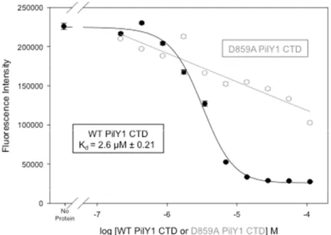
PilY1. Mutation of the calcium chelating residue Asp-859 to alanine eliminates specific calcium binding by the protein (Figure 2.2D). Removal of calcium either by Chelex-100 or by mutating a chelating residue did not change the overall structure (as measured by circular dichroism spectropolarimetry; Supplemental Figure 2.6) or melting temperature (Supplemental Figure 2.7) of the PilY1 CTD [23]. Additional mutations D851A, N853, and D855A were made to PilY1, all of which eliminated calcium binding and did not affect the overall secondary structure (Figure Ad 2.1A-C, 2.2A-C). Thus, calcium binding appears not to play a role in the overall structure of the PilY1 C-terminal domain.
2.3.3 Calcium Binding and Release by PilY1 are Essential for Functional Pili
19
PilY1 was previously shown to traffic to the surface T4P fraction [13] (Supplemental Figure 2.8), where it presumably functions through a direct association with the pilus fiber. To determine whether the defect in twitching observed with the D859A form of PilY1 was caused by disrupted pilus assembly/extension or increased pilus retraction, the PilY1 D859A mutant was expressed in a mutant strain lacking the PilT-retraction ATPase (PilTminus, PilY1minus strain). These bacteria regained the ability to produce normal levels of T4P, and the mutant PilY1 protein was capable of trafficking to the sheared pilus fraction (Figures 2.3C-F). This result indicates that the loss of surface pili observed with the PilY1 D859A mutant is caused by PilT-mediated pilus retraction. In the presence of PilT, the D859A form of PilY1 is incapable of antagonizing PilT-mediated pilus retraction despite normal trafficking of the protein. We conclude that the calcium-binding site in the PilY1 CTD is necessary for normal pilus function by antagonizing the retractile activity of PilT.
20
for the twitching motility defect. PilY1 D859K was capable of trafficking to the surface T4P fraction when expressed in both a pilY1 mutant background (Supplemental Figure 2.8) and a PilTminus, PilY1minus strain (Figures 2.3C-F). Thus, using the crystal structure as a guide, we produced a single-site mutation that dramatically alters pilus function without affecting pilus production. Based on these data, we conclude that the release of calcium by PilY1 is required for PilT-dependent fiber retraction and twitching motility.
Finally, we tested whether the calcium chelator EGTA impacts P. aeruginosa pilus production. We found that EGTA reduces the levels of surface pili present on bacteria expressing wild-type PilY1 while the non-retractile PilTminus strain is blind to calcium chelation (Figure 2.4A). Similarly, the D859K form of PilY1 is insensitive to the effects of EGTA and supports the production of wild-type levels of surface pili (Figure 2.4A). These data show that the PilY1 D859K mutant, mimicking the calcium bound state of PilY1, has increased capacity to antagonize PilT-mediated pilus retraction. Taken together, these cell-based studies support the conclusion that both calcium binding and calcium release by the unique site in PilY1 are essential for the proper regulation of pilus retraction dynamics and twitching motility.
2.4 DISCUSSION
21
twitching motility (Figure 2.4B, top). The D859A mutant form of PilY1 appears to mimic the calcium-free state of the protein, and is not capable of antagonizing PilT-mediated retraction; thus, the equilibrium is shifted toward retraction, similar to that seen with the PilY1minus strain (Figure 2.4B). Calcium chelation by EGTA also shifts the equilibrium toward retraction. In contrast, the D859K variant mimics the calcium-bound state, shifting the equilibrium toward pilus extension and producing an effect similar to that seen with the PilTminus strain (Figure 2.4B). The relatively moderate 2 µM calcium binding affinity exhibited by PilY1 is in the range where cycles of calcium binding and release would be expected to be physiologically relevant. Thus, we conclude that PilY1 acts as a calcium-dependent regulator of the opposing forces responsible for T4P-mediated twitching motility. However, we stress that this is a functional hypothesis; the physical contacts between PilY1 and other factors necessary to achieve this regulation have yet to be elucidated. It is interesting to note that calcium binding by bacterial pseudopilins has recently been shown to be critical for functional type II secretion, a process believed to be analogous to T4P biogenesis and function [24]. Thus, calcium may play an important role in several aspects of pilus biogenesis and regulation.
22
retracting, PilT-dominated state in which nearly nN forces are exerted [28,29]. The function of this mode would be to quickly draw the bacterial cell into close contact with host tissue cells. It is tempting to speculate that PilY1 may serve as the switch between twitching and rapid retraction. The sequence divergent N-terminal domains of PilY1 orthologues are proposed to perform target cell-specific adhesion; once that is achieved, perhaps the conserved C-terminal domains switch to the calcium-free state to facilitate rapid PilT-mediated retraction [14,30,31].
The C-terminal domain of P. aeruginosa PilY1 shares significant sequence identity with the equivalent regions of the Neisseria PilC’s (Figure 2.2C), and similar levels of identity in PilY1 homologues from other human, plant, and marine infectious bacteria [17]. We find that each PilY1 homologue contains a predicted calcium-binding site highly similar in sequence to that observed in the PilY1 C-terminal domain (see Supplemental Figure 2.2). Thus, calcium binding and release may play an equally important role in pilus biogenesis and motility in organisms that maintain a PilY1 homologue. Taken together, the structural and functional data we outline provide new insights into the complex functional regulation of the T4P essential to a range of human pathogenic bacteria.
2.5 METHODS
23
determination. L712M, L813M and L823M mutations were successfully generated, combined, expressed, purified and crystallized in PilY1 specimen sufficient for x-ray data collected. Diffraction data to 2.8 Å resolution were collected at the peak wavelength (0.979 Å) for selenium single-wavelength anomalous dispersion (SAD) phasing (Table 1), and indexed and scaled in space group C2 using HKL3000 [32,33]. Automated model building correctly built 30% of the final structure; complete building and refinement was accomplished using COOT and CNS, respectively [34,35]. Once the selenomethionine-substituted structure was refined at 2.8 Å resolution, it was used in molecular replacement to solve the 2.1 Å native structure, which was refined using COOT and CNS to the final statistics shown in Table 1 [34,35].
Calcium Binding and Biophysical Studies: Calcium was removed from purified PilY1 using Chelex-100 (BioRad) prior to biophysical studies [23]. A binding curve for Oregon Green® 488 BAPTA-5N was measured on a PHERAstar (BMGLabtech) at 488 nm and the Kd established at 10.1 µM. WT, D851A, N853A, D855A, D859A, and D859K PilY1 CTD was serial diluted 2 fold to calculate PilY1 CTD Kd for calcium binding, if applicable. For biophysical characterizations via circular dichroism spectropolarimetry, a wavelength scan from 195-260 was performed at 22 oC. Thermal denaturation protein samples were measured 210 nm at a range of 10 oC to 85 oC.
Isolation of Surface T4P: P. aeruginosa strains were spread on LB agar plates and grown until confluent. Bacteria were collected and suspended in 1 ml 0.15M NaCl/0.2% formaldehyde and vortexed vigorously for 1 minute to release surface T4P. Bacterial cells were removed by centrifugation at 12,000 x g for 5 minutes. T4P fractions were separated by SDS-PAGE (18% polyacrylamide) and pilin visualized by GelCode Blue stain (Pierce).
24
in a humidified incubator. Twitching motility was quantified by measuring the diameter of the subsurface zone of bacterial spread.
Western Blots: Whole cell lysates and T4P fractions were separated on 18% (pilin) or 7.5% (PilY1) SDS-polyacrylamide gels and transferred to nitrocellulose or PVDF membranes, respectively. Membranes were probed with PKL1 pilin monoclonal antibody or anti-PilY1 CTD rabbit serum. Horseradish peroxidase-conjugated secondary antibodies were used for the detection of specific antibody-antigen complexes. Blots were developed with chemiluminescence reagents and visualized via autoradiography.
2.6 CREDITS
25
2.7 FIGURE LEGENDS
Figure 2.1 Crystal structure of the P. aeruginosa PilY1 CTD. A. 2.1 Å resolution crystal structure of the P. aeruginosa PilY1 C-terminal domain, which begins at residue 644 and ends at 1148 and encompasses seven β-blades (I-VII; colored red, orange, yellow, green, blue, magenta and violet). B. Superposition of the seven-bladed PilY1 CTD β-propeller (red through violet) on the eight-bladed β-propeller of the quinohemoprotein alcohol dehydrogenase from Comamonas testosteroni (1KB0; grey).
Figure 2.2 PilY1 calcium-binding site. A. The PilY1 calcium binding site is composed of aspartic acids 851, 855 and 859, as well as asparagine 853, the main chain carbonyl oxygen of valine 857, and a water molecule (red sphere). Distances are in Å. B.
Superposition of the nine-residue calcium-binding site of PilY1 (green) on the canonical twelve-residue site in human calmodulin (CaM; cyan). The calcium atoms (Ca) and water molecules (W) are depicted as spheres. Viewed in roughly the same orientation as 2A. C.
Sequence conservation in the calcium binding sites of the PilY1 homologues PilC1 and PilC2 from Neisseria meningitidis (N.men.) and N. gonorrhoeae (N.gon.). D. The PilY1 CTD binds to calcium with a Kd of 2.6 µM (black), while the Asp-859-Ala mutant form of the CTD exhibits only non-specific calcium binding (grey). Error represents standard error of the mean.
Figure 2.3 The PilY1 calcium-binding site is essential for T4P production and function.
26
of pilin isolated from the bacterial surface. C-F. Abolishing T4P retraction allows conditional localization of PilY1 to the bacterial surface. C. Coomassie-stained gel of pilin isolated from the bacterial surface. D. Western blot of T4P-containing surface fractions probed with PilY1- specific antiserum. E. Western blot of whole cell lysates probed with a pilin-specific antibody.
F. Western blot of whole cell lysates probed with a PilY1-specific antiserum.
Figure 2.4 Calcium chelation impacts T4P production. A. Pilus preparations from a pilT
mutant or a pilY1 mutant expressing either PilY1, or mutant versions of PilY1 grown in the presence (+) or absence (-) of the calcium chelator EGTA. B. Summary of the impact specific mutants have on pilus state. Wild-type PilY1 appears to balance extension and retraction to produce twitching motility (boxed), while gene deletions, PilY1 site mutants, and the calcium chelator EGTA all significantly disrupt the equilibrium between pilus extension and retraction.
2.8 SUPPLEMENTAL FIGURE LEGENDS
Supplemental Figure 2.1 Topology diagram of the P. aeruginosa PilY1 CTD fold showing the positions of secondary structural elements (strands as arrows, helices as cylinders), disordered regions (dotted lines), the calcium binding site (green sphere), and the locations of the seven leucine-to-methionine mutations examined during structure determination. Colored as shown in Figure 1A.
Supplemental Figure 2.2 Sequence alignment of the PilY1 CTDs from three
27
(NEIGO). Secondary structure elements, disordered regions, the calcium binding site, and the leucine-to-methionine mutations are indicated as in Supplemental Figure 1.
Supplemental Figure 2.3 A. Seven-bladed β-propeller structure of WDR5 (purple) complexed with an H3 peptide containing lysine-4 (yellow). B. Seven-bladed β -propeller structure of the PilY1 CTD (cyan). C. Superposition of the β -propeller structures showing that the site employed by WDR5 for peptide binding is blocked by β helices 4 and 7 in PilY1.
Supplemental Figure 2.4 Comparison of the ligand-to-calcium atom distances in PilY1 and human calmodulin (CaM), as well as the sequences of the calcium binding sites in the two proteins.
Supplemental Figure 2.5 Calcium binding curve for purified P. aeruginosa PilY1 D859K C-terminal domain.
Supplemental Figure 2.6 Circular dichroism spectropolarimetry wavelength scans for purified P. aeruginosa PilY1 C-terminal domain proteins in the presence and absence of calcium.
Supplemental Figure 2.7 Melting temperature for purified P. aeruginosa PilY1 C-terminal domain proteins in different conditions as monitored by circular dichroism
spectropolarimetry.
28
specific antiserum. C. Western blot of whole cell lysates probed with a pilin-specific antibody. D. Western blot of whole cell lysates probed with PilY1-specific antiserum.
Supplemental Figure 2.9 Model of the Asp-859-Lys (D859K) mutation in the PilY1 C-terminal domain calcium binding site.
2.9 ADDITIONAL FIGURE LEGENDS
Additional Figure 2.1 A-C Calcium binding curve for purified P. aeruginosa PilY1 A. D851A B. N853A, and C. D855A C-terminal domain.
A
B
Figure 2.1
Figure 2.2
A
B
Figure 2.2 contd.
C
D
Figure 2.3
Figure 2.4
A
B
Supplemental Figure 2.1
Supplemental Figure 2.2
Supplemental Figure 2.3
A
C
B
Supplemental Figure 2.4
Supplemental Figure 2.5
Supplemental Figure 2.6
Supplemental Figure 2.7
Supplemental Figure 2.8
D859K
D859K
D855
N853
D851
V857
main
chain
Water
13
14
3.2
3.3
Supplemental Figure 2.9
D851A C-Terminal PilY1 Calcium Binding Curve
log [D851A PilY1]
-7 -6 -5 -4
Fl uo re sc ent S ig nal 0.0 5.0e+4 1.0e+5 1.5e+5 2.0e+5 2.5e+5
y = -21707.5897X + 7252.3782
N853A C-Terminal PilY1 Calcium Binding Curve
log [N853 PilY1]
-7 -6 -5 -4
F luor es cenc e Sig n al 0.0 5.0e+4 1.0e+5 1.5e+5 2.0e+5 2.5e+5
y = -14242.4516X + 4920.9358
D855A C-Terminal PilY1 Calcium Binding Curve
log [D855 PilY1]
-7 -6 -5 -4
Flour es cent Sig nal 0.0 5.0e+4 1.0e+5 1.5e+5 2.0e+5 2.5e+5 3.0e+5
y = -34144.70x + 9895.00
A.
B.
C.
Additional Figure 2.1
Additional Figure 2.2
A.
B.
C.
45
2.10REFERENCES
1. Craig L, Li J (2008) Type IV pili: paradoxes in form and function. Curr Opin Struct Biol 18: 267-277.
2. Merz AJ, So M, Sheetz MP (2000) Pilus retraction powers bacterial twitching motility. Nature 407: 98-102.
3. Skerker JM, Berg HC (2001) Direct observation of extension and retraction of type IV pili. Proc Natl Acad Sci U S A 98: 6901-6904.
4. Wolfgang M, Lauer P, Park HS, Brossay L, Hebert J, et al. (1998) PilT mutations lead to simultaneous defects in competence for natural transformation and twitching motility in piliated Neisseria gonorrhoeae. Mol Microbiol 29: 321-330.
5. Comolli JC, Hauser AR, Waite L, Whitchurch CB, Mattick JS, et al. (1999) Pseudomonas aeruginosa gene products PilT and PilU are required for cytotoxicity in vitro and virulence in a mouse model of acute pneumonia. Infect Immun 67: 3625-3630.
6. Mattick JS (2002) Type IV pili and twitching motility. Annu Rev Microbiol 56: 289-314. 7. Tang H, Kays M, Prince A (1995) Role of Pseudomonas aeruginosa pili in acute
pulmonary infection. Infect Immun 63: 1278-1285.
8. Chiang P, Sampaleanu LM, Ayers M, Pahuta M, Howell PL, et al. (2008) Functional role of conserved residues in the characteristic secretion NTPase motifs of the Pseudomonas aeruginosa type IV pilus motor proteins PilB, PilT and PilU. Microbiology 154: 114-126.
9. Jakovljevic V, Leonardy S, Hoppert M, Sogaard-Andersen L (2008) PilB and PilT are ATPases acting antagonistically in type IV pilus function in Myxococcus xanthus. J Bacteriol 190: 2411-2421.
10. Turner LR, Lara JC, Nunn DN, Lory S (1993) Mutations in the consensus ATP-binding sites of XcpR and PilB eliminate extracellular protein secretion and pilus biogenesis in Pseudomonas aeruginosa. Journal of bacteriology 175: 4962-4969.
11. Morand PC, Bille E, Morelle S, Eugene E, Beretti JL, et al. (2004) Type IV pilus retraction in pathogenic Neisseria is regulated by the PilC proteins. Embo J 23: 2009-2017.
12. Whitchurch CB, Hobbs M, Livingston SP, Krishnapillai V, Mattick JS (1991) Characterisation of a Pseudomonas aeruginosa twitching motility gene and evidence for a specialised protein export system widespread in eubacteria. Gene 101: 33-44. 13. Alm RA, Hallinan JP, Watson AA, Mattick JS (1996) Fimbrial biogenesis genes of
46
14. Rudel T, Scheurerpflug I, Meyer TF (1995) Neisseria PilC protein identified as type-4 pilus tip-located adhesin. Nature 373: 357-359.
15. Wolfgang M, Park HS, Hayes SF, van Putten JP, Koomey M (1998) Suppression of an absolute defect in type IV pilus biogenesis by loss-of-function mutations in pilT, a twitching motility gene in Neisseria gonorrhoeae. Proc Natl Acad Sci U S A 95: 14973-14978.
16. Kelley LA, Sternberg MJ (2009) Protein structure prediction on the Web: a case study using the Phyre server. Nat Protoc 4: 363-371.
17. Subramaniam S (1998) The Biology Workbench--a seamless database and analysis environment for the biologist. Proteins 32: 1-2.
18. Hendrickson WA, Horton JR, LeMaster DM (1990) Selenomethionyl proteins produced for analysis by multiwavelength anomalous diffraction (MAD): a vehicle for direct determination of three-dimensional structure. Embo J 9: 1665-1672.
19. Smith TF, Gaitatzes C, Saxena K, Neer EJ (1999) The WD repeat: a common architecture for diverse functions. Trends in Biochemical Sciences 24: 181-185. 20. Holm L, Kaariainen S, Rosenstrom P, Schenkel A (2008) Searching protein structure
databases with DaliLite v.3. Bioinformatics 24: 2780-2781.
21. Chattopadhyaya R, Meador WE, Means AR, Quiocho FA (1992) Calmodulin structure refined at 1.7 A resolution. J Mol Biol 228: 1177-1192.
22. Weber C, Lee VD, Chazin WJ, Huang B (1994) High level expression in Escherichia coli and characterization of the EF-hand calcium-binding protein caltractin. J Biol Chem 269: 15795-15802.
23. Suzuki H, Pabst MJ, Johnston RB, Jr. (1985) Enhancement by Ca2+ or Mg2+ of catalytic activity of the superoxide-producing NADPH oxidase in membrane fractions of human neutrophils and monocytes. J Biol Chem 260: 3635-3639.
24. Korotkov KV, Gray MD, Kreger A, Turley S, Sandkvist M, et al. (2009) Calcium is essential for the major pseudopilin in the type 2 secretion system. J Biol Chem 284: 25466-25470.
25. Maier B, Potter L, So M, Long CD, Seifert HS, et al. (2002) Single pilus motor forces exceed 100 pN. Proc Natl Acad Sci U S A 99: 16012-16017.
26. Maier B, Koomey M, Sheetz MP (2004) A force-dependent switch reverses type IV pilus retraction. Proc Natl Acad Sci U S A 101: 10961-10966.
47
28. Thomas W, Forero M, Yakovenko O, Nilsson L, Vicini P, et al. (2006) Catch-bond model derived from allostery explains force-activated bacterial adhesion. Biophys J 90: 753-764.
29. Sokurenko EV, Vogel V, Thomas WE (2008) Catch-bond mechanism of force-enhanced adhesion: counterintuitive, elusive, but ... widespread? Cell Host Microbe 4: 314-323. 30. Kirchner M, Heuer D, Meyer TF (2005) CD46-independent binding of neisserial type IV
pili and the major pilus adhesin, PilC, to human epithelial cells. Infect Immun 73: 3072-3082.
31. Kallstrom H, Liszewski MK, Atkinson JP, Jonsson AB (1997) Membrane cofactor protein (MCP or CD46) is a cellular pilus receptor for pathogenic Neisseria. Mol Microbiol 25: 639-647.
32. Otwinowski Z, Minor W (1997) Processing of X-ray diffraction data collected in oscillation mode. Methods in Enzymology 276: 307-326.
33. Minor W, Cymborowski M, Otwinowski Z, Chruszcz M (2006) HKL-3000: the integration of data reduction and structure solution--from diffraction images to an initial model in minutes. Acta Crystallogr D Biol Crystallogr 62: 859-866.
34. Cowtan PEaK (2004) Coot: Model-Building Tools for Molecular Graphics. Acta Crystallographica Section D - Biological Crystallography 60: 2126-2132.
1 Department of Biochemistry and Biophysics, University of North Carolina at Chapel Hill, Chapel Hill, North Carolina, United States of America, 2. Division of Plastic and Reconstructive Surgery,
Department of Surgery, Duke University Medical Center, Durham, North Carolina, United States of America, 3. Department of Microbiology and Immunology, University of North Carolina at Chapel Hill, Chapel Hill, North Carolina, United States of America, 4. Cystic Fibrosis/Pulmonary Research and Treatment Center, University of North Carolina at Chapel Hill, Chapel Hill, North Carolina, United States of America, 5. Department of Chemistry, University of North Carolina at Chapel Hill, Chapel Hill, North Carolina, United States of America.
Pending approval from PLoSONE
Michael D. L. Johnson contributed to all figures found in this manuscript. He also contributed additional figures, not found in the text, that were not originally part of this manuscript.
CHAPTER 3
Pseudomonas aeruginosa
PilY1 Binds Integrin in an RGD- and
Calcium-Dependent Manner
49
3.1 SUMMARY
PilY1 is a type IV pilus (tfp)-associated protein from the opportunistic pathogen
Pseudomonas aeruginosa that shares functional similarity with related proteins in infectious
50
3.2 INTRODUCTION
Pseudomonas aeruginosa is a Gram-negative, opportunistic pathogen prevalent in immunocompromised patients, burn victims, and people with cystic fibrosis. P. aeruginosa
can infect any part of the body, but typically targets the respiratory tissues, skin abrasions, and the urinary tract [1]. This pathogen accounted for ~11% of the hospital-acquired infections reported in the U.S in 2002 [2,3], and has also been shown to infect other mammalian species, as well as insects and plants [4,5]. P. aeruginosa uses a range of methods to resist the effects of antibiotics, including efflux pumps, adaptive mutagenesis, and protective biofilms [6,7,8]. As such, Pseudomonas aeruginosa presents a significant challenge to human health.
P. aeruginosa employs type IV pili (tfp) for twitching motility and infection. The precise mechanism of host cell attachment has remained unclear, although evidence exists that loops of the major pilus structural subunit PilA exposed at the tip of the pilus fiber bind to gangliosides GM1 and GM2 [9]. In contrast, some data indicate that several P. aeruginosa clinical isolates, as well as laboratory strains, do not employ GM1 and GM2 during host cell attachment [10,11]. As such, it has been proposed that other factors on the
Pseudomonas tfp are involved in binding to target cells [12,13].
Recent studies have suggested that host cell integrin proteins play a role in
Pseudomonas infection. Anti-integrin antibodies were shown to reduce P. aeruginosa
51
infection [18]. Integrins are responsible for a range of cellular processes, including cell-cell attachment, cellular signaling, and angiogenesis [19]. The most common ligands for integrins are proteins that contain an arginine-glycine-aspartic acid (RGD) sequence, although other short peptides have also been found to mediate integrin-protein interactions (e.g., leucine-aspartic acid-valine or LDV).
The PilC class of proteins in Neisseria gonorrhoeae, Neisseria meningitidis, and
Kingella kingae have been characterized as tfp biogenesis factors and as proteins involved in adhering to target tissues [20,21,22]. Generally, PilC N-terminal regions are associated with adhesion domains, while the C-terminal domains regulate tfp biogenesis. P. aeruginosa
PilY1 shares sequence homology with the C-terminal regions of the PilCs proteins, and this domain in PilY1 has been shown to be a calcium-mediated tfp biogenesis factor (Supplemental Table 3.1) [23]. Indeed, PilY1 is required for both twitching and swarming motility and adhesion to host cells [23,24,25]. The N-terminal domains of PilY1 and the PilCs share low sequence similarity, which might be expected if these domains are involved in target cell-specific activities. The target tissues of the Neisseria strains and Kingella are likely to be distinct from those targeted by P. aeruginosa. Here, we focus on putative regions of PilY1 involved in host cell attachment and we show that purified PilY1 binds to integrins in an RGD- and calcium-dependent manner in vitro.
3.3 RESULTS
3.3.1 PilY1 Contains a Conserved Integrin Binding Motif
52
proteins in P. aeruginosa contained an RGD or other putative integrin binding sequence [26,27]. One RGD motif was found in each of the PilY1s of P. aeruginosa strains of known sequence. For example, in the PilY1 from P. aeruginosa strain K (PAK), the RGD is located at residues 619-621; however, in the PAO1 P. aeruginosa strain PilY1, the RGD is at 657-659 (Figure 3.1B). Based on these observations, we hypothesized that PilY1 binds to integrin in an RGD-dependent manner.
3.3.2 PilY1 Binds Integrin in an RGD-Dependent Manner
![Figure 6.4 [Manganese] M0.00000.00020.00040.0006 0.0008 0.0010 0.0012Normalized Fluorescence0.00.20.40.60.81.01.2Kd = 3.06E-6 M](https://thumb-us.123doks.com/thumbv2/123dok_us/8251901.2186426/169.918.136.734.151.1120/figure-manganese-m-normalized-fluorescence-kd-e-m.webp)