Molecular Regulation of Preimplantation
Development of Farm Animals
Nasser Ghanem1,*; Romysa Samy1; MD Fakruzzamana2; Ibrahim Abdalla Hassan Barakat3,4; Kong IK5
1Animal Production Department, Faculty of Agriculture, Cairo University Research Park, Cairo University, Egypt. 2Department of Genetics and Animal Breeding, Faculty of Animal Science and Veterinary Medicine, Patuakhali Science
and Technology University, Out Campus, Khanpura, Babuganj, Barishal-8210, Bangladesh.
3Zoology Department, College of Science, King Saud University, Riyadh, Saudi Arabia. 4Cell Biology Department, National Research Center, Dokki, Giza, Egypt.
5Division of Applied Life Science (BK21), Graduate School of Gyeongsang National University, Jinju 660-701, Republic
of Korea.
*Correspondence to: Nasser Ghanem (Ph.D), Animal Production Department, Faculty of Agriculture, Cairo Univer-sity Research Park, Cairo UniverUniver-sity, Egypt.
Email: [email protected]
Chapter 5
Advances in Biotechnology
Background
Preimplantation period is the early series of events required to establish pregnancy. Therefore, it is one of the main research focus all over the world. The early classic studies was based on in vivo model to monitor the success of fertilization based on superovulation and
em-bryo flushing at different time points. However, application of in vitro embryo production has advanced our understanding on the molecular regulation of the events encompass this crucial
period. Indeed, different approaches and molecular techniques have been performed to get
deep insights into the molecular mechanisms controlling early events of preimplantation em-bryo development of several mammalian species. Semi- and quantitative real-time polymerase chain reaction was first introduced as gene-by-gene approach to study spatiotemporal regu -lation of well-known candidate genes and their involvement in determining the quality of oocytes and embryos. Another interesting focus of research was to identify the alterations of transcript abundance of candidate genes in response to various biological conditions like in vivo and in vitro culture conditions. The polyadenylation pattern during oocyte maturation and
www
.openaccessebooks.com
Ghanem N
embryonic development was the key molecular mechanism that was investigated deeply in many laboratories. �ecently, genome-wide assessment strategies of omics approaches �tran-�ecently, genome-wide assessment strategies of omics approaches �tran-scriptomics, mi�NAomics, proteomics, lipidomics and metabolomics) have been applied to
profile all detailed structural and functional changes of the whole genome in attempt to use this knowledge in downstream applications and practices. Although, the great efforts have been done in this field during the last decades, the applications are still in the infancy stage. In this chapter, more highlights will be focused on all attempts has been made in this field.
1. Introduction
The reproductive performance of a herd is the biggest factor affecting production
and product quality of livestock. Thus, a decline of either the female or the male fertility represents a dramatic economic loss in beef and dairy industries. Therefore, it would be of great
importance to define and manage the biological factors contributing to livestock fertility. In
large animal farm management, individual male could serve thousands of females worldwide
through application of artificial insemination. Therefore, a genetic mutation related to certain
phenotypic trait�s) of one male will transmitted to have a global economic impact. Thus, a single male has a larger impact on herd productivity than fertility of a single female. Additionally,
sperm viability is a determinant factor, which affects success rate of fertilization, and embryo
production [1]. Subsequently, the expression of compromised genetic information from the spermatozoa can impair embryo quality [2].A study done in bovine bulls has detected novel genetic loci harboring ITGB5 gene, which associated with the bull reproductive performance by controlling sperm-oocyte interaction, and resulted embryo development [3]. Expression
profile of selected genes was used as a good indicator for the physiochemical parameters of
semen collected from farm animals’ males of different species [4, 5 and 6]. This confirms the
involvement of sperm genes in various reproductive processes such as spermatogenesis, sperm motion [7]. fertilization of ova and subsequent preimplantation development [8].
On the female side, animal breeders normally select dairy cows based on milk record as the main phenotypic �productive) trait, which resulted in fertility decline of their progeny during next generations [9]. This attitude has resulted in compromising fertility of highly
milk producing which was measured by the reduction of first service conception rate from approximately 65% in 1951 to 40% in 1996 [10]. Buffalo breeding is faced with the low
fertility rate, which leads to high economic loss under heat stress conditions [11].In this regard,
cumulus-oocytes complexes are highly affected when the female is exposed to environmental
heat stress during follicular development, ovulation and in vivo events of oocyte maturation
[12, 13, 14]. Buffalo cumulus-oocyte complexes that were collected during hot season had a
high percentage of arrested oocytes in metaphase I stage of nuclear progression after in vitro maturation [15]. Interestingly, bovine oocytes that have been exposed to increased temperatures at 40.0°C and 41.0°C recorded lower rates of nuclear and cytoplasmic maturation that was
linked with reduced in vitro embryo development [16]. In support to previous observation, experimental exposure of COCs to heat shock during the maturation has revealed reduced cleavage rate as well as blastocyst development [17]. In vivo experiments have reported reduced pregnancy rate by 25% for each °C elevation in body temperature, which is due to the negative impact of heat stress on preimplantation development embryos [18]. The expression
of stress indictor gene known as HSP70 was increased in buffalo oocytes collected under heat
stress condition compared to those collected in cold season [19]. [20] have reported greater
expression profile of genes related to thermal stress (HSP 70.1 and HSP 70.2) and cell death inducing genes (CASPASE-3, BID and BAX) in buffalo embryos (8-16-cell and blastocyst
stages) exposed to 39.5°C and 40.5°C than that in control group. This implicates that exposure
of buffalo preimplantation stages of development to heat stress even for short duration reduced
embryo development rate and this was regulated by increase cell death genes and embryo try
to resist this negative effect by upregulation of heat shock genes.
The key biological factor that is behind the reduced fertility either in high milk producing cows and under heat stress is the poor oocyte, which compromise embryonic development qualities [21, 18]. It was established that the molecular mechanisms linked with low developmental potential of bovine oocytes is highly complex and may be reliant on many small changes in the �NA levels of many genes [22,23]. There is a clear relationship between altered transcript abundance patterns and some aspects of embryo quality �i.e. cryotolerance), which render the embryo capable of establishing a pregnancy, if transferred fresh, but incapable of withstanding cryopreservation [24,25]. The main factors implicated in embryo and fetal loss can be categorized as those of intrinsic genetic problems, environmental factors like heat stress, failures in maternal physiological environment, and endocrine related problems like unsuccessful embryo-maternal communication [26,27,28]. It has been stated, that aberrant gene expression either in the uterine endometrium [29] or in the embryo [29-32] is the major cause of pregnancy failure in cattle. Early embryogenesis depends on a tightly choreographed
succession of gene expression patterns involved in different biological processes, which define normal development [33, 34]. Even a defect in a single gene is sufficient to cause implantation failure [35]. In this regard, all HSP40 family genes were found to be up-regulated in degenerate embryos versus blastocysts [36]. In addition, heat shock protein gene Hsp70.1 was up-regulated in blastocysts produced in vitro compared to in vivo embryos [37]. Identification of
preimplantation gene regulation and functional analysis of key expressed molecular markers is crucial to understand and control important events ecompassed this period to improve farm animals’ fertility. Therefore, this chapter aims to highlights research has been done in the molecular regulation of preimplantation development.
1.1. Approaches of large-scale expression analysis in bovine
has advanced our understanding of cellular and molecular activity of certain biological
process. In particular, array technology is nowadays a powerful gene expression profile tool.
�esearchers have used gene-by-gene approach to identify gene expression before the availability of genome wide approaches such as microarray and next generation sequencing �NGS).
Traditional polymerase chain reaction (PCR) has been performed to profile the expression of housekeeping genes during in vitro maturation of buffalo oocytes [38, 39]. While, quantitative
real/time PC� was use to compare gene expression of key regulatory genes in bovine embryos produced either in vitro and in vivo [37]. Global approaches have been introduced to get deep
insights into the molecular regulation of specific event during preimplantation development. For example, differential display and suppressive subtractive hybridization were used to define
transcript abundance of genes associated with developmental competence of bovine oocytes
[40]. cDNA array approach was done to identify genes differentially regulated during bovine oocyte maturation using human specific array [41]. Nevertheless, significant research efforts
have been devoted to the development of cDNA resources in all major livestock species in the last few years.
Gene expression analysis using microarrays is a promising approach enabling global
gene profiling to define the big picture of early embryo development. However, mRNA analysis
in oocytes/embryos has to overcome many technical hurdles caused by the limited quantity of materials available and the biology of tissue studied [42]. Many universities and research
institutes have tried to generate their own specific array as platform for global gene expression
analysis. Alarge bovine microarray containing over 18,000 EST clones wasdeveloped [43].
Although this array covers a significant portionof the bovine genome, transcripts of oocyte origin may be under-represented,since the expressed sequence tags �ESTs) used for construction of this large array werederived from libraries of mixed tissue origin. A collaborative program
named GINGER (Gene Index for Gene Expression profiling in Ruminants) has constructed a
ruminant cDNA array with 1896 clones collected from non-normalised cDNA libraries of three tissues �muscle, embryo and mammary gland). This array was developed to be resource for gene
expression profiling in ruminant tissues involved in reproduction and production (milk and beef)
traits. Bovine cDNA array namely BlueChip containing ~2000 randomly selected clones was
constructed from four different subtraction suppressive hybridizations (SSH) between bovine embryos and somatic tissues [42] is one of the bovine preimplantation specific array. A cDNA
microarray with over 2000 randomly selected cDNA clones was generated from bovine oocyte library and was used to identify genes highlyexpressed in fetal ovary �an enriched source of oocytes) relativeto adult spleen and liver tissues [44]. Investigation in gene expression at the level of the m�NA in mammalian reproductive tract during early embryonic development may help to identify genes, which are involved in embryo-maternal communications [45]. In the last few years both cDNA and oligonucleotide microarray technology have been successfully applied to study endometrial gene expression �reviewed in Giudice 2003). In order to, identify
embryo induced transcriptome changes in bovine endometrium a combination of SSH and
cDNA microarray have been applied to compare the gene expression between uterine samples from pregnant and no pregnant heifers [46].
Functional genomics studies of oocyte competence was conducted using bovine cDNA array containing expressed sequence tags �ESTs) representing approximately 15 200 unique
genes [47]. Affymetrix bovine-specific DNA microarray is the biggest available array (with
>23,000 transcripts) was used for global transcription analysis in matured bovine oocytes and 8-cell embryos [48]. Global gene expression profiles of mouse and human preimplantation
embryos from the GV oocyte to blastocyst stages have been established via microarray analyses
using in vitro-transcribed antisense RNA as amplified target material [49]. In addition, cDNA
microarrays contained 932 bovine EST clone inserts has identified a range of mRNA transcripts that are differentially expressed between bovine blastocysts derived from in vitro versus in
vivo culture [50]. The transcriptome dynamics throughout preimplantation development o
bovine was done using NGS to find out distinct cluster of gene regulators [51]. Furthermore,
a multi-species cDNA microarray containing 3,456 transcripts from three distinct oocyte-libraries from bovine, mouse and Xenopus laevis were constructed to identify genes expressed in oocytes and conserved in these distant species [52]. �ecently, the integrated interaction of
ovarian miRNA and mRNA was performed in sheep to detect genes regulating prolificacy trait
using NGS [53, 54].
2. Sperm RNA Population
Sperm is a differentiated cell, has a specific function which deliver the haplotype to the
oocyte, it seems a simple mission, among species sperm phenotype shows a high degree of variation such as sperm size [55], studies documented that sperm not only delivers the DNA
to egg but also complex RNA which it is difficult to explain [56,57]. During the replacement
protamines with histones to compact DNA, a lot of changes occur in chromatin so �NA originates not from DNA transcription [56,58] and not dismiss from sperm formation process
for two reasons, firstly RNA stored in sperm cell used as a substrate to activate RNA translation
process [56], secondly �NA share during fertilization and embryo development [56,57,59].
The sperm has a large RNA population which is identified in many species including
insects [56] and plant [60] and mainly localized in sperm head �Johnson et al. 2015) including messenger �NA �m�NA), micro- �NA �mi�NA), interference �NA �i�NA), and antisense �NA [61]. and also can be characterized as coding �NAs, long non-coding �NAs �lnc�NAs) and small non-coding �NAs �snc�NAs) [62] �Das et al. 2013; Jodar et al. 2013; 2015, Sendler et al. 2013; Pantano et al. 2015; Selvaraju et al. 2017; Zhang et al. 2017). Sperm �NA transcripts heat shock proteins, cytochrome P450 aromatase and wide range of receptors [61]. Large number of non-coding �NAs has potential biological functions �Jodar et al. 2013), whereas
a large number of sperm-specific non-coding RNAs have been identified, including intron
retained regions and short expression regions �Jodar et al. 2013, Sendler et al. 2013, Selvaraju et al. 2017). Around 880 sperm lnc�NAs seem conserves between human and mouse, however their functions are unknown �Zhang et al. 2017).
The �NA-sequencing technology ��NA-seq) is a useful tool in studying of spermatozoa ribosomal �NAs �r�NA) and has revealed that ribosomal �NAs �r�NAs) represent in from human, pig, stallion and bull sperms �around 80% of total �NA), but in mice the r�NAs represent only 30% of total �NA �Das et al. 2013; Jodar et al. 2013; Johnson et al. 2015; Selvaraju et al. 2017; Gòdia et al. 2018a). The detection of mature sperm �NA explains the role of coding �NA fermentation in transcriptional shutdown during this stage. The spermatozoon’s intact transcripts selectively protected from degradation and it may have a vital role during spermatid maturation, fertilization and early embryogenesis �Sendler et al. 2013; Selvaraju et al. 2017).
2.1. Transcription and RNA storage during spermatogenesis
During spermatids maturation the absence of protein synthesis synchronize with a progressive decline in stored �NA. DNA transcription to �NA is also canceled in spermatids before and during the DNA condensation onto protamines [56], [58] because the DNA-protamine
is condensed at least six times efficient than DNA-histone (minimization the volume of the
nucleus) [63] and DNA toroid structures formation occur when protamine DNA becomes super condensed in later stage of spermatids formation. This toroid structure can be microscopically visualized approximately 50,000 base pairs of DNA [64] instead of the 146 base pairs of DNA for coiled twice [65] and linked to other toroids to form �DNase sensitive toroid) linked with region in which the DNA remains folding with histones [66]. The DNA toroids have not enough space to allow for �NA storage in opposite during spermatid chromatin condensation. The toroids links with the periphery of the nucleus and called �the peri-nuclear theca) [67]. These toroid structures protect the DNA from mutation, toxies and free radical [68]. The
DNA amount is wrapped to protamines showed a different percentage for most mammalian
species >98% of total DNA [69, 70], but for human sperm approximately 90% [68]. The super condensation minimize the volume leading to cellular process silencing and this is required for maintaining the sperm ability as a specialized cell deliver the haploid to the oocyte.
2.2. Biological roles of sperm molecules delivers to the oocyte
The microgamete sperm cell with delivers only the haploid genome to the oocyte which is called a macrogamete to contribute the cellular organelle required for embryogenesis as cytosol [71] and mitochondria because sperm structures are destroyed by the fertilized oocyte [72]. But it was observed that the meiosis II division activation in the fertilized oocyte [71] related to the sperm’s centrosome �non-genomic) which is critical factor for embryo development in mammals not in rodents [73], [74] and prove that the sperm can deliver more than genomic
material [75].
Also sperm cell transports specific phosphor-lipase C-z to the oocyte, which is present in
pre-nuclear theca of head and necessary for embryonic development activation [76], [77].
The increase of Ca2+ during fertilization induces the oocyte to complete meiosis and subsequently to begin in embryonic development. The signals required for oocytes calcium oscillations begin after fertilization and showed as PLC-z [76,78]. In addition, peri-nuclear
theca proteins coding other molecules working as a signals involved in different protein kinase
pathways but other transcriptional factors and structural proteins [79,80], activate the oocyte meiosis activation process and pronuclear development [57] need to be deeply studied. Some of sperm components are believed that slowly degrade such as mitochondria and others preserve until late embryonic stages [57,81].
2.3. RNA transfer to the oocyte and its functions
At early studies on sperm �NA, there was a belief that sperm �NA is non-functional and remain from the spermatid gene expression, on the other side, it was predicted that sperm �NA has a role after fertilization in formation of male pronucleus [82,83]. �ecently among
other cellular factors, is supposed that RNA affect on embryo development [84]. The first time that the three different types of sperm mRNA can be identified, the fertilizing sperm can
transfer m�NA into the oocyte, which can be intact for at least 3 h post-fertilization. At least
five sperm specific mRNA were recovered in oocytes post-fertilization, although they were
absent in unfertilized oocyte [85].
2.4. mRNA without function
Some mRNA is specific and only expressed during spermatid differentiation such as
m�NA encoding protamine-2 which expressed before, and during the DNA condensation phase when histones are replaced to protamine-2. Also protamine-2 role in chromatin
coiling synchronizes with spermatid differentiation by encoding mRNA could be involved in differentiation process. But the protamine mRNA degradation is very rapid in fertilized ova
explaining its deleterious expression after fertilization [86]. Other sperm cell m�NA may be residual from the last spermatid expression phase. �ecently, there is a thought that the analysis of sperm m�NA composition may be an indicator for male fertility [87, 88]. A group of genes known as GA17, COX5B and TFAM m�NAs exist in large quantities in all individual human ejaculates [89] but they are non-functional and cannot use in translation or trans-membrane in mature sperm [90]. GA17 coding for a putative fusogenic protein that may be important for sperm–oocyte interaction [91,92] COX5B is a subunit of the terminal mitochondrial respiratory transport enzyme and the mitochondrial transcription factor A �TFAM) genes are coded in nuclear but located in mitochondria. COX5B and TFAM m�NA imported to the mitochondria
matrix can be translated by the ribosomes then their products will be exported to the cytosol but this suggestion is not correct because the last described phenomena have not been occurred in the mitochondria of any cell type [93]. A more suitable explanation would be the presences of some remnant cytosolic ribosomes responsible for detecting the m�NA translation. �NA is absent in the sperm mitochondria and in the mid-piece [82]. This study agree with the suggestion that sperm mitochondria proteins do not involved in transcription and translation [94].
2.5. Sperm-egg binding
The oocyte plasma membrane consists of two regions, a microvilli-free region and a microvilli-rich region. The sperm fuses the oocyte in the microvilli-rich region [95,96]. The interaction between the sperm and oocyte involves sperm cell-oocyte cell adhesion, followed by the fusion of two gametes membranes [97]. The inner acrosomal membrane of the sperm comes into the oocyte membrane [98] through the equatorial region [99,100].
Many molecules present in sperm and oocytes and are crucial for successful gamete
binding as Fertilin α or ADAM1, fertilin β or ADAM2 and cyritestin or ADAM3. The role of ADAM1, ADAM2 and ADAM3 appear clearly in sperm oocyte binding [97]. Fertilin β is not
essential for plasma membrane binding and fusion but poor adhesion to zona pellucida was
observed in fertilin β and cyritestin knockout mice [101,102].
Oocytes integrins which found on the membrane surface are thought to be ADAMs
sperm receptors. Some studies discovered that α6β1 integrin is an oocyte receptor for fertilin β [103,104] but other studies revealed that α9β1 integrin is a receptor for fertilin β [105,106].
CD46 express in rodents on the sperm acrosomal membrane [107] and maintain the membrane
stability [108]. In human cells, CD 46 directly inter acts with β1 integrin and indirectly with
tetraspanins [109].
CD9 exists in oocytes membrane surface, one of tetraspanin protein family and is important for sperm-oocyte interactions [110]. The role of CD9 in sperm oocyte fusion has been detected in a numerous studies with CD9-null oocytes which the ability for strong sperm adhesion was reduced [111] the after fertilization reorganize microvilli distribution to enhance
membrane block (Żyłkiewicz et al., 2010). CD9 deficiency in mice caused a reduction in fertility [112,113] also CD81 is another member, expressed on the surface of oocyte, its specific
role is not fully studied until now but it interacts with CD9 [114]. CD81 gene silencing caused a reduction in fertility [115,116].
C�ISP1 is a sperm protein expressed by the cumulus cells surrounding the oocyte,
induces sperm direction through the modification of sperm hyper-activation and it may regulate
successful in-vivo insemination �Blasco, 2014).
IZUMO is a specific protein, expressed in sperm and essential for sperm-oocyte plasma
membrane binding and fusion [118]. IZUMO interacts directly with some oolema molecules.
Helical dimer of fragments of IZUMO N-terminus domain involved in sperm-oocyte fusion
[119]. Dimers are formed by the connection between IZUMO and other proteins via its N-terminal domain [118]. Moreover, IZUMO considered a key role for organizing and stabi-lizing of protein-like complex which is crucial for membrane fusion. Also protein, angiotensin protein which has the ability to convert enzyme 3 �ACE3) on the sperm acrosomal cap, capable of interacting with IZUMO [120]. On the oocyte membrane, Juno belongs to the folate receptor family and recognizes the sperm IZUMO to facilitate fertilization. It has been demonstrated that mice lacking Juno on the surface of their egg cells are infertile female mice with lacking Juno failed in normal sperm fusion. The rapid absence of Juno from the oocyte membrane immediately after fertilization implies the essential role of shedding Juno in the fertilization process to prevent poly-spermy occurrence in mammals [121].
Trypsin-like acrosin and spermosin proteases are two different molecules involved in the first physical contact of the oocyte and sperm, it was suggested that aproteasome system
participate in helping the sperm to penetrate the oocyte or in the process of sperm binding proteins [116]. Although sperm hyaluronidases are believed to be a limiting factor for fertilization
in mammals, and sperm-specific SPAM1 and HYAL5 hyaluronidase have been suggested to
paticipate in sperm-ZP binding in mice, the recent researches proved that hyaluronidases are not essential requirement for fertilization [122,123].
The oviduct tube which is the fertilization environment and secretions also play an essential role in the transport and interaction between male and female gametes. The lactoferrin expression in a human oviduct regulates polyspermy prevention process but in vitro inhibits
gametes interaction (Yoon, 2014), also causes a modification in sperm function by decreasing sperm α-D-mannose binding sites and increasing the tyrosine phosphorylation of sperm proteins (Zumoffen, 2013).
3. Oocyte Transcriptomics
In the natural reproductive cycle, around 80% of the ovulated oocytes will be fertiliyed and developed to blastocyst [124]. A sharp drop in development of bovine embryo occurred when in vivo recovered oocytes are continue matured under in vitro condition compared with their counterparts that are matured in vivo [125,126]. It is supposed that COCs that are matured in vivo have accumulated all molecules such as �NA, proteins that are required for
orchestrating early-cleaved embryos (Hyttel et al., 1997). During the initial cleavage divisions,
embryonic development is supported by maternal m�NAs and proteins synthesized and stored during oogenesis [44]. Oocyte-expressed genes are not only important for follicular growth
and development but also are crucial for early embryogenesis however, our understanding of composition of the oocyte transcriptome and the identity of key oocyte-expressed genes with important regulatory roles in folliculogenesis and early embryonic development is far from complete [44]. In addition, investigation on the molecular characteristics of oocytes of poor developmental competence is critical to form a foundation for the development of future
classification criteria for the selection of oocytes with superior developmental capacity [47]. Using oocyte-specific cDNA microarray, six genes were overexpressed in fetal ovary relative
to [ribosomal protein L7a, dynein light chain, Doc2α,calmodulin, leucine-rich protein, and the
novel gene clone (Begg20_H6)] [44]. Following cross-species hybridizations (bovine, mouse
and Xenopus laevis), 268 transcripts were reported to be preferentially expressed in the oocyte of these three species [52]. In this study, transcripts of SMFN �Small fragment nuclease), Spin
(Spindlin), and PRMT1 (Protein arginine methyltransferase 1) were identified in oocytes and
conserved in three evolutionarily distant species. �ecent report has evidenced that reduced transcript abundance for follistatin is associated with poor developmental competence of bovine oocyte [47].
Analysis of the relative abundance of transcripts during oocyte maturation will help to identify potential marker genes for bovine oocyte competence. Using heterologous human cDNA array, approximately 300 genes were expressed in the bovine oocyte, of which 70
were differentially expressed during meiotic maturation, the expressed genes were associated with cell cycle regulation (CCNB1 and CDC2), DNA transcription (TIF1 and GTF2H) and
apoptosis regulation �DAD1, CASP4, FASTK and BCL2L1) [41]. In array study conducted by [48], has showed that genes controlling DNA methylation �DNMT1 and DNMT2), transport
(IGF2R, VDP and ATP2B1) and metabolism (HSD11B2, MUT, SLC3A2 and PLCG1) were
up-regulated in matured oocytes compared to 8-cell stage embryos. Many genes �polyA, CPSF) and CPEB) related to cytoplasmic polyadenylation element �CPE)-dependent polyadenylation complex machinery were found to be expressed in bovine oocytes pre- and post-resumption
of meiotic maturation. Furthermore, the differential expression of the majority of these genes
further underlines the tight temporal control of protein synthesis required for oocyte maturation and in preparation of subsequent fertilization and early embryo development [127].
Previous studies have shown that development of early embryos to the blastocyst stage was greater when oocytes were obtained during follicular growth/stagnation phase �G/S) than in the dominance/regression phase �D/�) [58,128,125, 2,129,130]. The dominant follicle exerts
a direct inhibitory effect on the development of subordinate follicles in cattle [131]. causing
them to undergo atresia [132], which may lead to lower in vitro developmental competence compared to their counterparts at growth phase �as measured by blastocyst rate) [128]. Moreover, blastocysts derived from oocytes collected from both medium and small follicles at
in relative abundance of developmentally related transcripts (Cx43) [133]. However, the
molecular properties of these oocytes in bovine have not yet been investigated. Therefore, in one of our studies we have compared the transcript abundance of bovine oocytes retrieved from
small follicles at growth and dominance phases of the first follicular wave using custom cDNA
microarray. Comparative gene expression analysis of oocytes from growth and dominance
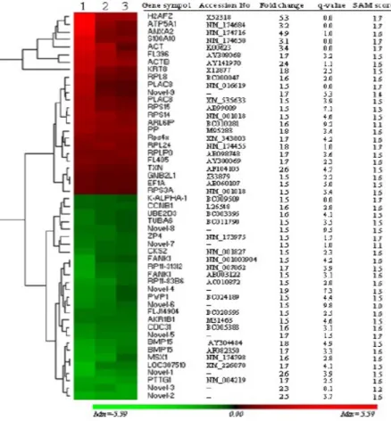
phases and subsequent data analysis using Significant Analysis of Microarray (SAM) revealed a total of 51 genes to be differentially regulated. Hierarchical clustering and heatmap was performed to show the general and magnitude expression of differentially regulated genes
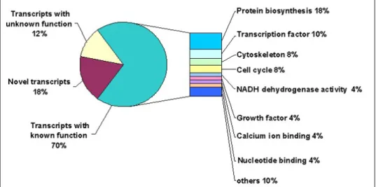
(Figure 1). Furthermore, gene ontology (GO) has classified expressed transcripts functionally into different clusters based on their molecular functions (Figure 2). Accordingly, differentially
regulated genes were found to represent transcripts with known function [70% �36/51)], with unknown function [12% �6/51)] and novel transcripts [18% �9/51)] (Figure 2). Transcripts with known function showed to be involved in protein biosynthesis �18%), transcription �10%),
cytoskeleton (8%), cell cycle (8%), NADH dehydrogenase activity (4%), calcium ion binding
�4%), nucleotide binding �4%) and other molecular functions �10%) (Figure 2). Quantitative
real-time PCR has confirmed the expression profile of 80% (8/10) in independent oocyte
populations from both growth and dominance phases to be in the same trend of microarray data
(Figure 3). The reported differences in developmental capacity of bovine oocytes derived from
small follicles at growth and dominance phases of follicular development are also accompanied
by differences in the relative abundance of transcripts related to the various molecular events
and processes governing oocyte growth and follicular development.
Figure 1: Hierarchical clustering and heatmap of 51 differentially expressed genes. The red blocks represent
Figure 2: Differentially expressed genes as classified based on the Gene Ontology Consortium classifications (http://
www.geneontology.org).
Figure 3: Quantitative real-time PCR validation of 10 differentially expressed genes in bovine oocytes recovered at growth phase (Day 3) vs. dominance phase (Day 7) as identified by microarray analysis (A, B, C). The relative abundance
of m�NA levels represents the amount of m�NA compared to the calibrator �with the lowest normalized value). Bars
with different superscripts (a, b) are significantly different at P < 0.05.
3.1. Transcriptome profile of cumulus cells
It is well established that integrated bilateral communication between the cumulus cells and its enclosed oocyte is key biological factor in acquisition of developmental competence to support preimplantation embryogenesis. Therefore, the molecular messages stored in cumulus cells are possible indicators of the further developmental fate of the oocytes until term.
Several studies have been conducted using farm animals and human granulosa cells to identify genomic signature�s) as molecular markers of developmental potential of oocyte to support embryogenesis and to establish pregnancy [134-136]. Suppressive subtractive hybridization combined with microarray were used to identify several potential cumulus cell markers of
bovine COCs quality that includes several growth differentiation factor GDF9 downstream target genes like GREM1, HAS2, PTGS2, and TNFAIP6 [134]. In addition, epidermal growth factor receptor (EGFR), inhibin beta a (INHBA), betacellulin (BTC) and CD44 molecule was
among candidate molecular markers that induce good potential of COCs. In another study, the transcript abundance of genes controlling of cumulus expansion process such as TNFAIP6
and nuclear maturation of oocyte like INHBA and FST were up-regulated in bovine in these
cells as potential predictors of COCs quality when matured under in vivo [136]. A study using prepubertal bovine calf oocytes as a model of poor oocyte competence; microarray analysis detected genes encoding the cathepsin family of cysteine proteinases �CTSB, CTSS and CTSZ) are linked with reduced competence of bovine oocytes [135]. In search of potential markers of
human granulosa cells, a study done by Feuerstein et al., (2007) has identified steroidogenic
acute protein �STA�), prostaglandin-endoperoxide synthase 2 �PTGS2), stearoyl-co-enzyme A desaturase 1 and 5 �SCD1 and SCD5) and amphiregulin �A�EG) are crucial regulators
of nuclear maturation and their profile are increased after meiosis resumption. Noteworthy,
reduced expression of connexin 43 �CX43) in cumulus is a good marker for nuclear maturation and further embryonic development upto blastocysts �Feuerstein et al. 2007).
3.2. Changes in gene expression during cleavage stages
In in vitro derived bovine embryos, estimates of total �NA content indicate that it declines from the mature oocyte to the morula stage, only to increase again at the blastocyst stage [137]. These estimates are based on Northern blot hybridization using probes for 28S and 18S r�NA, with the abundance of 5S r�NA following a similar pattern [137]. This pattern of �NA loss and reaccumulation mimics the patterns observed in the mouse although in the mouse the increase occurs by the 8- to 16-cell stage likely owing to the earlier onset of zygotic gene activation in this species �Piko and Clegg, 1982). In both species, the timing and the
increase in abundance of specific mRNA transcripts occurs in a transcript specific manner.
Examples of changes in steady state levels of various
During early development, the embryonic genome is inactive and the embryo relies on maternal messenger ribonucleic acid �m�NA) for protein synthesis �Thelie et al. 2009). The recruitment mechanisms by which dormant �NA is either targeted for translation or decay are still largely uncharacterized. The current model involves lengthening of the poly�A) tail, which triggers binding of the poly�A) binding protein and binding of translation initiation factors �Memili and First 2000; Groisman et al. 2002). �NA concentration is highest in the germinal vesicle stage oocyte and from then until the 8-cell stage, �NA is gradually depleted �Gilbert
et al. 2009; Vallée et al. 2009). Evidence in the mouse suggests that this decline is important for activation of the embryonic genome �Li et al. 2010). Depletion of maternal argonaute 2 �Ago2), which encodes a catalytic �NA hydrolase ��Nase), disrupts gene expression and the 2 cell embryo fails to become a blastocyst �Li et al. 2010). In the bovine, embryonic genome activation �EGA) occurs at the 8 to 16-cell stage �Memili and First 2000) through an unknown mechanism.
3.3. Embryo transcriptomics
In mammals, maternally inherited transcripts stored within the oocyte regulate the earliest stages of embryogenesis. After fertilization,the embryonic genome becomes transcriptionally active and the expressionsome of embryonic genes begins in a stage-specific manner,which contribute to the early development process [137]. In cattle, global embryonic genome activation �EGA) begins during the 8-cell to 16-cell stage and it is the most criticalevent in
early embryonic development. However, the identities of embryonic genes expressedand the
mechanism(s) of EGA are poorly defined in the bovine. In addition, understanding of EGA
will contribute to our understandingof nuclear reprogramming in somatic cellnuclear transfer
experiments. Transcripts expressed at the 8-cell stage (EGA) include regulators of different molecular functions like transcription (NFY and USF2), cell adhesion (DSC2 and COL12A1),
signal transducers �PTGE�4) and transporter�C�ABP1),metabolism �NEU3)and immune response �CXCL6) [48].
Accurate assessment of embryo viability is crucial for successful establishment and maintenance of pregnancy following embryo transfer. Evaluation of embryo quality is in
particular of high impact for in vitro-produced embryos as these embryos differ in many aspects
from their in vivo derived counterparts. The temporal or spatial and qualitative or quantitative shifts in the well-orchestrated expression patterns of developmentally important genes have been investigated in preimplantation bovine embryos following in vitro embryo manipulation.
So far, studies in bovine embryos indicate that many of the differences in quality of in vitro-
and in vivo-derived blastocysts can be related to culture environment-induced changes in
mRNA abundance. The post-fertilization embryo culture environment has a dramatic effect
on the pattern of gene expression in embryos, which in turn has serious implications for the normality of blastocyst development [138, 139]. This is the case, not only when one compares
in vitro and in vivo culture systems, but also comparing different in vitro culture systems [140].
�ecently, it was reported that in vitro-cultured embryos showed down-regulation of genes thatare involved in transcriptionand translation �CC�4-NOT, EEF1G, PABPN1, FOXO3A,
HMG2, GNB2L1 and DOT1L) events compared with in vivo counterparts, suggesting that in
vitro-derivedembryos are of inferior quality compared with in vivo-derivedembryos due to a
Imprinted genes appear to be more susceptible to alterations in epigenetic modifications [141]. especially after IVP of ovine [142] or bovine embryos [143]. Significant differences
in the expression of non-imprinted genes have also been reported in bovine IVP and somatic nuclear transferred �sNT) derived embryos compared to their in vivo counterparts [144].
Differences in gene expression patterns between IVP and sNT-derived embryos and their in
vivo counterparts may originate from all steps of the manipulation procedures, including in
vitro maturation, in vitro fertilization, and in vitro culture and, for sNT, from different somatic
nuclear transfer protocols [145]. In a recent study, it was found that there is a reduced levels of many genes required for the viability of every cell in the nuclear transferred �NT) embryo’s cellular machinery [146]. In this study, transcripts of mitochondria �12S and 16S r�NA and cytochrome C oxidase I), cytoskeletal �TUBB, TPX2, K�T18, TMSB4X and ACTB), protein
biosynthesis (EEF1A1, EFG2, BWZ1 and EIF5A) and ribosomal proteins (RPL4, RPL5, RPL21 and RPS20), protein binding/folding (GORASP2, HSPA9B, HSPA5 and LGALS3) and etabolism/biosynthesis (ADH5, PTGS2, SCP2, ACSL3, NDUFA1) were decreased in NT
compared to in vitro produced blastocysts.
There is a clear relationship between altered transcript abundance patterns and some aspects of embryo quality �i.e. cryotolerance), which render the embryo capable o f establishing a pregnancy, if transferred fresh, but incapable of withstanding cryopreservation [147], [25]. Various studies in both mice and bovine have shown that the in vitroproduction of embryos
under specific culture environments resultedin not only altered gene expression of transcripts related tometabolic and growth but also altered conceptus and fetal developmentfollowing transfer [148,149]. Despite the fact that data on transcriptional analysis of transferable blastocysts of various origins havebeen accumulated, so far no direct connection ofgene
expression and developmental competence has been established. However, a well-established
biopsy techniqueis needed to obtain cells from embryos before transfer without any lethaleffect
on the embryo during further development.For this, one study conducted in our laboratory to establish connection betweentranscriptional profile of embryos and the pregnancy success
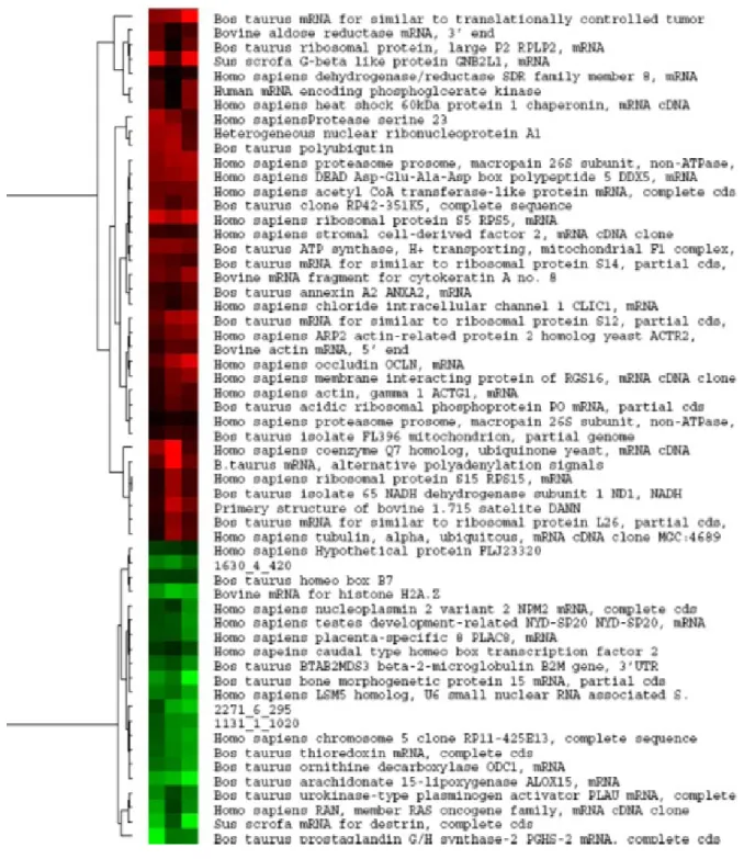
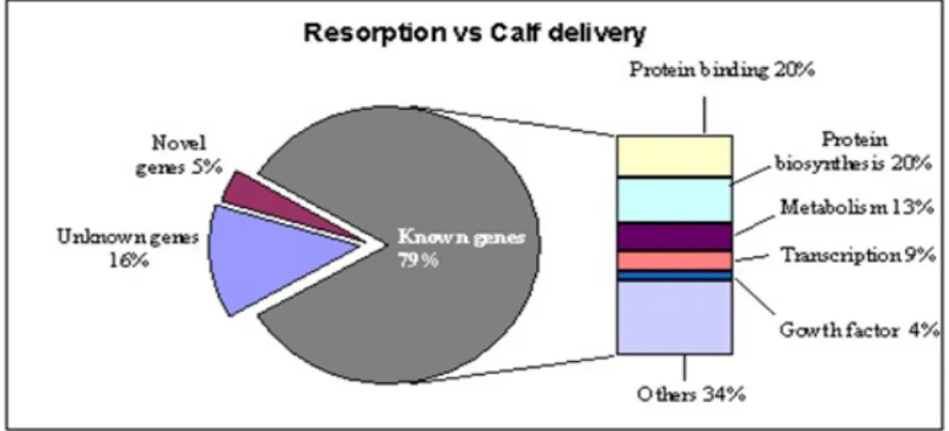
based on gene expression analysis of blastocyst biopsies takenprior to transfer to recipients [150]. Microarray data analysis revealed a total of 52 and 58 genes (Figure 4 and 5) were
differentially regulated during comparison between embryo biopsies resulting in no pregnancy
�G1) vs. calf delivery �G3) and those resulting in resorbed embryos �G2) vs. calf delivery �G3).
Ontological classification has showed different functional clusters of genes (Figure 6 and 7).
In addition, differentially regulated genes representgenes with known functions �77%), ESTs �11.5%), and novel transcripts�11.5%) �Figure 4 and 5). Biopsies resulted in calf delivery were enrichedwith genes necessary for implantation �COX2 and CDX2), carbohydratemetabolism �ALOX15), growth factor �BMP15), signal transduction (PLAU), and placenta-specific 8
�PLAC8). Biopsies from embryosresulting in resorption are enriched with transcripts involved protein phosphorylation �K�T8), plasma membrane �OCLN), andglucose metabolism �PGK1
and AK�1B1). Biopsies from embryosresulting in no pregnancy are enriched with transcripts involvedinflammatory cytokines (TNF), protein amino acid binding (EEF1A1),transcription factors �MSX1, PTTG1), glucose metabolism �PGK1,AK�1B1), and CD9, which is an inhibitor of implantation. The expressionof those unknown and novel ESTs showed profiles similar to
thoseof the annotated genes, as determined by tree hierarchical clusteringanalyses (Figure 4 and 5). Quantitative real-time PC� has validatedthe expression of 87% �13/15) of the genes generated from thearray hybridization. Thus, several genes identified in this experiment may
be associated withembryo loss or survival in blastocysts during preimplantationperiod.
Figure 4: Hierarchical clustering for the differentially expressed genes between biopsies derived from blastocysts
resulted in no pregnancy �G1) and calf delivery �G3). The columns represent the replicates. The rows represent 52 genes
Figure 5: Results of hierarchical clustering for the differentially expressed genes between biopsies derived from blastocysts resulted in resorption (G2) and calf delivery (G3) identified by microarray analysis. The columns represent the replicates.The rows represent 58 genes found to be differentially regulated between the two groups of biopsies.
Figure 6: Ontology classification for differentially expressed transcripts between biopsies derived from blastocysts resulted n no pregnancy and calf delivery. The known genes were classified functionally based on the Gene Ontology Consortium classification (http://www.geneontology.org)
Figure 7: Ontology classification for differentially expressed transcripts between biopsies derived from blastocysts resulted in resorption and calf delivery. The known genes were classified functionally based on the Gene Ontology Consortium classification (http://www.geneontology.org).
Figure 8: Quantitative real time PCR confirmation of selected transcripts between biopsies from blastocysts resulted in
calf delivery versus no pregnancy �A) and calf delivery versus resorption group �B) and those resulted in calf delivery versus resorption or no pregnancy �C).
4. Characterization and Functional Analysis of Candidate Genes
To correlate transcript level with corresponding protein level and to elucidate embryonic cellular function, detailed characterisation is necessary at the protein level. Oogenesin gene is expressed during oogenesis and early embryogenesis in mouse and its protein is synthesised from the oocyte to four-cell embryo stages, suggesting a possible role during oocyte maturation and/or embryonic genome activation [151].
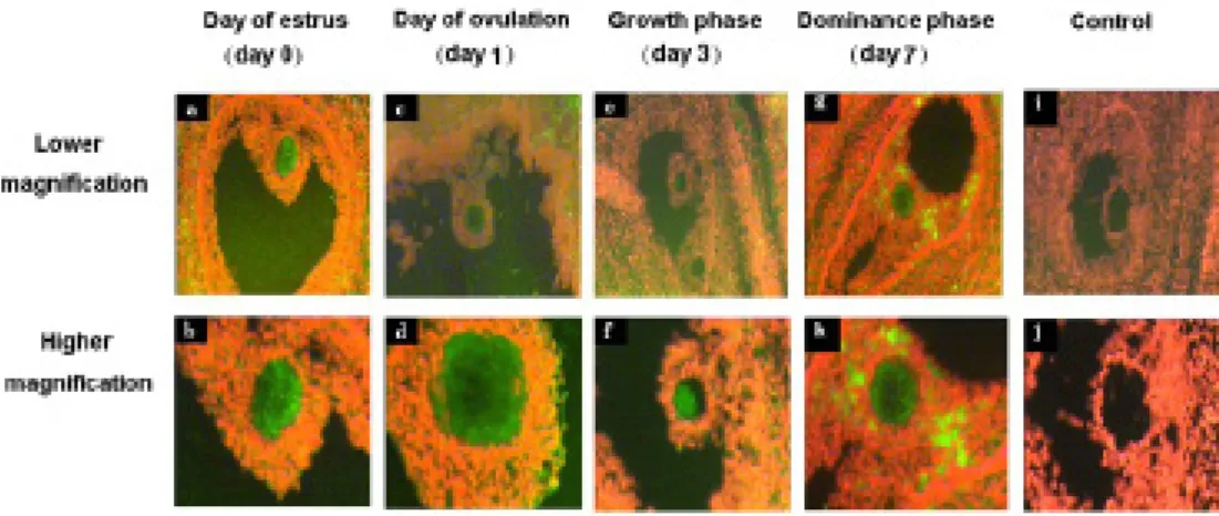
In one of our studies, MSX1 was selected as candidate gene for protein localization in early embryonic development and throughout follicular turnover. MSX1 protein was found to be localized at higher levels in the oocytes cytoplasm than in the surrounding cumulus cells
(Figure 9d, h, p) or other cellular layers of the growing follicle (Figure 9b, f, n) at all stages of follicular development except at growth phase (Figure 7j, l).
The MSX1 protein was found to be dispersed in the cytoplasm of immature and matured oocytes and early zygote stages (Figure 10a, b, c) but tends to be localized around the nucleus at advanced zygote, 2-cell, 4-cell and 8-cell cleavage stage embryos (Figure 10d, e, f, g).
Comparative analysis of protein signals between oocytes showed that fluorescence signals
were reduced after maturation (Figure 10b). Moreover, the in situ hybridization experiment results showed that MSX1 m�NA was localized in the oocytes, cumulus cells and follicular wall during the periods of follicular turnover under investigation (Figure 11).
Figure 9: Immunohistochemical localisation of MSX1 protein in bovine ovarian sections at day of estrus �b, d), day of ovulation �f, h), growth phase �j, l), dominance phase �m, q). Cumulus cells are marked with Cc and oocytes are marked with Oo. Negative controls were processed without addition of primary anti-MSX1 antibody �r, t). Sections were counterstained with toluidine blue �a, c, e, g, i, k, m, o, q and s). Images from the same ovarian sections were captured
with lower and higher magnification.
Figure 10: Subcellular localization of bovine MSX1 protein in bovine oocytes and early cleavage embryonic stages [immature oocyte �a), matured oocyte �b), zygote �c), advanced zygote �d), 2-cell �e), 4-cell �f) and 8-cell �g)]. Negative control �h) was processed without addition of primary anti-MSX1 antibody. Nuclei are stained with propidium iodide �red). Scale bars represent 20 µm.
Figure 11: Fluorescent in situ hybridization of MSX1 m�NA conducted with DIG labelled �NA antisense probe in bovine ovarian sections at day of estrus �A, B), day of ovulation �B, C), growth phase �D, E), dominance phase �F, G).
Cytoplasms of the oocytes (Oo) are darkly stained with green fluorescent compared to cumulus cells (Cc). Negative controls were hybridized with DIG labelled RNA sense probe (H, I). Images from the same ovarian sections were captured with lower and higher magnification.
5. References
1. Le Naour F, Rubinstein E, Jasmin C, Prenant M and Boucheix C: Severely reduced female fertility in CD9-deficient mice. Science 287: 319-321, 2000.
2. Leibfried-Rutledge ML (1999): Factors determining competence of in vitro produced cattle embryos. Theriogenology
51, 473-485.
3. Flesch FM, Gadella BM. Dynamics of the mammalian sperm plasma membrane in the process of fertilization.
Bio-chim Biophys Acta 2000; 1469: 197–235.
4. Kadivar A, Heidari Khoei H, Hassanpour H, Ghanaei H, Golestanfar A, Mehraban H et al. Peroxisome proliferator-activated receptors (PPARα, PPARγ and PPARβ/δ) gene expression profile on ram spermatozoa and their relation to the sperm motility. Vet Res Forum 2016; 7: 27-34.
5. Kim KU, Pang WK, Kang S, Ryu DY, Song WH, Rahman MS et al. Sperm solute carrier family 9 regulator 1 is cor
-related with boar fertility. Theriogenology 2019; 126:254-60.
6. Parthipan S, Selvaraju S, Somashekar L, Arangasamy A, Sivaram M, �avindra JP. Spermatozoal transcripts
expres-sion levels are predictive of semen quality and conception rate in bulls (Bos taurus). Theriogenology, 2017; 98:41-49. 7. Bissonnette N, Lévesque-Sergerie JP, Thibault C, Boissonneault G. Spermatozoal transcriptome profiling for bull sperm motility: a potential tool to evaluate semen quality. Reproduction 2009;138: 65–80.
8. Boerke A, Dieleman SJ, Gadella BM. A possible role for spermatozoa �NA in early embryo development.
Theriog-enology 2007; 68:147–55.
9. Macmillan KL, Lean IJ, Westwood CT (1996): The effects of lactation on the fertility of
10. Lucy, MC (2001): Reproductive loss in high-producing dairy cattle: Where will it end?.J Dairy Sci. 2001 Jun;84(6):1277-93.
11. Marai, I.F.M and Haeeb A.A.M. (2010). Buffalo's biological functions as affected by heat stress - a review. Livest. Sci.,(127):89 - 109.
12. Al-Katanani YM, Paula-Lopes FF, Hansen PJ. Effect of Season and Exposure to Heat Stress on Oocyte Competence in Holstein Cows J Dairy Sci. 2002; 85(2):390-6.
in transcript levels in the developing embryos associated with reduced developmental competence. Biol �eprod. 2012
Jan 16;86(1):1-9.
14. Sadeesh EM, Sikka P, Balhara AK, Balhara S. Developmental Competence and Expression Profile of Genes in Buf
-falo (Bubalus Bubalis) Oocytes and Embryos Collected Under Different Environmental Stress. Cytotechnology. 2016; 68(6):2271-2285.
15. Abdoon AS, Gabler C, Holder C, Kandil OM, Einspanier R.Seasonal variations in developmental competence and relative abundance of gene transcripts in buffalo (Bubalus bubalis) oocytes. Theriogenology. 2014 Nov;82(8):1055-67. 16. Maya-Soriano MJ, López-Gatius F, Andreu-Vázquez C, López-Béjar M. Bovine Oocytes Show a Higher Tolerance to Heat Shock in the Warm Compared With the Cold Season of the Year. Theriogenology. 2013; 79(2):299-305.
17. Edwards JL, Hansen PJ. Differential responses of bovine oocytes and preimplantation embryos to heat shock. Mol Reprod Dev. 1997; 46(2):138-45.
18. Nabenishi H, Ohta H, Nishimoto T, Morita T, Ashizawa K, Tsuzuki Y. The effects of cysteine addition during in vitro maturation on the developmental competence, ROS, GSH and apoptosis level of bovine oocytes exposed to heat stress. Zygote. 2012; 20(3):249-59.
19. Armon L, Ben-Ami I, Ron-El R and Eisenbach M: Human oocyte-derived sperm chemoattractant is a hydrophobic molecule associated with a carrier protein. Fertil Steril 102: 885-890, 2014.
20. Yadav A, Singh KP, Singh MK, Saini N, Palta P, Manik RS, Singla SK, Upadhyay RC, Chauhan MS. Effect of physi
-ologically relevant heat shock on development, apoptosis and expression of some genes in buffalo (Bubalus bubalis) embryos produced in vitro. Reprod Domest Anim. 2013 Oct;48(5):858-65.
21. Snijders SEM, Dillon P, O'Callaghan D, Boland MP (2000): Effect of genetic merit,
22. Donnison M, Pfeffer PL (2004): Isolation of genes associated with developmentally competent bovine oocytes and
quantitation of their levels during development. Biol �eprod 71, 1813-1821.
23. Ghanem N, Hölker M, Rings F, Jennen D, Tholen E, Sirard MA, Torner H, Kanitz W, Schellander K, Tesfaye D,
2007: Alterations in transcript abundance of bovine oocytes recovered at growth and dominance phases of the first fol
-licular wave. BMC Dev Biol 7, 90.
24. Rizos D, Ward F, Duffy P, Boland MP, Lonergan P (2002): Consequences of bovine
25. Rizos D, Gutiérrez-Adán A, Pérez-Garnelo S, de la Fuente J, Boland MP, Lonergan P (2003): Bovine embryo cul
-ture in the presence or absence of serum: implications for blastocyst development, cryotolerance, and messenger RNA
expression. Biol �eprod 68, 236-243.
26. Rodriguez-Zas S, Schellander K, Lewin H, 2008: Biological interpretations of transcriptomic profiles in mammalian
oocytes and embryos. �eproduction 135, 129-39.
27. Morris RL, Brown HM, Wright BD, Sharp DJ, Sullivan W, Scholey JM, 2001: Microinjection methods for analyzing
the functions of kinesins in early embryos. Methods Mol Biol 164,163-172.
28. Geisert RD, Morgan GL, Short EC Jr, Zavy MT, 1992: Endocrine events associated with endometrial function and
conceptus development in cattle. �eprod Fertil Dev 4, 301-305.
29. Salilew-Wondim D, Hölker M, Rings F, Ghanem N, Ulas-Cinar M, Peippo J, Tholen E, Looft C, Schellander K, Tesfaye D, 2010: Bovine pretransfer endometrium and embryo transcriptome fingerprints as predictors of pregnancy
success after embryo transfer. Physiol Genomics 42, 201-218.
30. El-Sayed A, Hoelker M, Rings F, Salilew D, Jennen D, Tholen E, Sirard M-A, Schellander K, Tesfaye D, 2006:
recipi-ents. Physiol. Genomics 28, 84-96.
31. Gad A, Besenfelder U, Rings F, Ghanem N, Salilew-Wondim D, Hossain MM, Tesfaye D, Lonergan P, Becker A, Cinar U, Schellander K, Havlicek V, Hölker M, 2011: Effect of reproductive tract environment following controlled
ovarian hyperstimulation treatment on embryo development and global transcriptome profile of blastocysts: implica
-tions for animal breeding and human assisted reproduction. Hum Reprod 26,1693-1707.
32. Ghanem N, Salilew-Wondim D, Gad A, Tesfaye D, Phatsara C, Tholen E, Looft C, Schellander K, Hoelker M, 2011:
Bovine blastocysts with developmental competence to term share similar expression of developmentally important
genes although derived from different culture environments. Reproduction 142, 551-564.
33. Niemann H, Carnwath J, Kues W, 2007: Application of DNA array technology to mammalian embryos. Theriogenol
-ogy 68, S165-177.
34. Ruddock-D'Cruz NT, Hall VJ, Tecirlioglu RT, French AJ, 2007: Gene expression analysis of single preimplantation
bovine embryos and the consequence for developmental potential. Soc �eprod Fertil Suppl 64, 341-363.
35. Copp AJ (1995): Death before birth: clues from gene knockouts and mutations. Trends Genet 11, 87-93.
36. Zhang B, Peñagaricano F, Driver A, Chen H, Khatib H, 2011: Differential expression of heat shock protein genes and
their splice variants in bovine preimplantation embryos. J Dairy Sci 94, 4174-4182.
37. Niemann H, and Wrenzycki C, 2000: Alterations of expression of developmentally important genes in preimplan
-tation bovine embryos by in vitro culture conditions: implications for subsequent development. Theriogenology 53,
21- 34.
38. Aswal APS, Raghav S, De S, Thakur M, Goswami SL, Datta TK, 2007a: Expression stability of two housekeeping
genes (18S rRNA and G3PDH) during in vitro maturation of follicular oocytes in buffalo (Bubalus bubalis). Anim Re
-prod Sci 103, 164–171.
39. Aswal APS, Datta TK, Raghav S, De S, Yadav P, Goswami SL, 2007b: Development of a competitive quantitative PCR strategy for evaluating the expression stability of 18s rRNA during in vitro maturation of buffalo (Bubalus bubalis)
follicular oocytes. �eprod Domest Anim 42, 195–201.
40. Robert C, Gagne D, Bousquet D, Barnes FL Sirard, MA (2001): Differential display and suppressive subtractive
hybridization used to identify granulosa cell messenger �NA associated with bovine oocyte developmental competence. Biol �eprod 64, 1812-1820.
41. Dalbiès-Tran �, Mermillod P �2003) Use of heterologous complementary DNA array screening to analyze bovine oocyte transcriptome and its evolution during in vitro maturation. Biol �eprod 68, 252-261.
42. Sirard MA, Dufort I, Vallee M, Massicotte L, Gravel C, Reghenas H, Watson AJ, King WA, Robert C (2005): Poten
-tial and limitations of bovine-specific arrays for the analysis of mRNA levels in early development: preliminary analysis
using a bovine embryonic array. �eprod Fertil Dev 17, 47-57.
43. Suchyta SP, Sipkovsky S, Kruska R, Jeffers A, McNulty A, Coussens MJ, Tempelman RJ, Halgren RG, Saama PM, Bauman DE, Boisclair YR, Burton JL, Collier RJ, DePeters EJ, Ferris TA, Lucy MC, McGuire MA, Medrano JF, Overton TR, Smith TP, Smith GW, Sonstegard TS, Spain JN, Spiers DE, Yao J, Coussens PM. (2003): Development and
testing of a high-density cDNA microarray resource for cattle. Physiol Genomics 15, 158-164.
44. Yao J, Ren X, Ireland JJ, Coussens PM, Smith TP, Smith GW (2004): Generation of a bovine oocyte cDNA library and microarray: resources for identification of genes important for follicular development and early embryogenesis.
Physiol Genomics 19, 84-92.
45. Wolf E, Arnold GJ, Bauersachs S, Beier HM, Blum H, Einspanier R, Fröhlich T, Herrler A, Hiendleder S, Kölle S, Prelle K, Reichenbach HD, Stojkovic M, Wenigerkind H, Sinowatz F 2003 Embryo-Maternal communication in bovine-
46. Bauersachs S, Ulbrich SE, Gross K, Schmidt SEM, Meyer HHD, Wenigerkind H, Vermehren M, Sinowatz F, Blum H, Wolf E 2006 Embryo-induced transcriptome changes in bovine endometrium reveal species-specific and common
molecular markers of uterine receptivity. �eproduction 132, 319-331.
47. Patel OV, Bettegowda A, Ireland JJ, Coussens PM, Lonergan P, Smith GW (2007): Functional genomics studies of oocyte competence: evidence that reduced transcript abundance for follistatin is associated with poor developmental
competence of bovine oocytes. �eproduction 133, 95-106.
48. Misirlioglu M, Page GP, Sagirkaya H, Kaya A, Parrish JJ, First NL, Memili E (2006): Dynamics of global transcrip
-tome in bovine matured oocytes and preimplantation embryos. Proc Natl Acad Sci 103, 18905-18910.
49. Kocabas AM, Crosby J, Ross PJ, Out HH, Beyhan Z, Can H, et al. 2006. The transcriptome of human oocytes. Proc Natl Acd Sci USA. 103:14027-32.
50. Corcoran D, Fair T, Park S, Rizos D, Patel OV, Smith GW, Coussens PM, Ireland JJ, Boland MP, Evans ACO, Lon
-ergan P (2006): Suppressed expression of genes involved in transcription and translation in in vitro compared to in vivo
cultured bovine embryos. �eproduction 131, 651-660.
51. Kues WA, Sudheer S, Herrmann D, Carnwath JW, Havlicek V, Besenfelder U, Lehrach H, Adjaye J, Niemann H. Genome-wide expression profiling reveals distinct clusters of transcriptional regulation during bovine preimplantation development in vivo. Proc Natl Acad Sci U S A. 2008 Dec 16;105(50):19768-73.
52. Vallée M, Robert C, Méthot S, Palin M-F, Sirard M-A.(2006): Cross-species hybridizations on a multi-species cDNA
microarray to identify evolutionarily conserved genes expressed in oocytes. BMC Genomics 7,113.
53. Hu X, Pokharel K, Peippo J, Ghanem N, Zhaboyev I, Kantanen J, Li MH (2016): Identification and characterization of miRNAs in the ovaries of a highly prolific sheep breed. Animal Genetics 47(2):234-239.
54. Pokharel K, Peippo J, Honkatukia M, Seppälä A, Rautiainen J, Ghanem N, Hamama TM, Crowe MA, Andersson M, Li MH, Kantanen J (2018) : Integrated ovarian mRNA and miRNA transcriptome profiling characterizes the genetic basis of prolificacy traits in sheep (Ovis aries).BMC Genomics, 29;19(1):104.
55. Pitnick, S. et al. (2009) Sperm morphological diversity. In Sperm Biology: An Evolutionary Perspective (Birkhead,
T.�. et al., eds), pp. 69–149, Academic Press
56. Fischer, B.E. et al. �2012) Conserved properties of Drosophila and human spermatozoal m�NA repertoires. Proc. �.
Soc. B 279, 2636–2644 8 Hecht, N.B. (1998) Molecular mechanisms of male germ cell differentiation. Bioessays 20,
555–561 9.
57. Miller, D. et al. �2005) The controversy, potential and roles of spermatozoal �NA. Trends Mol. Med. 11, 156–163
58. Hagemann LJ, Weilert LL, Beaumont S, Tervit HR (1998): Development of bovine embryos in single in vitro pro
-duction �sIVP) systems. Mol �eprod Dev 51, 143-147.
59. Boerke, A. et al. �2007) A possible role for sperm �NA in early embryo development. Theriogenology 68S, S147– S155
60. Bourc’his, D. and Voinnet, O. �2010) A small-�NA perspective on gametogenesis, fertilization, and early zygote development. Science 330, 617–622
61. Dadoune, J-P. (2009) Spermatozoal RNAs: what about their functions? Microsc. Res. Tech. 72, 536–551. dairy
cows. Aust vet J. 73, 141-147.
62. Krawetz SA. Paternal contribution: new insights and future challenges. Nat Rev Genet 2005; 6:633–42. 63. Gardener, A. and West, S.A. (2009) Green beards. Evolution 64, 25–38.
-phology and motility. Theriogenology 76, 1532–1539
65. Yan, W. et al. (2008) Birth of mice after intra cytoplasmic injection of single purified sperm nuclei and detection of
messenger �NAs and Micro�NAs in the sperm nucleus. Biol. �eprod. 78, 896–902
66. Liu, W-M. et al. (2012) Sperm-borne microRNA-34c is required for the first cleavage division in mouse. Proc. Natl.
Acad. Sci. U.S.A. 109, 490-494.
67. Gapp, K. et al. (2014) Implication of sperm RNAs in transgenerational inheritance of the effects of early trauma in mice. Nat. Neurosci. http:// dx.doi.org/10.1038/nn.3695
68. Hamilton, W.D. (1964) The genetical evolution of social behavior I & II. J. Theor. Biol. 7, 1–52.
69. Pomiankowski, A. (1999) Intragenomic conflict. In Levels of Selection (Keller, L., ed.), pp. 121–152, Princeton
University Press
70. Leigh, E.G. (1999) Levels of selection, potential conflicts, and their resolution: the role of the ‘common good’. In
Levels of Selection �Keller, L., ed.), pp. 15–30, Princeton University Press
71. Fisher, H.S. and Hoekstra, H.E. (2010) Competition drives cooperation among closely related sperm of deer mice.
Nature 463, 801–803.
72. Holman, L. and Snook, R.R. (2008) A sterile sperm caste protects brother fertile sperm from female-mediated death
in Drosophila pseudoobscura. Curr. Biol. 18, 292–296.
73. Higginson, D.M. and Pitnick, S. (2011) Evolution of intra-ejaculate sperm interactions: do sperm cooperate? Biol.
�ev. 86, 249–270.
74. Immler, S. et al. (2007) By hook or by crook? Morphometry, competition and cooperation in rodent sperm. PLoS
ONE 2, e170.
75. Moore, H.D. et al. (2002) Exceptional sperm cooperation in the wood mouse. Nature 418, 174–177.
76. Arnqvist, G. and Nilsson, T. (2000) The evolution of polyandry: multiple mating and female fitness in insects. Anim.
Behav. 60, 164-145.
77. Jennions, M.D. and Petrie, M. (2000) Why do females mate multiply? A review of the genetic benefits. Biol. Rev.
75, 21–64.
78. Hosken, D.J. and Stockley, P. (2003) Benefits of polyandry: a life history perspective. Evol. Biol. 33, 173–194.
79. Parker, G.A. �1970) Sperm competition and its evolutionary consequences in insects. Biol �ev. 45, 525–567. 80. Birkhead, T.�. and Moller, A.P. �1998) Sperm Competition and Sexual Selection, Academic Press.
81. Simmons, L.W. (2001) Sperm Competition and its Evolutionary Consequences in the Insects, Princeton University
Press.
82. Pessot CA, Brito M, Figueroa J, Concha II, Yanez A, Burzio LO. 1989 Presence of RNA in the sperm nucleus. Bio
-chem Biophys Res Commun 1989; 158: 272–8.
83. Swain JL, Stewart TA, Leder P. Parental legacy determines methylation and expression of an autosomal transgene: a molecular mechanism for parental imprinting. Cell 1987;50: 719–27.
84. Cummins JM. Cytoplasmic inheritance and its implications for animal biotechnology. Theriogenology 2001; 55:
1381–99.
RNA to the oocyte. Nature 2004; 429: 154.
86. Ziyyat A, Lefevre A. Differential gene expression in pre-implantation embryos from mouse oocytes injected with round spermatids or spermatozoa. Hum Reprod 2001; 16: 1449–56.
87. Ostermeier GC, Goodrich �J, DiamondMP, Dix DJ, Krawetz SA. Toward using stable spermatozoal �NAs for
prog-nostic assessment of male factor fertility. Fertil Steril 2005; 83: 1687–94.
88. Platts AE, Dix DJ, Chemes HE, Thompson KE, Goodrich R, Rockett JC, et al. Success and failure in human sper
-matogenesis as revealed by teratospermic RNAs. Hum Med Gen 2007; 16: 763–73.
89. Zhao Y, Li Q, Yao C, Wang Z, Zhou Y, Wang Y, et al. Characterization and quantification of mRNA transcripts in ejaculated spermatozoa of fertile men by serial analysis of gene expression. Hum Reprod 2006; 21: 1583–90.
90. Flesch FM, Gadella BM. Dynamics of the mammalian sperm plasma membrane in the process of fertilization.
Bio-chim Biophys Acta 2000; 1469: 197–235.
91. Miller D, Ostermeier GC. Towards a better understanding of RNA carriage by ejaculate spermatozoa. Hum Reprod Update 2006; 12: 757–67.
92. Miller* D, Ostermeier GC. Spermatozoal RNA: why is it there and what does it do? Gynecol Obstet Fertil 2006; 34: 840–6.
93. Gui LM, Joyce IM (2005): RNA interference evidence that growth differentiation factor-9 mediates oocyte regula
-tion of cumulus expansion in mice. Biol Reprod 72, 195-199.Gur Y, Breitbart H. Mammalian sperm translate nuclear-encoded proteins by mitochondrial-type ribosomes. Genes Dev 2006; 20: 411–6.
94. Sutovsky P, Moreno �D, �amalho-Santos J, Dominko T, Simerly C, Schatten G. Ubiquitinated sperm mitochondria,
selective proteolysis, and the regulation of mitochondrial inheritance in mammalian embryos. Biol Reprod 2000; 63:
582–90.
95. Ebensperger C and Barros C: Changes at the hamster oocyte surface from the germinal vesicle stage to ovulation. Gamete Res 9: 387-397, 1984.
96. Johnson MH, Eager D, Muggleton-Harris A and Grave HM: Mosaicism in organisation concanavalin A receptors on surface membrane of mouse egg. Nature 257: 321-322, 1975.
97. Evans JP and Florman HM: The state of the union: the cell biology of fertilization. Nat Cell Biol 4: s57-s63, 2002.
98. Huang TTF Jr and Yanagimachi R: Inner acrosomal membrane of mammalian spermatozoa: Its properties and pos
-sible functions in fertilization. Am J Anat 174: 249-268, 1985.
99. Yanagimachi R and Noda YD: Physiological changes in the postnuclear cap region of mammalian spermatozoa: A necessary preliminary to the membrane fusion between sperm and egg cells. J Ultrastruct Res 31: 486-493, 1970.
100. Bedford JM, Moore HDM and Franklin LE: Significance of the equatorial segment of the acrosome of the sperma
-tozoon in eutherian mammals. Exp Cell Res 119: 119-126, 1979.
101. Cho C, Bunch DO, Faure JE, Goulding EH, Eddy EM, Primakoff P and Myles DG: Fertilization defects in sperm from mice lacking fertilin β. Science 281: 1857-1859, 1998.
102. Nishimura H, Cho C, Branciforte DR, Myles DG and Primakoff P: Analysis of loss of adhesive function in sperm lacking cyritestin or fertilin beta. Dev Biol 233: 204-213, 2001.
103. Almeida EA, Huovila AP, Sutherland AE, Stephens LE, Calarco PG, Shaw LM, Mercurio AM, Sonnenberg A, Primakoff P, Myles DG and White JM: Mouse egg integrin α 6 β 1 functions as a sperm receptor. Cell 81: 1095-1104,
104. Chen H and Sampson NS: Mediation of sperm-egg fusion: Evidence that mouse egg α6β1 integrin is the receptor for sperm fertilin beta. Chem Biol 6: 1-10, 1999.
105. Eto K, Huet C, Tarui T, Kupriyanov S, Liu HZ, Puzon-McLaughlin W, Zhang XP, Sheppard D, Engvall E and Takada Y: Functional classification of ADAMs based on a conserved motif for binding to integrin α 9β 1: Implications for sperm-egg binding and other cell interactions. J Biol Chem 277: 17804-17810, 2002.
106. Zhu X and Evans JP: Analysis of the roles of RGD-binding integrins, α(4)/α(9) integrins, α(6) integrins, and CD9 in the interaction of the fertilin β (ADAM2) disintegrin domain with the mouse egg membrane. Biol Reprod 66:
1193-1202, 2002.
107. Mizuno M, Harris CL, Johnson PM and Morgan BP: Rat membrane cofactor protein (MCP; CD46) is expressed
only in the acrosome of developing and mature spermatozoa and mediates binding to immobilized activated C3. Biol
Reprod 71: 1374-1383, 2004.
108. Johnson PM, Clift LE, Andrlíková P, Jursová M, Flanagan BF, Cummerson JA, Stopka P and Dvorakova-Hortova
K: Rapid sperm acrosome reaction in the absence of acrosomal CD46 expression in promiscuous field mice (Apode
-mus). Reproduction 134: 739-747, 2007.
109. Lozahic S, Christiansen D, Manié S, Gerlier D, Billard M, Boucheix C and Rubinstein E: CD46 (membrane cofac
-tor protein) associates with multiple beta1 integrins and tetraspans. Eur J Immunol 30: 900-907, 2000.
110. Stein KK, Primakoff P and Myles D: Sperm-egg fusion: Events at the plasma membrane. J Cell Sci 117: 6269-6274,
2004.
111. Jégou A, Ziyyat A, Barraud-Lange V, Perez E, Wolf JP, Pincet F and Gourier C: CD9 tetraspanin generates fusion competent sites on the egg membrane for mammalian fertilization. Proc Natl Acad Sci USA 108: 10946-10951, 2011. 112. Kaji K, Oda S, Shikano T, Ohnuki T, Uematsu Y, Sakagami J, Tada N, Miyazaki S and Kudo A: The gamete fusion process is defective in eggs of Cd9-deficient mice. Nat Genet 24: 279-282, 2000.
113. Miyado K, Yamada G, Yamada S, Hasuwa H, Nakamura Y, Ryu F, Suzuki K, Kosai K, Inoue K, Ogura A, et al: Requirement of CD9 on the egg plasma membrane for fertilization. Science 287: 321-324, 2000.
114. Horváth G, Serru V, Clay D, Billard M, Boucheix C and Rubinstein E: CD19 is linked to the integrin-associated tetraspans CD9, CD81, and CD82. J Biol Chem 273: 30537-30543, 1998.
115. Takahashi Y, Bigler D, Ito Y and White JM: Sequence-specific interaction between the disintegrin domain of mouse ADAM 3 and murine eggs: Role of beta1 integrin-associated proteins CD9, CD81, and CD98. Mol Biol Cell 12:
809-820, 2001.
116. Rubinstein E, Ziyyat A, Prenant M, Wrobel E, Wolf JP, Levy S, Le Naour F and Boucheix C: Reduced fertility of female mice lacking CD81. Dev Biol 290: 351-358, 2006.
117. Ernesto JI, Weigel Muñoz M, Battistone MA, Vasen G, Martínez-López P, Orta G, Figueiras-Fierro D, De la Vega-Beltran JL, Moreno IA, Guidobaldi HA, et al: CRISP1 as a novel CatSper regulator that modulates sperm motility and orientation during fertilization. J Cell Biol 210: 1213-1224, 2015.
118. Inoue N, Ikawa M, Isotani A and Okabe M: The immunoglobulin superfamily protein Izumo is required for sperm to fuse with eggs. Nature 434: 234-238, 2005.
119. Inoue N, Hamada D, Kamikubo H, Hirata K, Kataoka M, Yamamoto M, Ikawa M, Okabe M and Hagihara Y: Mo
-lecular dissection of IZUMO1, a sperm protein essential for sperm-egg fusion. Development 140: 3221-3229, 2013.
120. Inoue N, Kasahara T, Ikawa M and Okabe M: Identification and disruption of sperm-specific angiotensin convert

![Figure 10: Subcellular localization of bovine MSX1 protein in bovine oocytes and early cleavage embryonic stages [immature oocyte �a), matured oocyte �b), zygote �c), advanced zygote �d), 2-cell �e), 4-cell �f) and 8-cell �g)]](https://thumb-us.123doks.com/thumbv2/123dok_us/9341589.2416361/19.892.167.728.887.1115/figure-subcellular-localization-protein-cleavage-embryonic-immature-advanced.webp)