Hepatocyte Tissue Factor Contributes to the Hypercoagulable
State in a Mouse Model of Chronic Liver Injury
Pierre-Emmanuel Rautou1,2,*, Kohei Tatsumi1,*, Silvio Antoniak1, A. Phillip Owens III1, Erica
Sparkenbaugh1, Lori A. Holle3, Alisa S. Wolberg3, Anna K. Kopec4, Rafal Pawlinski1, James P. Luyendyk4, and Nigel Mackman1
1Department of Medicine, Division of Hematology and Oncology, McAllister Heart Institute,
University of North Carolina at Chapel Hill, Chapel Hill, NC 27599, USA
2Service d’Hépatologie, Hôpital Beaujon, Assistance Publique-Hôpitaux de Paris, Clichy, France
3Department of Pathology and Laboratory Medicine, University of North Carolina at Chapel Hill,
Chapel Hill, NC 27599, USA
4Department of Pathobiology and Diagnostic Investigation, Michigan State University, East
Lansing, Michigan, USA
Summary
Background & Aims—Patients with chronic liver disease and cirrhosis have a dysregulated coagulation system and are prone to thrombosis. The basis for this hypercoagulable state is not completely understood. Tissue factor (TF) is the primary initiator of coagulation in vivo. Patients with cirrhosis have increased TF activity in white blood cells and circulating microparticles. The aim of our study was to determine the contribution of TF to the hypercoagulable state in a mouse model of chronic liver injury.
Methods—We measured levels of TF activity in the liver, white blood cells and circulating microparticles, and a marker of activation of coagulation [thrombinantithrombin complexes (TATc)] in the plasma of mice subjected to bile duct ligation for 12 days. We used wild-type mice, mice with a global TF deficiency (low TF mice), and mice deficient for TF in either myeloid cells (TFflox/flox, LysMCre mice) or in hepatocytes (TFflox/flox, AlbCre).
Corresponding Author. Pierre-Emmanuel Rautou, MD PhD, Service d’Hépatologie, Hôpital Beaujon, Assistance Publique-Hôpitaux
de Paris, Clichy, France, Telephone: +33 1 71 11 46 79, Fax: + 33 1 40 87 55 30, [email protected]. *Contributed equally to the study
Publisher's Disclaimer: This is a PDF file of an unedited manuscript that has been accepted for publication. As a service to our
customers we are providing this early version of the manuscript. The manuscript will undergo copyediting, typesetting, and review of the resulting proof before it is published in its final citable form. Please note that during the production process errors may be discovered which could affect the content, and all legal disclaimers that apply to the journal pertain.
Conflict of Interest
Authors have no direct and indirect conflicts of interest.
Author's contributions
HHS Public Access
Author manuscript
J Hepatol. Author manuscript; available in PMC 2017 January 01.
Published in final edited form as:
J Hepatol. 2016 January ; 64(1): 53–59. doi:10.1016/j.jhep.2015.08.017.
Author Manuscript
Author Manuscript
Author Manuscript
Results—Wild-type mice with liver injury had increased levels of white blood cell, microparticle TF activity and TATc compared to sham mice. Low TF mice and mice lacking TF in hepatocytes had reduced levels of TF in the liver and in microparticles and exhibited reduced activation of coagulation without a change in liver fibrosis. In contrast, mice lacking TF in myeloid cells had reduced white blood cell TF but no change in microparticle TF activity or TATc.
Conclusions—Hepatocyte TF activates coagulation in a mouse model of chronic liver injury. TF may contribute to the hypercoagulable state associated with chronic liver diseases in patients.
Keywords
Liver injury; coagulation; microparticle; thrombosis
Introduction
Patients with chronic liver disease, and particularly cirrhosis, have a dysregulated coagulation system [1–2]. The traditional view of coagulation disorders in patients with chronic liver disease has changed from concerns about bleeding to thrombosis [1–2]. Routine diagnostic tests of coagulation, such as the prothrombin time and the activated partial thromboplastin time, are frequently prolonged in patients with chronic liver disease suggesting that these patients would be prone to bleeding [1–2]. However, an increasing number of studies indicate that these abnormal findings in routine coagulation tests do not necessarily predict an increased bleeding tendency in patients with chronic liver disease [1– 6]. Rather, recent findings indicate a thrombotic risk in these patients [7–10]. Likewise, thrombin-generation tests performed in the presence of the anticoagulant thrombomodulin or snake-venom extract (Protac, Pentapharm) have shown that plasma from patients with cirrhosis generate similar, or even greater, amounts of thrombin than plasmas from healthy subjects [1, 3–6]. The basis for this hypercoagulable state in patients with chronic liver disease is not completely understood [1–2]. It has been suggested that it may be due, in part, to increased levels of the procoagulant factor VIII (FVIII) and von Willebrand factor and reduced levels of the anticoagulant protein C and antithrombin [1–2, 11]. However, this hypercoagulable state is likely to be more complex than simply changes in a few proteins.
Tissue factor (TF) is the transmembrane receptor for FVII/VIIa and the TF:FVIIa complex functions as the primary initiator of coagulation in vivo [12]. It is essential for hemostasis. TF can be found in low- (also called encrypted) and high-activity (also called de-encrypted) states, which is thought to be due to differences in the conformation of TF [13]. TF is constitutively expressed by cells within and surrounding the blood vessel wall, such as vascular smooth muscle cells, pericytes and adventitial fibroblasts [12]. In addition, TF has been implicated in thrombosis associated with a variety of diseases [12, 14–16]. Several studies have shown that monocyte TF expression and circulating microparticle (MP) TF activity are increased in patients with cirrhosis [17–20]. MPs are submicron membrane vesicles derived from apoptotic and/or activated cells [21]. TF-positive MPs are highly procoagulant [21–22]. Recently, we reported that hepatocytes constitutively express TF in an inactive state, but this TF is rapidly activated during acute hepatocellular injury and activates the coagulation system [23]. The contribution of different cellular sources of TF to the activation of coagulation in chronic liver disease has not been evaluated.
Author Manuscript
Author Manuscript
Author Manuscript
In this study, we investigated the role of TF in the activation of coagulation in a mouse model of chronic liver disease. In addition, we determined the source of TF responsible for the activation of coagulation. We found that TF expression by hepatocytes activates coagulation in this model.
Material and Methods
Mice
Wild-type C57BL/6J male mice were obtained from The Jackson Laboratory (Bar Harbor, ME). Transgenic low TF male mice on a C57BL/6J background were generated as previously described [24]. These mTF−/−, hTF+/+ mice express no mouse TF but have low levels of human TF expressed from a minigene (~1% of levels compared with wild-type mice) in all tissues [24]. Littermate controls containing the same human transgene and expressing either 50% (mTF+/−, hTF+/+ mice, hereafter referred to as TF+/− mice) or 100% of levels of murine TF (mTF+/+, hTF+/+ mice, hereafter referred to as TF+/+ mice) were used as controls. The generation of the TFflox/flox, lysozyme (LysM) Cre recombinase mice, which deletes the TF gene in myeloid cells by ~90%, has been described [15–16]. The generation of the TFflox/flox, AlbCre mice, with a deletion of the TF gene in hepatocytes, has been described [23]. Mice were fed a normal laboratory diet and given water ad libitum. All mouse studies were performed with the approval of the University of North Carolina at Chapel Hill Institutional Animal Care and Use Committee (IACUC).
Bile Duct Ligation (BDL)
Male mice between the ages of 9 and 17 weeks were anesthetized with 2.5% inhaled isoflurane. The abdomen was then shaved and prepared utilizing sterile technique. An upper-midline laparotomy incision was made, and the common bile duct was ligated with 6-0 silk suture (Unify® sutures, AD Surgical). The muscle layer and skin were closed with absorbable and non-absorbable suture material, respectively (Unify® sutures, AD Surgical). All steps, excluding ligation of the bile duct, were performed for sham operations. Criteria for successful BDL at the time of animal sacrifice included jaundiced soft tissues, patchy liver discoloration, and biliary tree dilation. The success rate for the different BDL surgeries was 59%, 65%, 66%, and 75% in wild type mice, low TF mice, TFflox/flox, LysMCre and
TFflox/flox, AlbCre mice and their respective littermate controls, respectively.
Plasma and Serum Preparation
Mice were sedated with 3% isoflurane and blood was collected from the inferior vena cava into syringes pre-filled with 3.8% sodium citrate (1 volume of citrate for 9 volumes of blood). Mice were then euthanized. Mouse blood was centrifuged at 4,000 × g for 15 minutes and then at 13,000 × g for 2 minutes to prepare platelet-free plasma. Plasma was stored at −80°C until use, while the blood pellet was processed, as stated below. The same procedure was applied using syringes without citrate to obtain serum.
Plasma Levels of Thrombin-Antithrombin Complexes (TATc)
Mouse plasma levels of TATc complexes were measured using the Enzygnost TAT micro kit (Siemens Healthcare, Marburg, Germany), according to the manufacturer’s instructions.
Author Manuscript
Author Manuscript
Author Manuscript
Prothrombin Time and Activated Partial Thromboplastin Time
For measuring the prothrombin time (PT) of plasma, 50 µL of PT reagent (Thromboplastin-D, Pacific Hemostasis, Middletown, VA) was added to 25 µL of plasma and the clotting time was measured using a STart4 coagulation analyzer (Diagnostica Stago, Parsippany, NJ). For measuring the activated partial thromboplastin time (aPTT) of plasma, 25 µL of aPTT reagent (TriniCLOT aPTT S, Tcoag, Wicklow, Ireland) was mixed with 25µL of plasma and incubated. Then 25 µL of 0.02 mol/L CaCl2 was added and the clotting time was
measured using a STart4 coagulation analyzer.
Measurement of Plasma Thrombin Generation
Plasma thrombin generation was measured in 6-fold diluted murine plasma by calibrated automated thrombography as described [25]. Reactions were performed in the absence and presence of rabbit thrombomodulin (20 nmol/L, Haematologic Technologies, Essex
Junction, VT) to detect alterations in procoagulant and anticoagulant (protein C/S) pathways [3, 6, 26]. Thrombin generation parameters were calculated using Thrombinoscope software version 3.0.0.29 (Thrombinoscope BV, Maastricht, Netherlands).
MP TF Activity Assay
MP TF activity was measured as described [27]. Briefly, plasma MPs were pelleted at 20,000 g for 30 minutes at 4°C, washed three times and resuspended in HBSA buffer (137 mmol/L NaCl, 5.38 mmol/L KCl, 5.55 mmol/L glucose, 10 mmol/L HEPES, 0.1% bovine serum albumin, pH 7.5). For measurement of TF-specific activity, samples were then incubated with either an inhibitory rat anti-mouse TF monoclonal antibody (1H1, 100 µg/mL, kindly provided by Dr. Daniel Kirchhofer, Genentech), or a rat IgG control (Sigma Aldrich) for 15 minutes at room temperature. Next, 4.88 nmol/L mouse FVIIa, 146 nmol/L human FX, and 10 mmol/L CaCl2, were added to the sample and incubated for 1 hour at
37°C in a 96-well plate. FXa levels were determined using the chromogenic substrate, Pefachrome FXa 8595 (4 mmol/L; Pentapharm, Basel, Switzerland). Absorbance (at 405 nm) was determined using a SpectraMax M5 and analyzed using Softmax Pro v 5.2C software (Molecular Devices, Sunnyvale, CA). TF activity was determined from a standard curve generated with recombinant human relipidated TF (0–14 pg/mL, Innovin®, Dade Behring). TF-specific activity was determined by subtracting the activity in the presence of the blocking antibody from the activity in the presence of the IgG control.
White blood cell (WBC) TF Activity
WBC were isolated from blood as described [14]. The cell pellet was resuspended in 200 µL of HBSA buffer and diluted 1:20 before adding to the TF activity assay described above for the MP TF assay and incubated for 15 minutes.
Liver Procoagulant Activity
The procoagulant activity of liver tissue lysates was measured using a 1-stage clotting assay with a Start4 coagulation analyzer as described [16]. Briefly, frozen liver tissue was homogenized in 15 mmol/L n-Octyl-β-D-glycopyranoside and 25 mmol/L HEPES buffer (10 mg tissue/ 100 µL buffer) for 30 seconds. Samples were incubated at 37°C for 15
Author Manuscript
Author Manuscript
Author Manuscript
minutes. For the clotting assay, 25 µL of sample was incubated with 25 µL of pooled mouse plasma for 1 minute, then 25 µL of CaCl2 (20 mmol/L) was added and clotting time was
measured. The procoagulant activity of the sample was calculated by reference to a standard curve generated using recombinant human relipidated TF Innovin (Dade Behring), then normalized to the total protein concentration determined using the DC protein assay (Bio-Rad, Hercules, CA). We have previously found that the anti-mouse TF antibody 1H1 (50 µg/mL) reduced the procoagulant activity by ~90% [28]. It should be noted that MP, WBC and liver TF activity in the different mouse strains were measured over a period of 2 years with different batches of reagents and this may explain some of the observed variations between experiments.
Western Blotting
Liver tissue samples were homogenised in RIPA lysis puffer supplemented with proteinase and phosphatase inhibitors (Roche; Santa Cruz Biotechnologies). Thirty µg of total liver tissue protein was diluted in NuPage LDS sample Buffer (Life Technologies), boiled for 5 minutes, and then applied to 4%–20% Tris-Glycine gels (Bio-Rad Laboratories) to separate proteins using electrophoresis. Proteins were transferred to PVDF membranes (Millipore), and membranes were blocked for 1 hour with Odyssey blocking buffer (LI-COR
Biosciences). Primary antibody against mouse TF (1:500 dilution, clone AF3178, R&D systems) was incubated overnight at 4°C. Primary antibody was detected by fluorescence-labelled secondary antibodies (1:10,000 dilution) for 1 hour and membranes analysed using an Odyssey Infrared Imaging System (LI-COR Biosciences).
Interleukin 6 ELISA
Mouse interleukin 6 (IL-6) levels were determined using mouse IL-6 Quantikine ELISA Kit (RnD Systems), according to the manufacturer’s instructions.
Assessment of Liver Injury and Fibrosis
Measurements of alanine aminotransferase (ALT) and aspartate aminotransferase (AST) levels were performed at the Animal Clinical Laboratory Core Facility, University of North Carolina at Chapel Hill, by using an automated blood chemical analyzer VT350 (Ortho-Clinical Diagnostics Company, Rochester NY). Hepatic collagen content was analyzed by Sirius red staining of paraffin-embedded sections. The area of Sirius red staining was determined in a cross-section of the entire left lateral liver lobe using virtual microscopy and automated analysis with Image J, as described [29].
Gene Expression Analysis
cDNA synthesis was performed with iScript™ RT Supermix (Bio-Rad, Hercules, CA). PCR was performed on a realplex2 Mastercycler (Eppendorf, Hamburg, Germary) with the use of SSoFast™ Probes Supermix (Bio-Rad). Mouse Ribosomal Protein L4 (Rpl4) was used to normalize sample amplification. The primers used were purchased from Integrated DNA Technologies (Coralville, IA) and described in Supplementary Table 1. Relative expression was calculated using the 2-delta-delta CT method.
Author Manuscript
Author Manuscript
Author Manuscript
Fibrin staining
Paraffin sections (4 µm thick) were subjected to antigen retrieval (10 mmol/L citrate buffer, pH 6.0) for 25 minutes at 95°C. Endogenous peroxidase and endogenous biotin were blocked using hydrogen peroxide (3%) and avidin/biotin blocking kit (Vector Laboratories), respectively. Sections were incubated with mouse IgG per the mouse-on-mouse biotinylated anti-Mouse IgG reagent (Vector Laboratories), and staining for fibrin was performed using mouse anti-human fibrinogen antibody (dilution 1:1000 ; gift from Dr. C Esmon, Oklahoma Medical Research Foundation) [30]. Subsequently, sections were incubated with the mouse-on-mouse biotinylated anti-Mouse IgG reagent, followed by Vectastain ABC kit reagents (Vector Laboratories). Slides were developed using ImmPACT DAB Peroxidase substrate (Vector Laboratories) and counterstained with hematoxylin (Dako). Fibrin staining was graded in a blinded fashion for fibrin deposition by using a scale from 0 (no deposition) to 4 (abundant fibrin signal).
Statistics
Quantitative variables were expressed as median (interquartile range) and categorical variables as absolute and relative frequencies. Comparisons between 2 groups of
independent quantitative variables were performed using non parametric Mann-Whitney U test. Comparisons between 3 groups of independent quantitative variables were performed using one-way analysis of variance, and the Student- Newman-Keuls post hoc test. All tests were two-sided and significance was assigned by P < 0.05. Data handling and analysis were performed with SPSS 17.0 (SPSS Inc., Chicago, IL).
Results
Activation of Coagulation in BDL Mice
We analysed mice 12 days after BDL based on our data from time course experiments and previous studies [31–32]. The serum AST and ALT levels were markedly elevated in the BDL-injured mice (Supplementary Fig. 1A and B), indicating hepatic parenchymal cell injury. We observed evidence of chronic injury in the livers of BDL-injured mice with a significant increase in expression of the fibrogenic-related genes transforming growth factor beta 1, collagen-1α1 and collagen-3α1 and in collagen deposition (Supplementary Fig. 1 C– G). Furthermore, BDL-injured mice had increased levels of serum IL-6 compared to sham mice, indicating an increase in inflammation (Supplementary Fig. 1H).
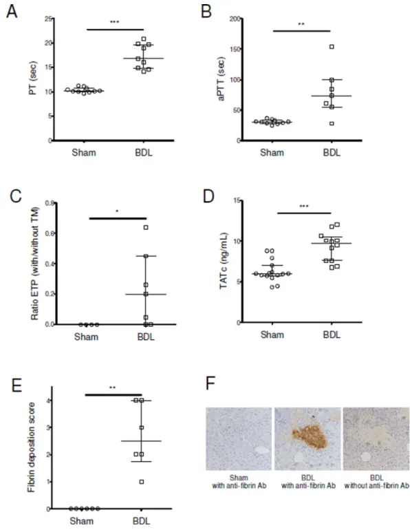
Next, we analyzed coagulation changes in these mice. BDL mice had prolonged PT, aPTT (Fig. 1A–B). In the absence of thrombomodulin, endogenous thrombin potential was not different between BDL and sham mice [728 (663–958) vs. 944 (453–1294) nM*min; p=0.93]. However, a resistance to thrombomodulin was observed in BDL mice (Fig. 1C). Liver injury was associated with an activation of coagulation demonstrated by a significant increase in TATc levels (Fig. 1D). There was also an increased fibrin deposition in the liver in BDL mice, mainly in areas of hepatic necrosis (Fig. 1E–F).
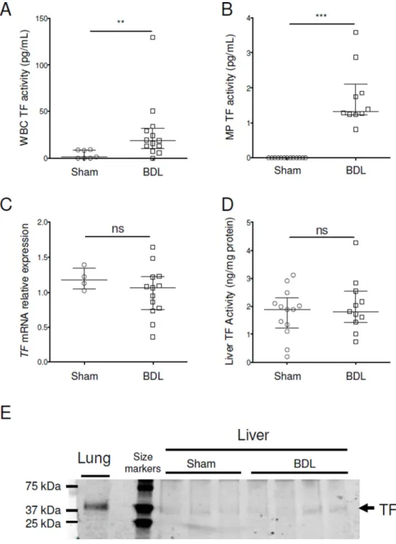
We also measured levels of WBC TF as well as MP TF activity in the plasma. We found that WBC and MP TF activities were both increased in mice subjected to liver injury (Fig.
Author Manuscript
Author Manuscript
Author Manuscript
2A and B). We also determine the TF activity per WBC because the WBC count increases after BDL. The increase in WBC TF activity was also significant when expressed as TF activity per WBC (data not shown). Finally, we measured TF mRNA, activity and protein in the liver. BDL did not increase TF mRNA expression, TF activity or protein (Fig. 2C–E). However, it should be noted that the activity assay cannot distinguish between encrypted and de-encrypted TF in vivo since it uses detergent to de-encrypt TF in the liver.
TF Contributes to the Activation of Coagulation in BDL-injured Mice
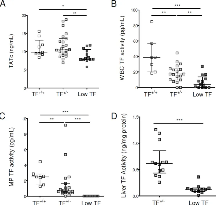
We next investigated the role of TF in the activation of coagulation after BDL using low TF mice. Importantly, activation of coagulation was significantly attenuated in BDL-injured low TF mice compared with TF+/− and TF+/+ mice (Fig. 3A). In TF+/− and low TF mice, the
median WBC TF activity was 45% and 10% of TF+/+ mice, respectively (Fig. 3B). MP TF
activity was undetectable in low TF mice and was 26% in TF+/− mice compared with the
level observed in TF+/+ mice (Fig. 3C). The median liver TF activity in low TF mice was
18% of TF+/− mice (Fig. 3D). We did not observe any difference in the serum levels of AST
or ALT, or in expression of profibrogenic genes or in hepatic collagen deposition or in serum levels of IL-6 (Supplementary Fig. 2A–G). These data indicate that TF plays a major role in the activation of coagulation in BDL-injured mice. However, a deficiency of TF does not affect inflammation, liver injury or fibrosis in this model.
Myeloid Cell TF does not Contribute to the Activation of Coagulation in BDL-injured Mice
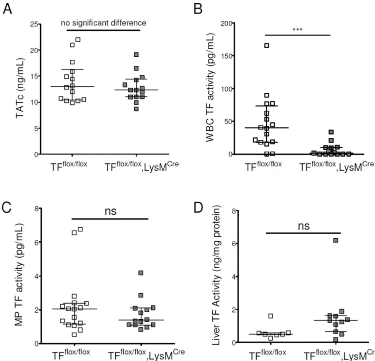
Monocytes from patients with cirrhosis express TF [17–19] and WBC TF activity is increased in BDL-injured mice (Fig. 2A). Therefore, we determined the role of myeloid cell TF in the activation of coagulation in the BDL model. We performed BDL on TFflox/flox,
LysMCre and on TFflox/flox (control) mice. We did not observe any difference in coagulation
activation (Fig. 4A) but, as anticipated, TFflox/flox, LysMCre mice had reduced WBC TF
activity (Fig. 4B). However, MP TF activity was not decreased in TFflox/flox, LysMCre mice
(Fig. 4C). There was no change in liver TF activity in TFflox/flox, LysMCre mice (Fig. 4D).
Serum aminotransferase activity was slightly higher in TFflox/flox, LysMCre than in
TFflox/flox mice, but there was no difference in liver fibrosis (Supplementary Fig. 3A–C). In
addition, we did not observe any difference in the serum levels of IL-6 (Supplementary Fig. 3D). These results indicate that myeloid cell TF does not play a major role in the activation of coagulation in the BDL model.
Hepatocyte TF Contributes to the Activation of Coagulation in BDL-injured Mice
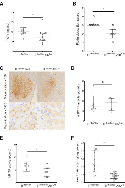
We next investigated the role of hepatocyte TF in the activation of coagulation after BDL using TFflox/flox, AlbCre and TFflox/flox (control) mice. Importantly, activation of coagulation
was significantly attenuated in BDL-injured TFflox/flox, AlbCre mice compared with
TFflox/flox mice (Fig. 5A). In addition, TFflox/flox, AlbCre mice had reduced liver fibrin
deposition, MP TF activity and liver TF activity (Fig. 5B, C, E and F). As anticipated, we did not find any difference in WBC TF activity in TFflox/flox, AlbCre mice (Fig 5D). We did
not observe any difference in the serum levels of AST, ALT, or in expression of fibrogenic-related genes or in the liver fibrosis or in serum levels of IL-6 (Supplementary Fig. 4A–G).
Author Manuscript
Author Manuscript
Author Manuscript
These data indicate that hepatocyte TF plays a major role in the activation of coagulation in BDL-injured mice, without affecting inflammation or liver injury in this model.
Discussion
A major finding of this study was that TF contributes to the activation of coagulation in a model of chronic liver disease. Although TF is the main initiator of the coagulation cascade, its role in the hypercoagulable state associated with chronic liver disease had not been studied in detail. Indeed, the concept of hypercoagulable state in chronic liver disease is based on thrombin-generation tests on plasma samples performed in the presence of
thrombomodulin or snake-venom extract (Protac, Pentapharm) [1, 3–6]. This assay triggered a rethinking of the alternations in the coagulation system associated with chronic liver disease. However, there are several limitations with this approach. This assay uses platelet-free plasma. Therefore, it does not measure TF in blood cells or TF expressed by cells in organs, such as hepatocytes [17]. Moreover, exogenous TF is added to the plasma to trigger coagulation and, although at low concentration, this likely overwhelms any endogenous MP TF activity in the plasma [33]. The novelty of our approach is the use of genetically modified mice to examine the role of TF in vivo in a mouse model that reproduces
coagulation changes associated with chronic liver disease and cirrhosis, including prolonged PT and aPTT, resistance to thrombomodulin, activation of coagulation and increases in WBC and MP TF activities [1, 17–19, 34]. A practical consequence of these findings could be that strategies aimed at reducing TF deencryption may decrease thrombosis risk in patients with advanced cirrhosis [35].
A second major finding of this study was that hepatocyte TF is the major source for activation of coagulation in chronic liver disease (Fig. 6). In wild-type mice, liver TF activity was not different between BDL-injured and sham mice. This result was anticipated because the assay uses a cell lysis step that activates all the liver TF [23]. Therefore, the assay does not distinguish between encrypted (non-active) and deencrypted (active) TF. We found here that genetically reducing TF expression in all cells, and selectively in
hepatocytes, reduced liver TF activity and activation of coagulation in BDL-injured mice. Although the liver expresses relatively low levels of TF compared to other organs/tissues, [36] it is a prominent source of procoagulant TF in chronic liver injury, as observed in acute liver diseases [23, 37]. We have previously demonstrated that hepatocytes express TF that lacks detectable procoagulant activity in the healthy liver [23]. However, hepatocyte injury triggers hepatocyte TF-dependent activation of coagulation in mice. Of importance, activation of coagulation by hepatocyte TF would not require increased TF expression. Rather, this could occur via molecular activation of the existing TF:FVIIa complex. This is consistent with our finding that total liver TF activity did not increase after BDL.
BDL-injured mice displayed elevated WBC TF activity, recapitulating the increase in monocyte TF activity reported in patients with cirrhosis [17–19]. However, we found that a deficiency of TF in myeloid cells did not reduce activation of coagulation after BDL. This lack of effect of myeloid TF on activation of coagulation is in agreement with a previous study showing that myeloid cell TF deficiency does not affect coagulation activation after
Author Manuscript
Author Manuscript
Author Manuscript
acute cholestatic liver injury [38]. These results suggest that WBC TF activity is not a prominent contributor to the activation of coagulation in chronic liver disease.
The third major finding of this study was that hepatocytes are the source of the increase in MP TF activity observed in BDL-injured mice. This observation indicates that injured hepatocytes could be a prominent source of procoagulant MPs in patients with chronic liver disease. We previously found that MP TF activity is elevated in patients with cirrhosis and increases with cirrhosis severity [34]. In patients, we could not determine the cellular origin of TF+ MP because the low levels of TF present on the MPs could not be detected by flow cytometry. Nevertheless, we examined the correlations between MP TF activity and plasma levels of the specific MP subtypes in patients with cirrhosis. MP TF activity correlated with circulating levels of hepatocyte MPs (Cytokeratine-18+ MPs) (Spearman r = 0.377; p = 0.031; n=33), but not with leukocyte MPs levels (unpublished data). These unpublished data obtained in patients with cirrhosis support our findings obtained in mice. Hepatocytederived circulating MPs are also observed in acute liver injury [37]. We can speculate that an increase in hepatocyte-derived, TF-positive MPs may predict venous thrombosis events, including portal vein thrombosis in patients with chronic liver disease and cirrhosis.
We did not observe any effect of TF deficiency on expression of profibrogenic genes or on hepatic collagen deposition in the BDL model. This allowed us to investigate the mechanism of activation of coagulation associated with chronic liver disease without being biased by an effect on fibrosis. We were initially surprised that there was not a reduction in fibrosis in low TF mice since a previous study found that heterozygous TF deficiency exhibited reduced hepatic fibrosis in experimental biliary hyperplasia induced by α
-naphthylisothiocyanate [39]. However, despite the observation that protease activated receptor 1 inhibition reduces liver injury after BDL [40], other thrombin-driven processes, including platelet activation and fibrin deposition, have been shown to exert
hepatoprotective effects in the context of cholestatic liver damage [29, 41]. These results suggest that the association of TF-driven coagulation with liver pathology is dependent on the experimental model.
In conclusion, our results demonstrate that hepatocyte TF contributes to the activation of coagulation in a mouse model of chronic liver disease. These data suggest that TF may contribute to the hypercoagulable state and thrombotic events in patients with chronic liver disease.
Supplementary Material
Refer to Web version on PubMed Central for supplementary material.
Acknowledgments
We thank Martin Baunacke, Rebecca Lee, Jian-Guo Wang, and Ying Zhang for their skilled technical assistance. We would like to thank Daniel Kirchhofer for providing the 1H1 rat anti–mouse TF antibody (Genentech, Inc.). We thank Yacine Boulaftali and Dominique Valla for helpful discussions.
Financial Support
Author Manuscript
Author Manuscript
Author Manuscript
P.-E.R. was supported by the Philippe Foundation and by an American Heart Association Mid-Atlantic postdoctoral fellowship (12POST11970008). K.T was supported by the Uehara Memorial Foundation, Japan. A.P.O.III was supported by an NIH F32 NRSA postdoctoral fellowship (1F32-HL099175-03). This work was supported by the Société Nationale Française de Gastroentérologie (bourse Robert Tournut), the Association Française pour l’Étude du Foie, the National Institutes of Health (grant T32-HL007149-37, E.S.; grant ES017537, J.L.; grant HL095096, N.M.; grant P50HL120100, ASW), the FDA Center for Tobacco Products (CTP) (ASW).
List of Abbreviations
ALT alanine aminotransferase
aPTT Activated Partial Thromboplastin Time
AST aspartate aminotransferase
BDL bile duct ligation
F factor
IL-6 interleukin 6
LysM Lysozyme
MP microparticle
PT Prothrombin Time
TAT thrombin-antithrombin complexes
TF tissue factor
WBC white blood cell
References
1. Tripodi A, Mannucci PM. The coagulopathy of chronic liver disease. N Engl J Med. 2011; 365:147– 156. [PubMed: 21751907]
2. Lisman T, Caldwell SH, Burroughs AK, Northup PG, Senzolo M, Stravitz RT, et al. Hemostasis and thrombosis in patients with liver disease: the ups and downs. J Hepatol. 2010; 53:362–371.
[PubMed: 20546962]
3. Tripodi A, Primignani M, Chantarangkul V, Dell'Era A, Clerici M, de Franchis R, et al. An imbalance of pro- vs anti-coagulation factors in plasma from patients with cirrhosis. Gastroenterology. 2009; 137:2105–2111. [PubMed: 19706293]
4. Tripodi A, Salerno F, Chantarangkul V, Clerici M, Cazzaniga M, Primignani M, et al. Evidence of normal thrombin generation in cirrhosis despite abnormal conventional coagulation tests. Hepatology. 2005; 41:553–558. [PubMed: 15726661]
5. Tripodi A, Primignani M, Lemma L, Chantarangkul V, Dell'Era A, Iannuzzi F, et al. Detection of the imbalance of procoagulant versus anticoagulant factors in cirrhosis by a simple laboratory method. Hepatology. 2010; 52:249–255. [PubMed: 20578143]
6. Gatt A, Riddell A, Calvaruso V, Tuddenham EG, Makris M, Burroughs AK. Enhanced thrombin generation in patients with cirrhosis-induced coagulopathy. J Thromb Haemost. 2010; 8:1994–2000. [PubMed: 20546119]
7. Northup PG, McMahon MM, Ruhl AP, Altschuler SE, Volk-Bednarz A, Caldwell SH, et al. Coagulopathy does not fully protect hospitalized cirrhosis patients from peripheral venous thromboembolism. Am J Gastroenterol. 2006; 101:1524–1528. quiz 1680. [PubMed: 16863556] 8. Sogaard KK, Horvath-Puho E, Gronbaek H, Jepsen P, Vilstrup H, Sorensen HT. Risk of venous
thromboembolism in patients with liver disease: a nationwide population-based case-control study. Am J Gastroenterol. 2009; 104:96–101. [PubMed: 19098856]
Author Manuscript
Author Manuscript
Author Manuscript
9. Gulley D, Teal E, Suvannasankha A, Chalasani N, Liangpunsakul S. Deep vein thrombosis and pulmonary embolism in cirrhosis patients. Dig Dis Sci. 2008; 53:3012–3017. [PubMed: 18443906] 10. Tripodi A, Anstee QM, Sogaard KK, Primignani M, Valla DC. Hypercoagulability in cirrhosis:
causes and consequences. J Thromb Haemost. 2011; 9:1713–1723. [PubMed: 21729237]
11. Tripodi A, Primignani M, Lemma L, Chantarangkul V, Mannucci PM. Evidence that low protein C contributes to the procoagulant imbalance in cirrhosis. J Hepatol. 2013
12. Mackman N. The role of tissue factor and factor VIIa in hemostasis. Anesth Analg. 2009; 108:1447–1452. [PubMed: 19372318]
13. Bach RR. Tissue factor encryption. Arterioscler Thromb Vasc Biol. 2006; 26:456–461. [PubMed: 16397140]
14. Owens AP 3rd, Passam FH, Antoniak S, Marshall SM, McDaniel AL, Rudel L, et al. Monocyte tissue factor-dependent activation of coagulation in hypercholesterolemic mice and monkeys is inhibited by simvastatin. J Clin Invest. 2012; 122:558–568. [PubMed: 22214850]
15. Pawlinski R, Tencati M, Holscher T, Pedersen B, Voet T, Tilley RE, et al. Role of cardiac myocyte tissue factor in heart hemostasis. J Thromb Haemost. 2007; 5:1693–1700. [PubMed: 17663739] 16. Pawlinski R, Wang JG, Owens AP 3rd, Williams J, Antoniak S, Tencati M, et al. Hematopoietic and nonhematopoietic cell tissue factor activates the coagulation cascade in endotoxemic mice. Blood. 2010; 116:806–814. [PubMed: 20410508]
17. Saliola M, Lorenzet R, Ferro D, Basili S, Caroselli C, Santo AD, et al. Enhanced expression of monocyte tissue factor in patients with liver cirrhosis. Gut. 1998; 43:428–432. [PubMed: 9863491] 18. Ferro D, Basili S, Pratico D, Iuliano L, FitzGerald GA, Violi F. Vitamin E reduces monocyte tissue
factor expression in cirrhotic patients. Blood. 1999; 93:2945–2950. [PubMed: 10216089] 19. Panasiuk A, Zak J, Panasiuk B, Prokopowicz D. Increase in expression of monocytic tissue factor
(CD142) with monocytes and blood platelet activation in liver cirrhosis. Blood Coagul Fibrinolysis. 2007; 18:739–744. [PubMed: 17982314]
20. Rautou PE, Vion AC, Luyendyk JP, Mackman N. Circulating microparticle tissue factor activity is increased in patients with cirrhosis. Hepatology. 2014; 60:1793–1795. [PubMed: 24470301] 21. Owens AP 3rd, Mackman N. Microparticles in hemostasis and thrombosis. Circ Res. 2011;
108:1284–1297. [PubMed: 21566224]
22. Rautou PE, Mackman N. Microvesicles as risk markers for venous thrombosis. Expert Rev Hematol. 2013; 6:91–101. [PubMed: 23373784]
23. Sullivan BP, Kopec AK, Joshi N, Cline H, Brown JA, Bishop SC, et al. Hepatocyte tissue factor activates the coagulation cascade in mice. Blood. 2013; 121:1868–1874. [PubMed: 23305736] 24. Parry GC, Erlich JH, Carmeliet P, Luther T, Mackman N. Low levels of tissue factor are
compatible with development and hemostasis in mice. J Clin Invest. 1998; 101:560–569. [PubMed: 9449688]
25. Dargaud Y, Spronk HM, Leenders P, Hemker HC, Ten Cate H. Monitoring platelet dependent thrombin generation in mice. Thromb Res. 2010; 126:436–441. [PubMed: 20843543] 26. Lisman T, Bakhtiari K, Pereboom IT, Hendriks HG, Meijers JC, Porte RJ. Normal to increased
thrombin generation in patients undergoing liver transplantation despite prolonged conventional coagulation tests. J Hepatol. 2010; 52:355–361. [PubMed: 20132999]
27. Wang JG, Manly D, Kirchhofer D, Pawlinski R, Mackman N. Levels of microparticle tissue factor activity correlate with coagulation activation in endotoxemic mice. J Thromb Haemost. 2009; 7:1092–1098. [PubMed: 19422446]
28. Luyendyk JP, Cantor GH, Kirchhofer D, Mackman N, Copple BL, Wang R. Tissue factor-dependent coagulation contributes to alphanaphthylisothiocyanate- induced cholestatic liver injury in mice. Am J Physiol Gastrointest Liver Physiol. 2009; 296:G840–G849. [PubMed: 19179621] 29. Joshi N, Kopec AK, O'Brien KM, Towery KL, Cline-Fedewa H, Williams KJ, et al.
Coagulation-driven platelet activation reduces cholestatic liver injury and fibrosis in mice. J Thromb Haemost. 2015; 13:57–71. [PubMed: 25353084]
30. Hui KY, Haber E, Matsueda GR. Monoclonal antibodies to a synthetic fibrinlike peptide bind to human fibrin but not fibrinogen. Science. 1983; 222:1129–1132. [PubMed: 6648524]
Author Manuscript
Author Manuscript
Author Manuscript
31. Yang YY, Liu H, Nam SW, Kunos G, Lee SS. Mechanisms of TNFalpha-induced cardiac dysfunction in cholestatic bile duct-ligated mice: interaction between TNFalpha and endocannabinoids. J Hepatol. 2010; 53:298–306. [PubMed: 20626112]
32. Georgiev P, Jochum W, Heinrich S, Jang JH, Nocito A, Dahm F, et al. Characterization of time-related changes after experimental bile duct ligation. Br J Surg. 2008; 95:646–656. [PubMed: 18196571]
33. Ollivier V, Wang J, Manly D, Machlus KR, Wolberg AS, Jandrot-Perrus M, et al. Detection of endogenous tissue factor levels in plasma using the calibrated automated thrombogram assay. Thromb Res. 2010; 125:90–96. [PubMed: 19345399]
34. Rautou PE, Mackman N. A key role for tissue factor in activation of coagulation in cirrhosis. J Hepatol. 2013; 58:S245–S245.
35. Langer F, Ruf W. Synergies of phosphatidylserine and protein disulfide isomerase in tissue factor activation. Thromb Haemost. 2014; 111:590–597. [PubMed: 24452853]
36. Mackman N, Sawdey MS, Keeton MR, Loskutoff DJ. Murine tissue factor gene expression in vivo. Tissue and cell specificity and regulation by lipopolysaccharide. Am J Pathol. 1993; 143:76– 84. [PubMed: 8317556]
37. Stravitz RT, Bowling R, Bradford RL, Key NS, Glover S, Thacker LR, et al. Role of procoagulant microparticles in mediating complications and outcome of acute liver injury/acute liver failure. Hepatology. 2013; 58:304–313. [PubMed: 23389887]
38. Luyendyk JP, Flanagan KC, Williams CD, Jaeschke H, Slusser JG, Mackman N, et al. Tissue factor contributes to neutrophil CD11b expression in alpha-naphthylisothiocyanate-treated mice. Toxicol Appl Pharmacol. 2011; 250:256–262. [PubMed: 21070799]
39. Sullivan BP, Weinreb PH, Violette SM, Luyendyk JP. The coagulation system contributes to alphaVbeta6 integrin expression and liver fibrosis induced by cholestasis. Am J Pathol. 2010; 177:2837–2849. [PubMed: 21037076]
40. Fiorucci S, Antonelli E, Distrutti E, Severino B, Fiorentina R, Baldoni M, et al. PAR1 antagonism protects against experimental liver fibrosis. Role of proteinase receptors in stellate cell activation. Hepatology. 2004; 39:365–375. [PubMed: 14767989]
41. Kodama T, Takehara T, Hikita H, Shimizu S, Li W, Miyagi T, et al. Thrombocytopenia exacerbates cholestasis-induced liver fibrosis in mice. Gastroenterology. 2010; 138:2487–2498. 2498 e2481–2498 e2487. [PubMed: 20206174]
Author Manuscript
Author Manuscript
Author Manuscript
Figure 1. Activation of coagulation in mice with chronic liver injury
Bile duct ligation (BDL) or sham surgery was performed in C57BL/6 mice. (A–C) Twelve days after surgery we measured prothrombin time (PT) and activated partial thromboplastin time (aPTT), as well as the ratio of the values of endogenous thrombin potential (ETP) obtained without and with thrombomodulin (TM) We also measured plasma levels of thrombin-antithrombin complexes (TATc) (D) and liver fibrin deposition (E, quantification; F, representative images).
Data are given as median (horizontal bar), 25th and 75th percentile (error bar).
Author Manuscript
Author Manuscript
Author Manuscript
*, p<0.05; **, p<0.01; ***, p<0.001.
Author Manuscript
Author Manuscript
Author Manuscript
Figure 2. Tissue factor expression in mice with chronic liver injury
White blood cell (A) and MP (B) TF activities were determined in C57BL/6 mice after BDL or sham surgery. We also measured TF mRNA, activity and protein in the liver of these mice (C–E). For western blot (E), we used lung as a positive control.
Data are given as median (horizontal bar), 25th and 75th percentile (error bar).
Abbreviations: BDL, Bile Duct Ligation; MP, microparticle; ns, not significant; TF, tissue factor; WBC, white blood cell.
**, p<0.01; ***, p<0.001.
Author Manuscript
Author Manuscript
Author Manuscript
Figure 3. Tissue factor contributes to activation of coagulation in mice with chronic liver injury BDL was performed in mice expressing 1% (low TF mice), 50% (TF+/− mice) and 100% (TF+/+) of wild-type TF levels. Plasma levels of TATc were measured (A). White blood cell (B), MP (C) and liver (D) TF activities were determined.
Data are given as median (horizontal bar), 25th and 75th percentile (error bar).
Abbreviations: BDL, Bile Duct Ligation; MP, microparticle; TATc, thrombinantithrombin complexes; TF, tissue factor; WBC, white blood cell.
*, p<0.05; **, p<0.01; ***, p<0.001. Only significant differences are shown.
Author Manuscript
Author Manuscript
Author Manuscript
Figure 4. Myeloid tissue factor does not contribute to activation of coagulation in mice with chronic liver injury
BDL was performed in TFflox/flox, LysMCre and in TFflox/flox mice. Plasma levels of TATc were measured (A). White blood cell (B), MP (C) and liver (D) TF activities were
determined.
Data are given as median (horizontal bar), 25th and 75th percentile (error bar).
Abbreviations: BDL, Bile Duct Ligation; MP, microparticle; ns, not significant; TATc, thrombin-antithrombin complexes; TF, tissue factor; WBC, white blood cell. ***, p<0.001.
Author Manuscript
Author Manuscript
Author Manuscript
Figure 5. Hepatocyte tissue factor contributes to activation of coagulation in mice with chronic liver injury
BDL was performed in TFflox/flox, AlbCre and in TFflox/flox mice. Plasma levels of TATc were measured (A). Liver fibrin deposition was assessed (B, quantification; C,
representative images). White blood cell (D), MP (E) and liver (F) TF activities were determined.
Data are given as median (horizontal bar), 25th and 75th percentile (error bar).
Abbreviations: BDL, Bile Duct Ligation; MP, microparticle; ; ns, not significant; TATc, thrombin-antithrombin complexes; TF, tissue factor; WBC, white blood cell.
Author Manuscript
Author Manuscript
Author Manuscript
*, p<0.05; **, p<0.01; ***, p<0.001.
Author Manuscript
Author Manuscript
Author Manuscript
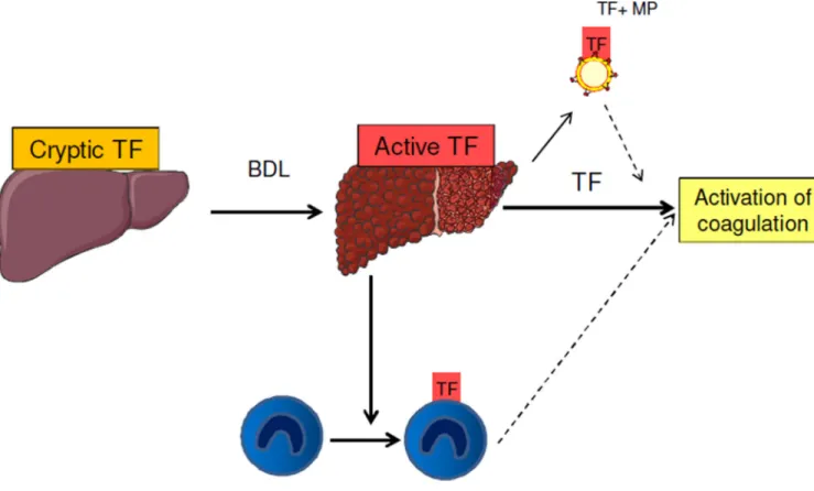
Figure 6. Potentital sources of tissue factor (TF) that may activate coagulation in chronic liver disease
We propose that under normal condition, hepatocyte TF is cryptic. In chronic liver diseases, hepatocyte TF is actived and contributes to the activation of coagulation and liver fibrin deposition. Myeloid cell TF activity is increased in liver injury, but is not a prominent contributor to the systemic activation of coagulation. Microparticle (MP) TF activity is increased in cirrhosis. However, we cannot determine the relative contribution of hepatocyte TF versus MP TF to the activation of coagulation.
Full and dashed lines indicate likely and potential links, respectively.