A HEAVY-CHAIN-ONLY CAR-T CELL THERAPY ELICITS COMPARABLE ANTITUMOR RESPONSE COMPARED WITH CANONICAL CAR-T CELLS
Guanmeng Wang
A thesis submitted to the faculty at the University of North Carolina at Chapel Hill in partial fulfillment of the requirements for the degree of Master of Science in the Joint Department of Biomedical
Engineering in the School of Medicine
Chapel Hill 2019
Approved by: Gianpietro Dotti Barbara Savoldo David Zaharoff
iii ABSTRACT
Guanmeng Wang: a heavy-chain-only CAR-T cell therapy elicits comparable antitumor response compared with canonical CAR-T cell
(Under the direction of Gianpietro Dotti)
iv
v
ACKNOWLEDGMENTS
I would like to thank Dr. Gianpietro Dotti for providing me with the opportunity to work in one of the leading labs of CAR-T therapy in the world. It is my great honor and privilege to work with Dr. Dotti and the lab members. I am thankful for all the help they gave me in the past year. I am also thankful for Dr. Barbara Savoldo and Dr. David Zaharoff for joining my thesis committee and the help they offered.
I would also like to appreciate all the assistance during my study provided by the biomedical engineering department. I especially appreciate Vilma Berg, graduate program coordinator of biomedical engineering department, for her support.
vi
TABLE OF CONTENTS
LIST OF TABLES ... vii
LIST OF FIGURES ... viii
LIST OF ABBREVIATIONS ... ix
CHAPTER 1: INTRODUCTION ... 1
1.1 Cancer Immunotherapy ... 1
1.2 Chimeric Antigen Receptor (CAR)-T cell Immunotherapy ... 1
1.3 Limitation of CAR-T cells therapy for solid tumors ... 2
1.4 Safety issues of CAR-T cell therapy ... 3
1.5 Objective... 4
CHAPTER 2: HEAVY-CHAIN-ONLY CAR-T CELL ... 5
2.1 Materials and Methods ... 5
2.2 Result ... 8
2.3 Discussion... 15
vii
LIST OF TABLES
viii
LIST OF FIGURES
Figure 1 -Conventional PSMA.CAR (J591) and heavy chain only PSMA.CAR (HuVh) are equally
expressed by T cells ... 9
Figure 2 -T cells expressing J591 or HuVh show comparable activities in vitro ... 10
Figure 3 - T cells expressing J591 or HuVh show similar antitumor activities in vitro ... 12
Figure 4 -T cells expressing J591 or HuVh show comparable antitumor activities in vivo ... 13
ix
LIST OF ABBREVIATIONS
ACT Adoptive cell transfer ANOVA Analysis of variance
BLI Bioluminescence
CAR Chimeric antigen receptor CAR-Ts Chimeric antigen receptor T cells CRS Cytokine release syndrome FDA Food and Drug Administration
Fv Variable fragment
HLA Human leukocyte antigen HuVh Human variant heavy chain
IACUC Institutional Animal Care Use Committee IFNγ Interferon gamma
IL-2/6/15 Interleukin-2/6/15
i.v. Intravenous
mAb Monoclonal antibody
MHC Major histocompatibility complex MDSCs Myeloid derived suppressor cells
NSG NOD/SCID/γcnull
PD1 Programmed cell death 1 PGE2 prostaglandin E2
1
CHAPTER 1: INTRODUCTION
1.1 Cancer Immunotherapy
Immunotherapy utilizes human’s own immune system to cure diseases. Cancer immunotherapy, in particular, has gained much attention in recent years as it demonstrated promising tumor regressions in many forms of late stage cancers that cannot be cured by conventional chemotherapy and radiotherapy (Borghaei et al., 2009; Hodi et al., 2010; Kantoff et al., 2010). Cancer immunotherapies can be classified as active and passive immunotherapies: active immunotherapies activate endogenous immune system, while passive immunotherapies relay on transferring components of the immune systems to patients (Ito and E.Chang, 2013). Regardless of the type of immunotherapies, cancer cells can be recognized and killed by the patient’s own immune system which generally does not identify cancer cells. Monoclonal antibody (mAb) and adoptive cell transfer (ACT) therapy are the two major immunotherapies tested in clinical trials and some are approved by the Food and Drug Administration (FDA).
1.2 Chimeric Antigen Receptor (CAR)-T cell Immunotherapy
2
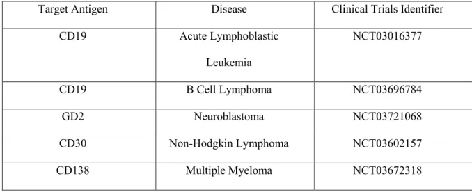
in which two costimulatory domains have been included and are in clinical studies (June et al., 2018). Most of the clinical success comes from the treatment of hematological malignancies. CD19 is a very effective target for B cell malignancies due its consistent and exclusive expression in B cell lineage (van Zelm et al., 2006). In clinical trials in acute lymphoblastic leukemia and lymphoma up to 90% of the patients achieved complete remission (Davila et al., 2014; Kochenderfer et al., 2015; Porter et al., 2015; Turtle et al., 2016). FDA approved two CD19.CAR-T cell therapies, Kymriah and Yescarta, in 2017 and 2018 to treat B-cell acute lymphoblastic leukemia (ALL) and B-cell non-Hodgkin lymphoma (NHL). Based on these promising clinical results, multiple clinical studies are currently ongoing in several institutions. The following table summarizes active clinical trials of CAR-T cells at UNC.
Target Antigen Disease Clinical Trials Identifier
CD19 Acute Lymphoblastic
Leukemia
NCT03016377
CD19 B Cell Lymphoma NCT03696784
GD2 Neuroblastoma NCT03721068
CD30 Non-Hodgkin Lymphoma NCT03602157
CD138 Multiple Myeloma NCT03672318
Table 1. Ongoing clinical trials of CAR-T immunotherapy at UNC Lineberger Comprehensive Center
1.3 Limitation of CAR-T cells therapy for solid tumors
3
escape due to lack or loss of antigen expression (Newick et al., 2017). Trafficking of the CAR-T cells into the solid tumors is also impaired due to the lack of chemokine and receptor mismatch between tumor cells and T cells, especially CXCR3hi CD8+ T cells (Harlin et al., 2009). The immunosuppressive tumor
microenvironment (TME) of solid tumors creates both a physical and metabolic obstacle for CAR-T cells. The sinuous structure of tumor stroma, high pressure inhibits extravasation, low concentration of oxygen or hypoxia, and lack of nutrient like glucose all jeopardize T cell effector functions (Fischer et al., 2007; Hatfield et al., 2015). The immunosuppressive TMEs also secrete cytokines such as TGFβ, IL-4 and
soluble factors like prostaglandin E2(PGE2) which inhibit T cell proliferation and antitumor responses (Goodwin et al., 1977; Massagué, 2008). Lastly, the immunosuppressive immune cells residing in the TME such as regulatory T cells (Tregs), myeloid derived suppressor cells (MDSCs) and tumor-associated macrophages (TAMs) produce immunosuppressive cytokines, express immune checkpoints, and finally down-regulate T cell functions (Gabrilovich and Nagaraj, 2009; Sakaguchi et al., 2010).
1.4 Safety issues of CAR-T cell therapy
4
observed following CAR-T cell treatment with unknown mechanism. Known symptoms include headaches, confusion, hallucination, seizures, and dysmetria (Grupp et al., 2013; Kochenderfer et al., 2012; Kochenderfer et al., 2010; Lee et al., 2014). It is speculated that elevated levels of cytokines are responsible for the toxicities, leading to increased permeability of central nervous system parenchyma (June et al., 2018).
1.5 Objective
Compared with traditional scFv CAR, a heavy-chain-only CAR offers a small size advantage, which allows to incorporate other transgenes into the construct, has limited immunogenicity and avoids the aggregation issue in scFv(Vu et al., 1997). In this work, a heavy-chain-only CAR construct was developed. The heavy-chain-only CAR-T cells achieve comparable antitumor response compared with traditional scFv CAR-T cells both in vitro and in vivo. This proof-of-concept experiment lays the
5
CHAPTER 2: HEAVY-CHAIN-ONLY CAR-T CELL
2.1 Materials and Methods
CAR construction. Conventional PSMA.CAR (J591), heavy chain only PSMA.CAR (HuVh), conventional MSLN.CAR (scFv), heavy chain only MSLN.CAR(HuVh) were assembled with the CD8α hinge (H), transmembrane domain (TM), the CD28 costimulatory domain (CD28) and CD3ζ intercellular signaling domain and cloned into the SFG retroviral vector(Diaconu et al., 2017). A FLAG-tag was incorporated after both heavy chains to detect the expression of CARs by anti-FLAG Ab through flow cytometry.
Cell lines. Tumor cell lines PC-3, C4-2(prostate cancer) and Aspc-1(pancreatic cancer) were
purchased from ATCC. All tumor cell lines were cultured with RPMI-1640(Gibco) supplemented with 10% FBS (Sigma), 2 mM GlutaMax (Gibco) and penicillin (100 units/mL) and streptomycin (100 µg/mL; Gibco). All cells were cultured at 37°C with 5% CO2. PC-3 cell line was transduced with retroviral vector encoding PSMA to make PC-3-PSMA and both PC-3-PSMA and Aspc-1 were transduced with retroviral vectors encoding Firefly- Luciferase-eGFP (FFluc-eGFP) gene(Diaconu et al., 2017).
6
CAR-T cell generation. Buffy coats from healthy donors (Gulf Coast Regional Blood Center) were processed with Lymphoprep density separation (Fresenius Kabi Norge) to isolate peripheral blood mononuclear cells, which were then activated on plates coated with 1 µg/mL CD3 (Miltenyi Biotec) and 1 µg/mL CD28 (BD Biosciences) mAbs. Two days later, activated T cells were transduced with retroviral supernatants on 24-well plates coated with retronectin with7 µg/mL (Takara Bio Inc.) , T cells were collected three days after transduction and expanded in 40% RPMI-1640(Gibco) and 40% Click’s medium (Irvine Scientific), 10% HyClone FBS (GE healthcare), 2 mM GlutaMAX(Gibco), 100 unit/mL of Penicillin and 100 mg/mL of streptomycin(Gibco) with 10 ng/mL IL-7(PeproTech) and 5 ng/mL IL-15 (PeproTech). T cells were collected for functional assays 12-14 days after activation.
Flow cytometry. mAbs for human CD3(APC-H7; SK7; 560176), CD4(BV711; SK3; 563028), CD8(APC; SK1; 340584), CD45RA (PE; HI100; 555489), CD45RO (BV786; UCHL1; 564290), CD69 (FITC; L78; 347823), CCR7 (FITC; 150503; FAB197F-100), PD-1(PE-Cy7; EH12.1;561272),
Lag3(PE;T47-530;565616), FLAG (APC; L5; 637308), Granzyme-B(PE;GB11;561142) from BD biosciences and BioLegend were used. Samples were acquired with BD FACSCanto II or BD LSRFortessa. A minimum of 10,000 events were acquired for each sample and were analyzed using FlowJo 10(FlowJo LLC).
Western blot analysis. CAR-T cells were incubated with 2 µg anti-FLAG Ab in 100 µL PBS for 20 mins on the ice and then with 2 µg goat anti-mouse secondary Ab for another 20 mins on the ice. Cells were then incubated in the 37°C water bath for the selected time points and then lysed with 2x Laemelli buffer for 10 mins. Cell lysates were then separated in 4% to 15% 10 well SDS-PAGE gels and
transferred to polyvinylidene difluoride (PVDF) membranes at 75V for 120 mins (Bio-Rad). Blots were examined for human CD3ζ (Santa Cruz Biotechnology), p-Y142 CD3ζ (Abcam), pan-ERK (BD
7 instructions.
Proliferation assay. T cells were labeled with 1.5 mM carboxyfluorescein diacetate succinimidyl ester (CFSE; Invitrogen) and plated with tumor cells at an E:T ratio of 1:1. CFSE signal dilution from gated T cells on day 5 was measured using flow cytometry(Diaconu et al., 2017).
In vitro cytotoxicity assay. Tumor cells were seeded in 24-well plates at a concentration of
0.5x10⁵ cells/well overnight. CAR-T cells were added to the plate at an E:T ratio of 1:5 without
exogenous cytokines. Cocultures were analyzed 5-7 days following coculture to measure residual tumor cells and T cells by flow cytometry. Dead cells were recognized by Zombie Aqua Dye (Biolegend) staining while CAR-T cells were identified by CD3 staining and tumor cells by GFP.
Activation and exhaustion marker analysis. Tumor cells were seeded in 24-well plates at a concentration of 0.4x10⁵ cells/well overnight. CAR-T cells were added to the plate at an E:T ratio of 1:2 without exogenous cytokines. CD69, PD-1 and Lag3 expression were measured by flow cytometry from day 0 to day 5 each day after coculture. Dead cells were recognized by Zombie Aqua Dye (Biolegend) staining while CAR-T cells were identified by CD3 staining and tumor cells by GFP. For the granzyme-B staining, Golgi protein inhibitor (BD Biosciences) was added on day 1 of coculture for 6 hours.
Cocultures were then first stained with Zombie Aqua Dye (Biolegend) and CD3 Ab followed by fixation/permeabilization solution (BD Biosciences). Intracellular staining of granzyme-B was then conducted.
Cytokine analysis. 1 x 10⁵ CAR-T cells were cocultured with 5x10⁵ tumor cells in 24-well plates without exogenous cytokines. Supernatant after 24 hours was collected and cytokines (IFN-γ and IL-2) were measured with duplicates using ELISA kits (R&D system) following manufacturer’s instructions.
Xenograft murine models. 6-8 weeks old female NSG mice were injected intravenously through tail vein with PC-3-PSMA-FFluc-eGFP of 1x10⁶ per mice. Fourteen days later, CAR-T cells were
8 imaging system (PerkinElmer).
Statistical analysis. All data was calculated and represented as mean with standard deviation. One-way ANOVA with Turkey post hoc analysis was used to compare multiple groups. Two-tailed t test was used to compared two groups. A p value of less than 0.05 was significant. All calculations and figures were achieved by GraphPad Prism7 (La Jolla,CA).
2.2 Result
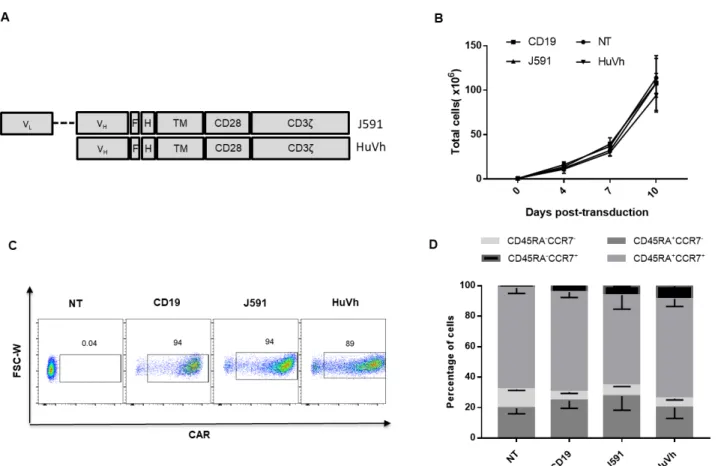
We constructed the J591.CAR and HuVh.CAR using the scFv from the PSMA-specific J591 Ab and the Vh from the PSMA-specific HuVh Ab, respectively joined to the CD8α stalk, CD28
costimulatory domain and CD3ζ intracellular domain. A flag tag was incorporated into the cassettes to
9
Figure 1 Conventional PSMA.CAR (J591) and heavy chain only PSMA.CAR (HuVh) are equally expressed by T cells. Schematic diagram of J591 and HuVh CAR constructs. (B) In vitro expansion of CD19.CAR-T cells, J591.CAR-T cells, HuVh.CAR-T cells and non-transduced (NT) T cells; error bars represent SD, (n = 4). (C) Representative flow cytometry plots illustrating CAR expression of the CD19.CAR-T cells, J591.CAR-T cells and HuVh.CAR-T cells. (D) Immunophenotypic composition of CD19.CAR-T cells, J591.CAR-T cells, HuVh.CAR-T cells and NT cells at day 14 of culture; error bars represent SD (n = 4).
10
(Figure 2B). We also analyzed the kinetics of T cell activation upon CAR crosslinking by measuring CD69 expression. As shown in Figure 2C and 2D, equal upregulation and subsequent down regulation of CD69 was observed in J591.CAR-T cells and HuVh.CAR-T cells. Similarly, upregulation upon antigen stimulation and down regulation after antigen removal were observed for Lag-3 and PD-1 in both J591.CAR-T cells and C5.CAR-T cells (Figure 2D).
Figure 2 T cells expressing J591 or HuVh show comparable activities in vitro. Western blots detecting phosphorylation of CD3ζ, Akt and ERK of CAR-T cells activated via CAR crosslinking with
11
CD69 and Lag-3 expression of T cells expressing either J591 or HuVh. Blue light and red histograms illustrate isotype control and the specific marker, respectively. (D) Summary of CD69, PD-1 and Lag-3 kinetic expression (n = 4).
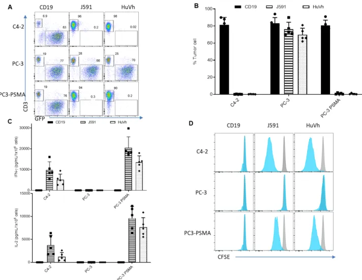
To examine and compare the antitumor effect of HuVh.CAR-T cells with J591.CAR-T cells in vitro, we measured the remaining percentage of tumor cells left after 5 days of coculture with both CAR-T cells. HuVh.CAR-CAR-T cells are able to specifically eliminate PSMA-positive target cells to the same extent as conventional scFv CAR-T cells and do not demonstrate off-target effect while CD19.CAR-T cells, as a control, did not kill the tumor cells (Figure 3A and 3B). We also measured cytokine secretion of IFN-γ and IL-2 after 24 hours of coculture. HuVh.CAR-T cells display a comparable release of IFN-γ and IL-2 compared with J591.CAR-T cells, which also confirms a comparable antitumor effect in vitro (Figure 3C). Further, we investigated proliferation of HuVh.CAR-T cells and J591.CAR-T cells through
12
Figure 3 T cells expressing J591 or HuVh show similar antitumor activities in vitro. Representative flow cytometry plots showing coculture with C4-2-eGFP (positive control), PC-3-eGFP (negative control) and PC-3-PSMA-eGFP (target cell). CD19.CAR-T cells, J591.CAR-T cells and HuVh.CAR-T cells were cocultured with C4-2-eGFP, PC-3-eGFP and PC-3-PSMA-eGFP cell lines at an E:T ratio of 1:5 for 6 days(B) Summary of coculture of CD19.CAR-T cells, J591.CAR-T cells and HuVh.CAR-T cells with C4-2-eGFP, PC-3-eGFP and PC-3-PSMA-eGFP cell lines; error bars represent SD(n=4). (C) IFN-γ and IL-2 release from the coculture supernatant with CD19.CAR-T cells, J591.CAR-T cells and HuVh.CAR- T cells as measured by ELISA (n=4). (D) Representative flow cytometry figures showing CFSE proliferation assay. Blue and gray histograms represent activated and control groups
13
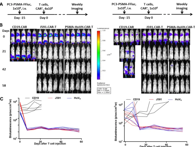
Next, an in vivo model is used to investigate the antitumor effect of PSMA-HuVh.CAR-T cells in vivo. a high dosage group and a low dosage group experiments are independently conducted in NSG mice by inoculation of PSMA-positive tumor model and transfusion of CAR-T cells (Figure 4A). Both high dosage group and low dosage group showed a complete tumor eradication as measured by tumor
bioluminescence (Figure 4B and 4C). The high dosage group stopped weekly imaging on day 60 due to the graft-versus-host disease (GVHD) and the experiment was suspended. The low dosage group however did not show significant GVHD. Heavy-chain-only CAR-T cells therefore demonstrated comparable antitumor effect compared with scFv-based CAR-T cells in vivo.
14
Kinetics of tumor growth in (B) measured by bioluminescence with 5 mice per group for high dosage (left) and low dosage (right) groups.
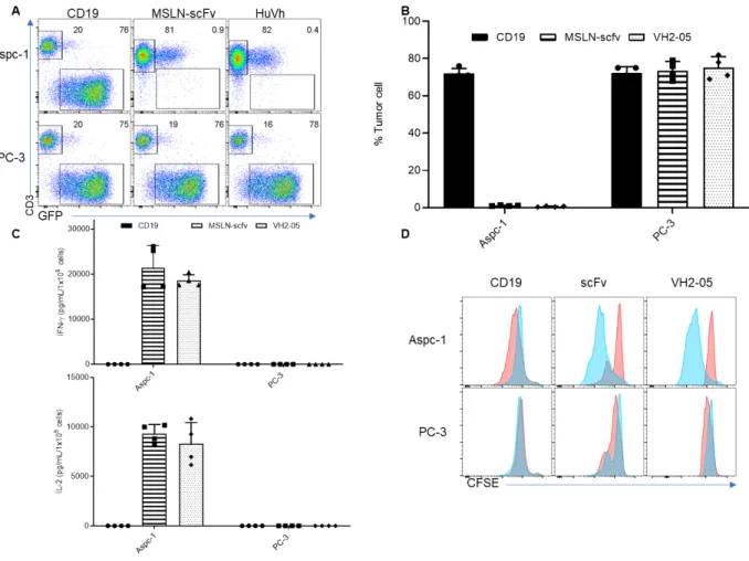
Next, we move onto another target MSLN and construct the conventional scFv CAR-T cells and HuVh.CAR-T cells respectively in the same backbone as J591.CAR-T cell. In a similar fashion, we examined the in vitro cytotoxicity of HuVh.CAR-T cell through tumor coculture, cytokine release assay and proliferation assay. As shown in Figure 5A and Figure 5B, both CAR T cells selectively lead to complete elimination of MSLN-positive tumor cells without off-target response. They well proliferate upon antigen recognition and have a similar cytokine release profile, demonstrating a comparable in vitro cytotoxicity with scFv CAR-T cell (Figure 5C and 5D).
15
cocultured with PC-3-eGFP and Aspc-1-eGFP cell lines at an E:T ratio of 1:5 for 6 days. (B) Summary of coculture of CD19.CAR-T cells, MSLN.scFv.CAR-T cells and MSLN.HuVh.CAR-T cells with PC-3-eGFP and Aspc-1- PC-3-eGFP cell lines; error bars represent SD (n=4). (C) IFN-γ and IL-2 release from the coculture supernatant with CD19.CAR-T cells, MSLN.scFv.CAR-T cells and MSLN.HuVh.CAR-T cells as measured by ELISA(n=4). (D) Representative flow cytometry figures illustrating CFSE proliferation assay. Blue and gray histograms represent activated and control groups respectively(n=4).
2.3 Discussion
16
17 REFERENCES
Barber, A., Meehan, K.R., and Sentman, C.L. (2011). Treatment of multiple myeloma with adoptively transferred chimeric NKG2D receptor-expressing T cells. Gene Ther 18, 509-516.
Barber, A., Zhang, T., and Sentman, C.L. (2008). Immunotherapy with chimeric NKG2D receptors leads to long-term tumor-free survival and development of host antitumor immunity in murine ovarian cancer. J Immunol 180, 72-78.
Borghaei, H., Smith, M.R., and Campbell, K.S. (2009). Immunotherapy of cancer. Eur J Pharmacol 625, 41- 54.
Brentjens, R., Davila, M., Riviere, I., Wang, X.Y., Bartido, S., Park, J., Bouhassira, D., Curran, K., Chung, S., Stefanski, J., et al. (2014). Efficacy and Toxicity Management of 19-28z CAR T Cell Therapy in B Cell Acute Lymphoblastic Leukemia. Mol Ther 22, S295-S296.
Chen, Y., Sun, C., Landoni, E., Metelitsa, L., Dotti, G., and Savoldo, B. (2019). Eradication of
Neuroblastoma by T Cells Redirected with an Optimized GD2-Specific Chimeric Antigen Receptor and Interleukin-15. Clin Cancer Res 25, 2915-2924.
Davila, M.L., Riviere, I., Wang, X., Bartido, S., Park, J., Curran, K., Chung, S.S., Stefanski, J., Borquez- Ojeda, O., Olszewska, M., et al. (2014). Efficacy and toxicity management of 19-28z CAR T cell therapy in B cell acute lymphoblastic leukemia. Sci Transl Med 6, 224ra225.
Diaconu, I., Ballard, B., Zhang, M., Chen, Y., West, J., Dotti, G., and Savoldo, B. (2017). Inducible Caspase- 9 Selectively Modulates the Toxicities of CD19-Specific Chimeric Antigen Receptor-Modified T Cells. Mol Ther 25, 580-592.
Fischer, K., Hoffmann, P., Voelkl, S., Meidenbauer, N., Ammer, J., Edinger, M., Gottfried, E., Schwarz, S., Rothe, G., Hoves, S., et al. (2007). Inhibitory effect of tumor cell-derived lactic acid on human T cells. Blood 109, 3812-3819.
Gabrilovich, D.I., and Nagaraj, S. (2009). Myeloid-derived suppressor cells as regulators of the immune system. Nature reviews immunology 9, 162.
Geiger, T.L., and Jyothi, M.D. (2001). Development and application of receptor-modified T lymphocytes for adoptive immunotherapy. Transfus Med Rev 15, 21-34.
Goodwin, J.S., Bankhurst, A.D., and Messner, R.P. (1977). Suppression of human T-cell mitogenesis by prostaglandin. Existence of a prostaglandin-producing suppressor cell. J Exp Med 146, 1719-1734. Grupp, S.A., Kalos, M., Barrett, D., Aplenc, R., Porter, D.L., Rheingold, S.R., Teachey, D.T., Chew, A., Hauck, B., Wright, J.F., et al. (2013). Chimeric antigen receptor-modified T cells for acute lymphoid leukemia. N Engl J Med 368, 1509-1518.
Harlin, H., Meng, Y., Peterson, A.C., Zha, Y., Tretiakova, M., Slingluff, C., McKee, M., and Gajewski, T.F. (2009). Chemokine expression in melanoma metastases associated with CD8+ T-cell recruitment. Cancer Res 69, 3077-3085.
18
S., Philbrook, P., Ko, K., Cannici, R., et al. (2015). Immunological mechanisms of the antitumor effects of supplemental oxygenation. Sci Transl Med 7, 277ra230.
Hodi, F.S., O'Day, S.J., McDermott, D.F., Weber, R.W., Sosman, J.A., Haanen, J.B., Gonzalez, R., Robert, C., Schadendorf, D., Hassel, J.C., et al. (2010). Improved survival with ipilimumab in patients with metastatic melanoma. N Engl J Med 363, 711-723.
Hollyman, D., Stefanski, J., Przybylowski, M., Bartido, S., Borquez-Ojeda, O., Taylor, C., Yeh, R., Capacio, V., Olszewska, M., Hosey, J., et al. (2009). Manufacturing Validation of Biologically
Functional T Cells Targeted to CD19 Antigen for Autologous Adoptive Cell Therapy. J Immunother 32, 169-180.
Ito, F., and E.Chang, A. (2013). Cancer Immunotherapy. Surgical Oncology Clinics of North America 22, 765-783.
June, C.H., O'Connor, R.S., Kawalekar, O.U., Ghassemi, S., and Milone, M.C. (2018). CAR T cell immunotherapy for human cancer. Science 359, 1361-1365.
Kantoff, P.W., Higano, C.S., Shore, N.D., Berger, E.R., Small, E.J., Penson, D.F., Redfern, C.H., Ferrari, A.C., Dreicer, R., Sims, R.B., et al. (2010). Sipuleucel-T immunotherapy for castration-resistant prostate cancer. N Engl J Med 363, 411-422.
Kochenderfer, J.N., Dudley, M.E., Feldman, S.A., Wilson, W.H., Spaner, D.E., Maric, I.,
Stetler-Stevenson, M., Phan, G.Q., Hughes, M.S., Sherry, R.M., et al. (2012). B-cell depletion and remissions of malignancy along with cytokine-associated toxicity in a clinical trial of anti-CD19 chimeric-antigen-receptor-transduced T cells. Blood 119, 2709-2720.
Kochenderfer, J.N., Dudley, M.E., Kassim, S.H., Somerville, R.P., Carpenter, R.O., Stetler-Stevenson, M., Yang, J.C., Phan, G.Q., Hughes, M.S., Sherry, R.M., et al. (2015). Chemotherapy-refractory diffuse large B- cell lymphoma and indolent B-cell malignancies can be effectively treated with autologous T cells expressing an anti-CD19 chimeric antigen receptor. J Clin Oncol 33, 540-549.
Kochenderfer, J.N., Wilson, W.H., Janik, J.E., Dudley, M.E., Stetler-Stevenson, M., Feldman, S.A., Maric, I., Raffeld, M., Nathan, D.A., Lanier, B.J., et al. (2010). Eradication of B-lineage cells and regression of lymphoma in a patient treated with autologous T cells genetically engineered to recognize CD19. Blood 116, 4099-4102.
Kong, S., Sengupta, S., Tyler, B., Bais, A.J., Ma, Q., Doucette, S., Zhou, J., Sahin, A., Carter, B.S., Brem, H., et al. (2012). Suppression of human glioma xenografts with second-generation IL13R-specific
chimeric antigen receptor-modified T cells. Clin Cancer Res 18, 5949-5960.
Lee, D.W., Gardner, R., Porter, D.L., Louis, C.U., Ahmed, N., Jensen, M., Grupp, S.A., and Mackall, C.L. (2014). Current concepts in the diagnosis and management of cytokine release syndrome. Blood 124, 188- 195.
Lee, D.W., Kochenderfer, J.N., Stetler-Stevenson, M., Cui, Y.K., Delbrook, C., Feldman, S.A., Fry, T.J., Orentas, R., Sabatino, M., Shah, N.N., et al. (2015). T cells expressing CD19 chimeric antigen receptors for acute lymphoblastic leukaemia in children and young adults: a phase 1 dose-escalation trial. Lancet 385, 517- 528.
19
Enhanced Cancer Immunotherapy by Chimeric Antigen Receptor-Modified T Cells Engineered to Secrete Checkpoint Inhibitors. Clin Cancer Res 23, 6982-6992.
Massagué, J. (2008). TGFβ in cancer. Cell 134, 215-230.
Murad, J.M., Graber, D.J., and Sentman, C.L. (2018). Advances in the use of natural receptor-or ligand-based chimeric antigen receptors (CARs) in haematologic malignancies. J Best Practice Research Clinical Haematology 31, 176-183.
Newick, K., O'Brien, S., Moon, E., and Albelda, S.M. (2017). CAR T Cell Therapy for Solid Tumors. Annu Rev Med 68, 139-152.
Paszkiewicz, P.J., Frassle, S.P., Srivastava, S., Sommermeyer, D., Hudecek, M., Drexler, I., Sadelain, M., Liu, L., Jensen, M.C., Riddell, S.R., et al. (2016). Targeted antibody-mediated depletion of murine CD19 CAR T cells permanently reverses B cell aplasia. J Clin Invest 126, 4262-4272.
Pegram, H.J., Lee, J.C., Hayman, E.G., Imperato, G.H., Tedder, T.F., Sadelain, M., and Brentjens, R.J. (2012). Tumor-targeted T cells modified to secrete IL-12 eradicate systemic tumors without need for prior conditioning. Blood 119, 4133-4141.
Philip, B., Kokalaki, E., Mekkaoui, L., Thomas, S., Straathof, K., Flutter, B., Marin, V., Marafioti, T., Chakraverty, R., Linch, D., et al. (2014). A highly compact epitope-based marker/suicide gene for easier and safer T-cell therapy. Blood 124, 1277-1287.
Porter, D.L., Hwang, W.T., Frey, N.V., Lacey, S.F., Shaw, P.A., Loren, A.W., Bagg, A., Marcucci, K.T., Shen, A., Gonzalez, V., et al. (2015). Chimeric antigen receptor T cells persist and induce sustained remissions in relapsed refractory chronic lymphocytic leukemia. Sci Transl Med 7, 303ra139.
Sakaguchi, S., Miyara, M., Costantino, C.M., and Hafler, D.A. (2010). FOXP3+ regulatory T cells in the human immune system. Nat Rev Immunol 10, 490-500.
Somerville, R.P.T., Devillier, L., Parkhurst, M.R., Rosenberg, S.A., and Dudley, M.E. (2012). Clinical scale rapid expansion of lymphocytes for adoptive cell transfer therapy in the WAVE (R) bioreactor. J Transl Med 10.
Turtle, C.J., Riddell, S.R., and Maloney, D.G. (2016). CD19-Targeted chimeric antigen receptor-modified T- cell immunotherapy for B-cell malignancies. Clin Pharmacol Ther 100, 252-258.
van Zelm, M.C., Reisli, I., van der Burg, M., Castaño, D., van Noesel, C.J., van Tol, M.J., Woellner, C., Grimbacher, B., Patiño, P.J., and van Dongen, J.J. (2006). An antibody-deficiency syndrome due to mutations in the CD19 gene. New England Journal of Medicine 354, 1901-1912.
Vera, J.F., Brenner, L.J., Gerdemann, U., Ngo, M.C., Sili, U., Liu, H., Wilson, J., Dotti, G., Heslop, H.E., Leen, A.M., et al. (2010). Accelerated Production of Antigen-specific T Cells for Preclinical and Clinical Applications Using Gas-permeable Rapid Expansion Cultureware (G-Rex). J Immunother 33, 305-315. Vu, K.B., Ghahroudi, M.A., Wyns, L., and Muyldermans, S. (1997). Comparison of llama VH sequences from conventional and heavy chain antibodies. Mol Immunol 34, 1121-1131.