THE ADHESION MOLECULE PECAM-1 DIRECTS ENDOTHELIAL NITRIC OXIDE SYNTHASE ACTIVITY AND CARDIAC FUNCTION TO REGULATE CARDIOVASCULAR
HOMEOSTASIS
Margaret Elizabeth McCormick
A dissertation submitted to the faculty of the University of North Carolina at Chapel Hill in partial fulfillment of the requirements for the degree of Doctor of Philosophy in the
Curriculum of Cell and Molecular Physiology
Chapel Hill 2012
ABSTRACT
MARGARET ELIZABETH MCCORMICK: The adhesion molecule PECAM-1 directs endothelial nitric oxide synthase activity and cardiac function to regulate cardiovascular
homeostasis
(Under the direction of Ellie Tzima)
The mammalian vascular system is integral to the regulation of homeostasis within an organism. This system is composed of a vast network of blood vessels that transport oxygen and nutrients throughout the body. Blood vessels are lined by endothelial cells (ECs) that provide an interface between circulating blood and the underlying tissue. Based on their unique location, it is unsurprising that ECs are master regulators of numerous physiological processes. ECs are equipped with receptors that interact with their surrounding environment. Among these molecules is Platelet Endothelial Cell Adhesion Molecule-1, or PECAM-1. Since its identification in the early 1990s, PECAM-1 has gained increasing attention for its multi-faceted roles in vascular biology. In this dissertation, I describe two novel roles for PECAM-1 which advance our understanding for how this receptor regulates EC signaling and function.
One of the most important cellular signaling molecules in the vasculature is nitric oxide (NO) generated by the enzyme endothelial nitric oxide synthase (eNOS). NO is required for vasodilation and maintenance of the endothelium. Reduced NO production can lead to endothelial dysfunction, the first step in a host of diseases, including atherosclerosis (hardening of the arteries). Previous studies have suggested a role for PECAM-1 in
regulates the expression of a trafficking protein that influences eNOS localization and activity.
In the second half of my dissertation, I focus in vivo using wild type and PECAM-1
-/-mice. Our lab has shown that PECAM-1-mediated signaling facilitates activation and expression of molecules that participate in cellular crosstalk. Furthermore, it is well documented that cell crosstalk is central to proper cardiac function. Therefore, we hypothesized that genetic deletion of PECAM-1 may have deleterious effects on heart function. In agreement with this hypothesis, we are the first group to report that PECAM-1
-/-mice display dilated cardiomyopathy. Work described in this dissertation defines the mechanism leading to these defects in function. Taken together, the work presented in this thesis further expands the evolving roles of PECAM-1 in cardiovascular biology and
ACKNOWLEDGEMENTS
First and foremost I would like to thank my mentor, Ellie Tzima, for all of her
guidance and support over the last 5 years. I came to UNC knowing that I wanted to work in her lab and never once have I regretted this decision. I immensely respect her opinion and scientific foresight, her pursuit of cutting edge science and ideas, and her fearless endeavor into new areas of research. She has pushed me to have confidence in my ideas and myself. When my thesis work took us into the cardiac field, she never once shied away from
pursuing it. I hope to take what I’ve learned from Ellie and expand on it as I continue to mature as a scientist.
I would also like to thank everyone in the Tzima lab, past and present: Dan Sweet, Caitlin Collins, Chris Givens, Zhongming Chen, Young Greenberg, Meghan Childs, Sruthi Cherkur, Meredith Brown and Yunhao Liu. Their unwavering support over the last 5 years has meant the world; most of this would not have been possible without them. To Dan, Caitlin and Chris, I look forward to being colleagues and friends for many years to come. Dan, for loving Pennsylvania as much as I do, and challenging me daily in the best way possible to make my projects more complete. Caitlin for reading everything I’ve ever written and for hearing every thought I’ve had and still being my colleague and friend (she knows how hard that is…). Zhongming Chen, AKA the mouse man, TGIF. And Chris, you’ve got some big shoes to fill (insert ha!), but I’m confident you can and will make us all proud.
The research environment at UNC has benefited my time as a graduate student immensely. I have relied on so many amazing people for their insight, expertise and
without his flexibility and positive attitude, my work would have been far less successful and significantly less enjoyable. A big thanks to the physiology department. I joined this
department in 2007 because it felt like a supportive home to spend 5 years of school and it has been. Adriana Tavernise, Vicki Morgan and the rest of the staff have made my life, both scientifically and otherwise, run incredibly smoothly.
It is hard to put into words what my friends at UNC and in Chapel Hill have meant to me. They have all contributed in ways big and small to my well-being as a graduate
student. Gray Camp is my role model for a thoughtful, kind, brilliant person. I truly cannot imagine what life here would have been like without him. Lauren Strickland for being such a positive influence in my life and for showing me what North Carolina is all about – hollerin’. Katy Liu for her non-judgmental friendship and support and a fantastic roommate (Juj included!). And thank you to Anne-Marie Neiser for being up for anything and always having a smile on your face. Also, a special thank you to my F32 roommates (Morgan, Tricia, and Jill) for keeping me social when science threatened to overwhelm me.
And of course I want to thank my parents, Bob and Melinda. My parents brought my brothers and I up to observe, question and think about the world around us. They exposed us to new places and experiences and instilled in us the curiosity they had for the world and all things nature. I believe all of that combined to put me where I am today and for that I am eternally grateful. I would also like to thank my brothers, Rob and Brian, for their love and support and being above all else, my friends.
PREFACE
The work presented in Chapter II work was previously published in Arteriosclerosis, Thrombosis and Vascular Biology in March 2011. My roles in this project included helping design and perform the experiments, data analysis and writing the manuscript. Reema Goel and Debra Newman performed the knockdown studies and performed the corresponding data analysis (Figure S2.1). David Fulton performed the in vivo NO assays (Figure 2.1). Ellie Tzima was the principal investigator and helped design experiments, analyze data and write the manuscript.
The citation for the manuscript is as follows: McCormick ME, Goel R, Fulton D, Newman D, Tzima E. Platelet-Endothelial Cell Adhesion Molecule-1 Regulates Endothelial NO Synthase Activity and Localization Through Signal Transducers and Activators of Transcription 3-dependent NOSTRIN Expression. Arterioscler Throm Vasc Biol.(2011); 3:643-649.
Work described in Chapter III has currently been submitted for publication. My roles in this project included helping design and perform experiments as well as data analysis and writing the manuscript. Gianluigi Pironti and Howard Rockman performed the isolated
cardiomyocyte experiments (Table 3.1). Mauricio Rojas performed the conscious echos, pressure-volume loops and TAC surgeries. Monte Willis performed conscious
TABLE OF CONTENTS
LIST OF TABLES... xii
LIST OF FIGURES………. ... xiii
LIST OF ABBREVIATIONS AND SYMBOLS……… ... xv
CHAPTERS I. INTRODUCTION ... 1
THE VASCULAR ENDOTHELIUM ... 1
PECAM-1 STRUCTURE AND FUNCTION ... 2
eNOS AND NO SIGNALING IN ENDOTHELIAL CELLS ... 3
NOSTRIN REGULATES eNOS TRAFFICKING AND ACTIVITY ... 5
CELL-CELL CROSSTALK AND THE EFFECTS ON CELL AND ORGAN FUNCTION ... 6
CARDIAC FUNCTION AND DILATED CARDIOMYOPATHY ... 8
RESEARCH PRESENTED IN THIS DISSERTATION……… ... 9
Chapter II: Determine the role of PECAM-1 in regulating basal eNOS activity ... 9
Chapter III: Determine the role of PECAM-1 in regulating cardiac function... 9
Chapter IV: Conclusions and Perspectives ... 10
REFERENCES... 15
II. PECAM-1 REGULATES eNOS ACTIVITY AND LOCALIZATION THROUGH STAT3-DEPENDENT NOSTRIN EXPRESSION... 19
INTRODUCTION... 20
METHODS ... 21
Animals... 21
Cell Culture and Reagents ... 21
Immunoprecipitation and Western Blotting... 22
Immunofluorescence ... 22
Cellular Fractionation ... 22
In Vitro and In Vivo NO Measurements ... 23
RNA Isolation and Quantitative PCR ... 24
Chromatin Immunoprecipitation Assays ... 24
Quantification and Statistical Analysis... 24
RESULTS... 24
PECAM-1 Regulates Basal eNOS Activity ... 24
Differential Association of eNOS and Caveolin-1 in the Absence of PECAM-1 ... 25
Role of PECAM-1 in eNOS Localization/Trafficking ... 26
PECAM-1 Mediates STAT3-Dependent NOSTRIN Expression in ECs ... 27
Binding of STAT3 to the NOSTRIN Promoter in ECs... 28
DISCUSSION ... 29
SUPPLEMENTAL MATERIAL ... 39
REFERENCES... 45
III. A MECHANOSENSOR MEDIATES CROSSTALK BETWEEN ENDOTHELIAL CELLS AND CARDIOMYOCYTES TO REGULATED CARDIAC FUNCTION ... 49
OVERVIEW ... 49
Animals... 51
Cell Culture and Reagents ... 51
Echocardiography measurements... 51
Transverse Aortic Constriction ... 51
RNA Isolation and Quantitative PCR ... 51
Preparation of Lysates and Immunoblot Analysis ... 52
Histological analysis and immunohistochemistry ... 52
Cardiomyocyte isolation/Functional Assays ... 53
Transmission Electron Microscopy... 53
Quantitation and Statistical Analysis ... 53
RESULTS... 53
PECAM-1-/- mice exhibit both systolic and diastolic dysfunction indicative of dilated cardiomyopathy ... 53
Phenotypic characterization of PECAM-1-/- mice... 54
Increased systolic dysfunction in PECAM-1-/- mice after biomechanical stress ... 55
Cardiomyocytes isolated from PECAM-1-/- hearts display normal contractility ... 57
Impaired Neuregulin-ErbB signaling in the PECAM-1-/- mice ... 58
DISCUSSION ... 59
SUPPLEMENTAL MATERIAL ... 71
REFERENCES... 79
IV. CONCLUSIONS AND PERSPECTIVES ... 85
OVERVIEW ... 85
CHAPTER II: PECAM-1 REGULATES eNOS ACTIVITY AND LOCALIZATION THROUGH STAT3-DEPENDENT NOSTRIN EXPRESSION ... 86
PECAM-1 regulation of JAK/STAT activity ... 88
CHAPTER III: A MECHANOSENSOR MEDIATES CROSSTALK BETWEEN ENDOTHELIAL CELLS AND CARDIOMYOCYTES TO REGULATE CARDIAC FUNCTION ... 90
Shear stress signaling in the heart ... 91
Which endothelial cells are important for regulating cardiac function? ... 92
INTEGRATING IN VITRO AND IN VIVO RESEARCH ... 94
Balance of Signaling ... 94
Contribution of alternative signaling pathways ... 95
The signaling relationship between PECAM-1 and eNOS/NO in the heart ... 97
Implications for cardiovascular biology... 98
LIST OF TABLES
Supplemental Table 2.1 PCR and ChIP primers ... 43 Supplemental Table 2.2 Predicted STAT3 binding sites within the
NOSTRIN promoter ... 44 Table 3.1 Contractile parameters in adult cardiomyocytes isolated from
WT and PECAM-1 mice... 68 Supplemental Table 3.1 Echocardiographic data from WT and PECAM-1-/-
mice pre- and post-TAC... 72 Supplemental Table 3.2 Echocardiographic data from aging WT and
PECAM-1-/- mice ... 73
LIST OF FIGURES
Figure 1.1 PECAM-1 localization and structure in ECs ... 11
Figure 1.2 Multi-domain structure of NOSTRIN ... 12
Figure 1.3 Intimate association of ECs and CMs in the heart... 13
Figure 1.4 Overview of research presented in this dissertation ... 14
Figure 2.1 Basal eNOS phosphorylation and NO production in the PECAM-1 KO ... 33
Figure 2.2 Differential association of the negative eNOS binding partner, caveolin-1, in the absence of PECAM-1 ... 34
Figure 2.3 PECAM-1 regulates eNOS subcellular localization ... 35
Figure 2.4 Reduced NOSTRIN expression in ECs lacking PECAM-1 ... 36
Figure 2.5 PECAM-1 regulates NOSTRIN expression in a STAT3-dependent manner... 37
Figure 2.6 Model of PECAM-1-mediated NOSTRIN expression and eNOS trafficking... 38
Supplemental Figure 2.1 PECAM-1 knockdown in HUVECs... 41
Supplemental Figure 2.2 PECAM-1 is not required for ionomycin-induced eNOS activation ... 42
Figure 3.1 PECAM-1-/- mice have increased chamber size with systolic and diastolic dysfunction... 63
Figure 3.2 Normal cellular architecture in PECAM-1-/- hearts ... 65
Figure 3.3 PECAM-1-/- mice have impaired response to hemodynamic stress... 66
Figure 3.4 Impaired cellular activation and remodeling after TAC in the PECAM-1-/- mice ... 67
Figure 3.5 Misregulated NRG-1/ErbB2 signaling in PECAM-1-/- hearts... 69
Supplemental Figure 3.1 Mean arterial pressure (MAP) in PECAM-1-/- mice is similar to WT ... 74
Supplemental Figure 3.2 No evidence for hypoxia in PECAM-1-/- hearts ... 75
Supplemental Figure 3.4 Adult mouse cardiomyocytes do not express
LIST OF ABBREVIATIONS AND SYMBOLS
ANP Atrial Natriuretic Peptide
αSka α-skeletal actin
BAEC Bovine Aortic Endothelial Cell
βMHC β-Myosin Heavy Chain BMP Bone Morphogenic Protein
Ca2+ Calcium
Cav1 Caveolin-1
cGMP cyclic Guanosine Monophosphate ChIP Chromatin Immunoprecipitation DCM Dilated Cardiomyopathy
EC Endothelial Cell
EF Ejection Fraction
eNOS endothelial Nitric Oxide Synthase Erk1/2 Extracellular related kinase 1/2
FS Fractional Shortening
FSS Fluid Shear Stress
HUVEC Human Umbilical Vein Endothelial Cell ITAM Immunoreceptor Tyrosine Activation Motif ITIM Immunoreceptor Tyrosine Inhibitory Motif
JAK Janus Kinase
L-NAME L-NG-Nitroarginine Methyl Ester
LV Left Ventricle
NRG1 Neuregulin-1
NO Nitric Oxide
NOSIP eNOS Interacting Protein NOSTRIN eNOS Traffic Inducer
PECAM-1 Platelet Endothelial Cell Adhesion Molecule-1
PKA Protein Kinase A
PLN Phospholamban
PV Pressure-Volume
qPCR quantitative Polymerase Chain Reaction
ROS Reactive Oxygen Species
SERCA2 Sarco/Endoplasmic Reticulum Ca2+-ATPase
STAT3/5 Signal Transducer and Activator of Transcription 3/5 TAC Transverse Aortic Constriction
TEM Transmission Electron Microscopy VE-Cadherin Vascular Endothelial Cadherin VEGF Vascular Endothelial Growth Factor
VEGFR2 Vascular Endothelial Growth Factor Receptor 2 VSMC Vascular Smooth Muscle Cell
CHAPTER I. Introduction
The vascular endothelium
The vascular system plays a vital role in regulating homeostasis. Defects in vascular function lead to the development of diseases such as atherosclerosis and coronary artery disease. In addition to transporting oxygen and nutrients, the vasculature is also
fundamental for the delivery of important hormonal/neurohormonal signals that control organ system function. An illustration of this is the hormone vasopressin, which is released by the posterior pituitary and travels through the vasculature to the renal tubules to increased blood volume and ultimately blood pressure.
William Harvey first described the presence of circulating blood in the early 1600s. Shortly after this observation, it was recognized that blood is separated from tissue by a series of vessels 1. It took over a century to realize that a thin layer of cells, termed endothelial cells (ECs), line the blood vessel wall. Despite this observation, as recently as the 1960s, ECs were thought of simply as “the cellophane wrapper” of the vasculature 2. A tremendous amount of work done in the following decades revealed that ECs play a
the brain have tight junctions and a thick basement membrane to prevent the mixing of blood with cerebrospinal fluid. Conversely, ECs of the glomerulus have specialized filtration slits to permit filtration of fluids and small molecules into the renal tubules.
The endothelium is lined with receptors and adhesion molecules that provide a key interface between ECs and the extracellular environment. These proteins also mediate intracellular signaling and facilitate complex interactions with neighboring cell types. EC input comes in many varieties, including circulating growth factors (vascular endothelial growth factor, VEGF), hormones (endothelin-1), metabolites (nitric oxide, NO) and
mechanical forces (blood flow). Due to their integral role, it is important to have a thorough understanding of how ECs are able to integrate information provided by their environment.
PECAM-1 Structure and Function
One important EC adhesion molecule is platelet endothelial cell adhesion molecule-1 (PECAM-1). In addition to being expressed on the surface of ECs, it is also found on
platelets and leukocytes. PECAM-1 was initially identified for its role in leukocyte transendothelial migration, the process by which immune cells cross the vessel wall to underlying sites of inflammation 3. More recently, it has been recognized as a multifaceted molecule in both endothelial and vascular biology.
When tyrosine residues Y663 and Y686 within these domains are phosphorylated, PECAM-1
recruits Src-homology 2 (SH2) domain-containing proteins 6. This is critical for PECAM-1 biology, as it allows the protein to participate in a number of important signaling pathways despite the lack of intrinsic kinase activity. PECAM-1 becomes tyrosine phosphorylated in response to a number of signals, including homophilic binding and shear stress. It has been previously shown that fluid shear stress (FSS) is a potent stimulus for PECAM-1
phosphorylation by the Src-family kinase Fyn 7, 8. PECAM-1 mediates a number of shear stress-responsive pathways, including the activation of phosphoinositide-3 kinase and Akt. Importantly, PECAM-1 null ECs display impaired responses to shear stress 9.
The PECAM-1-/- mouse was generated, in part, to address its role in leukocyte transendothelial migration (TEM) 10. Somewhat surprisingly, these animals were viable and had normal vasculogenesis despite previous studies implicating a role for PECAM-1 in angiogenesis 11, 12. Initially, only minor defects in leukocyte TEM were identified in the PECAM-1-/- mouse. However, subsequent studies using other stress models, such as
endotoxic induced-LPS shock, identified further defects in immunologic and angiogenic responses 13-‐15. Additionally, our lab has demonstrated that PECAM-1 deficiency results in impaired flow-mediated vascular remodeling 16. Furthermore, PECAM-1 null mice exhibit blunted arteriogenesis and reduced collateral remodeling due to deficits in shear stress-induced endothelial nitric oxide synthase (eNOS) and nuclear factor kappa B (NF-κB) signaling 17.
eNOS and NO Signaling in endothelial cells
endothelium, eNOS catalyzes the formation of NO and citrulline through the oxidation of L-arginine. NO regulates vascular tone through activation of guanylyl cyclase and subsequent cGMP production in vascular smooth muscle cells (VSMCs). In this cell type, cGMP acts a second messenger to mediate relaxation and vasodilation. Numerous stimuli activate eNOS (via phosphorylation) to generate NO, including humoral factors, such as VEGF, bradykinin and estrogen, as well as mechanical forces such as shear stress, which is one of the most potent activators of eNOS 19, 20.
The responsiveness of eNOS to diverse types of stimuli requires dynamic regulation of the enzyme. eNOS activity is regulated by several different mechanisms including protein-protein interactions (Ca2+/Calmodulin (CaM), caveolin-1, andHsp90); post-translational
regulation (phosphorylation, acylation); cofactors and substrates (BH4); and subcellular
localization (plasmamembrane caveolae, Golgi, and cytosolic compartments) 21, 22. Basally, eNOS is kept inactive via association with the inhibitory protein caveolin-1 (Cav1), which typically occurs at the plasma membrane 18. Following stimulation, either through growth factors such as VEGF or shear stress, Ca2+/CaM is able to interact with eNOS. This helps
displace eNOS from binding Cav1 and subsequently activates the enzyme. Additionally, FSS enhances the eNOS-Hsp90 interaction, thereby facilitating phosphorylation of Ser1179 by Akt 23, 24. Phosphorylation of this serine residue plays a critical role in the early NO production in response to shear stress 19.
Subcellular localization is another important aspect of eNOS regulation. There are three pools of eNOS located within the cell: at the perinuclear Golgi complex; at the plasma membrane; and a cytosolic compartment 22. eNOS is localized to the plasma membrane primarily in caveolae through acylation, including myristoylation and palmitoylation 18. The plasma membrane pool of eNOS is highly responsive to increases in intracellular Ca2+
Akt-mediated activation 25. The proteins NOSIP (eNOS interacting protein) 26 and NOSTRIN (eNOS traffic inducer) 27 mediate trafficking of eNOS between the plasma membrane and Golgi complex. Interestingly, the exact mechanisms that regulate this trafficking are not clearly established.
Misregulation of eNOS signaling has profound consequences on the health of the blood vessel. Endothelial dysfunction, present in a variety of pathologic conditions including atherosclerosis, is generally characterized by impaired NO production. It is therefore critical to characterize the mechanisms governing eNOS signaling to further understand how these processes contribute to vessel health and disease. eNOS-/- mice have been generated to address these important questions. Not surprisingly, these animals suffer from hypertension (through misregulated NO production), increased atherosclerotic lesions, and a heightened injury response 28, 29.
In Chapter II, I describe a novel mechanism of basal eNOS regulation. Previous studies have demonstrated a role for PECAM-1 in shear-induced eNOS activation. Here I show that PECAM-1 mediates basal eNOS activity through a pathway involving the eNOS trafficking protein NOSTRIN.
NOSTRIN regulates eNOS trafficking and activity
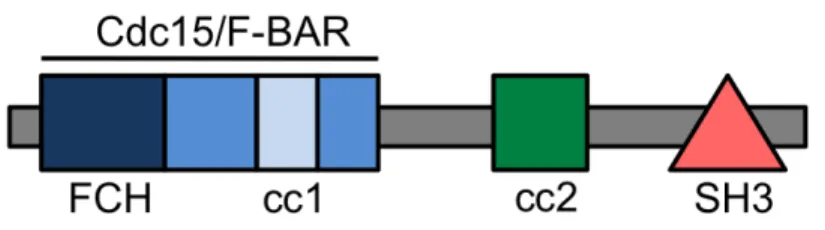
One of the more recently described regulators of eNOS activity and localization is the cytoplasmic protein NOSTRIN. This protein was discovered a decade ago in a yeast two-hybrid screen using the oxygenase domain of human eNOS as bait 27. NOSTRIN is a 58-kD protein containing an N-terminal FCH region, a coiled-coil domain and C-terminal SH3 domain (Figure 1.2). The FCH region is followed by a coiled-coil stretch that composes the F-BAR domain, making it a member of the F-BAR (Fes/CIP4 homology and
cytoskeletal interactions 30 and serve as adaptor proteins. The SH3 domain is required for binding eNOS 27 and the second coiled-coil domain facilitates oligomerization. NOSTRIN is expressed in highly vascularized tissues such as heart, lung and kidney 27. Much of the focus on NOSTRIN has been its role as a member of the complex eNOS trafficking
machinery 27. It is able to independently bind both caveolin-1 and eNOS, forming a ternary complex important for eNOS localization 31. Changes in NOSTRIN expression can alter eNOS activity and localization; when NOSTRIN is overexpressed, eNOS activity is significantly decreased 31, 32.
More recent studies have revealed additional functions for NOSTRIN in endothelial cells. In vivo studies using zebrafish have identified a novel role for NOSTRIN in mediating angiogenesis during development and in the adult 33. The authors demonstrate that
NOSTRIN forms a complex with Rac1 and Sos1, important for FGFR1-mediated Rac1 activation 33. In addition to the full-length protein a shortened isoform, NOSTRINβ, was recently described 34. Unlike NOSTRIN, this variant is primarily expressed in the nucleus and thought to regulate expression of the NOSTRIN gene. The importance of NOSTRIN in EC physiology and pathophysiology is currently the topic of ongoing investigation.
Cell-cell crosstalk and implications in cell and organ function
Cell-cell crosstalk, defined as communication through paracrine/autocrine factors released from one cell that affect the behavior and function of another cell, is an integral biological concept. This phenomenon is essential both during development and is critical for maintaining homeostasis within the adult. Crosstalk occurs through a number of
However, the research presented in this dissertation specifically focuses on the crosstalk between ECs and underlying cells.
During development tissues release signaling molecules that influence migration, proliferation, and differentiation of constituent cells. For example, most tissues release the signaling protein VEGF that signals to ECs to induce the growth of new blood vessels. This provides oxygen and nutrients to the newly forming tissue. VEGF-mediated signaling is also necessary for the patterning and maintenance of the nascent blood vessels 36. Other
examples of cytokines important in crosstalk during development include platelet-derived growth factor (PDGF), fibroblast growth factor (FGF), and bone morphogenic protein (BMP) 37. Studies using genetic ablation of PDGF and its receptor PDGFR have demonstrated an important role for this signaling pathway in neural and vascular development 38. In vascular development, PDGF is primarily secreted by ECs of the emerging vessel sprouts, while the receptor is expressed by pericytes and VSMCs. Here, EC-derived PDGF signaling
stimulates both proliferation and migration of these neighboring cells to surround and stabilize developing blood vessels 38.
In the adult, cell crosstalk is essential for homeostasis. For instance, NO released from ECs signals to underlying VSMCs to regulate vessel tone. Misregulation of this signaling can lead to EC dysfunction and as mentioned previously, hypertension.
Additionally, signaling from ECs to VSMCs can affect the growth and proliferation of VSMCs and their contractile properties. Cell crosstalk can also be facilitated through direct
The heart is an organ whose function relies heavily on the crosstalk between cell types; primarily the crosstalk between ECs and cardiomyocytes (CMs, heart muscle cells). ECs exist in close proximity to CMs, providing a microenvironment that facilitates EC-CM communication (Figure 1.3A-D). Cardiac ECs are thought to release a number of paracrine factors, including neuregulin-1 (NRG1), VEGF, and NO that influence CM function.
Interestingly, crosstalk is reciprocated, as CMs release VEGF and angiopoietin to affect EC growth and function 40. Characterizing the regulation of these signaling pathways is
physiologically relevant, as abnormal signaling can result in cardiac pathologies, such as cardiac hypertrophy.
Cardiac function and dilated cardiomyopathy
The heart is a fascinating organ; it beats continuously throughout our lifetime and is able to continuously adapt to changes in demand. In order to function properly, it requires coordination of signaling pathways and electrical signals. Imbalance of either of these components can lead to dramatic and deleterious changes in heart structure or function. Diseases that are characterized by disruptions to structure or function are termed
cardiomyopathies. Dilated cardiomyopathy is the most common cardiomyopathy worldwide and represents one of the most common causes of heart failure. In this disorder, dilatation and impaired contraction of the ventricles develops and can lead to heart failure and sudden death. Although myocardial injury, as well as abnormalities in the function and integrity of CMs, can lead to dilated cardiomyopathy, cardiac endothelial dysfunction has also been implicated in progression of the disease 41. In Chapter III, I describe a mechanism by which PECAM-1, expressed in ECs, is able to regulate signaling pathways in CMs that are
RESEARCH PRESENTED IN THIS DISSERTATION
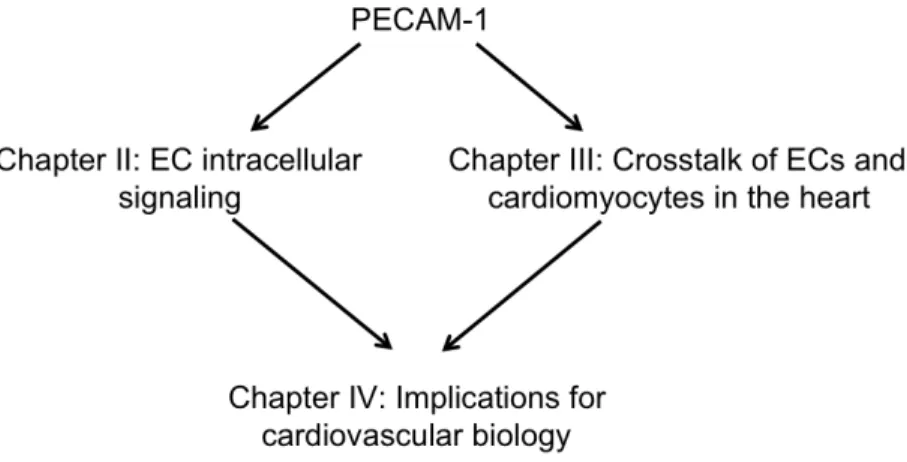
As described in the following chapters, the goals of this thesis are as follows (Figure 1.4):
Chapter II. Determine the role of PECAM-1 in regulating basal eNOS activity eNOS plays an important role in vascular biology. Misregulation can lead to endothelial dysfunction and cardiovascular disease. Previous studies have demonstrated a role for PECAM-1 in shear stress-induced eNOS activation. However, my preliminary data showed that PECAM-1 knockout ECs have higher basal pSer1179eNOS compared to PECAM-1 expressing cells. This observation suggests that PECAM-1 is not only required for shear-induced eNOS activation but also regulates basal eNOS activity. In this chapter I will i) characterize the influence of PECAM-1 expression in basal eNOS activity and ii) determine
the mechanism by which PECAM-1 regulates basal eNOS activity. I show that PECAM-1 mediates basal eNOS activation, in part, by regulating the expression of the eNOS trafficking protein NOSTRIN.
Chapter III. Determine the role of PECAM-1 in regulating cardiac function
Previous studies have shown a requirement for PECAM-1 in regulating vascular function and remodeling. Because the vascular system is important for cardiac function, I
hypothesized that cardiac function would be impaired in PECAM-1-/- animals. To address this possibility, we first performed echocardiography and found that the PECAM-1-/- hearts
had impaired function relative to wild-type hearts. Using biochemical and ex vivo
Chapter IV. Conclusions and Perspectives
Figures
Figure 1.1. Structure and localization of PECAM-1.
A. Mouse endothelial cells stained for PECAM-1 (green), acetylated LDL (red) and nucleus (DAPI). PECAM-1 staining is concentrated as cell-cell junctions. The accumulation of acetylated LDL within these cells is a marker of ECs. B. Representative structure of PECAM-1. The protein has 6 extracellular Ig-like domains, a single pass transmembrane domain and a relatively short cytoplasmic tail which contains ITIM residues Y663 and Y686
Figure 1.2. Multi-domain structure of NOSTRIN
Figure 1.3. Intimate relationship between cardiac endothelial cells and cardiomyocytes.
REFERENCES
1. Fishman AP. Endothelium: a distributed organ of diverse capabilities. Ann N Y Acad Sci. 1982; 401: 1-8.
2. Florey. The endothelial cell. Br Med J. 1966; 2: 487-490.
3. Muller WA, Weigl SA, Deng X, Phillips DM. PECAM-1 is required for transendothelial migration of leukocytes. J Exp Med. 1993; 178: 449-460.
4. Sun J, Williams J, Yan HC, Amin KM, Albelda SM, DeLisser HM. Platelet endothelial cell adhesion molecule-1 (PECAM-1) homophilic adhesion is mediated by immunoglobulin-like domains 1 and 2 and depends on the cytoplasmic domain and the level of surface
expression. J Biol Chem. 1996; 271: 18561-18570.
5. Sun QH, DeLisser HM, Zukowski MM, Paddock C, Albelda SM, Newman PJ. Individually distinct Ig homology domains in PECAM-1 regulate homophilic binding and modulate receptor affinity. J Biol Chem. 1996; 271: 11090-11098.
6. Newman PJ, Newman DK. Signal transduction pathways mediated by PECAM-1: new roles for an old molecule in platelet and vascular cell biology. Arterioscler Thromb Vasc Biol. 2003; 23: 953-964.
7. Osawa M, Masuda M, Kusano K, Fujiwara K. Evidence for a role of platelet endothelial cell adhesion molecule-1 in endothelial cell mechanosignal transduction: is it a
mechanoresponsive molecule? J Cell Biol. 2002; 158: 773-785.
8. Chiu YJ, McBeath E, Fujiwara K. Mechanotransduction in an extracted cell model: Fyn drives stretch- and flow-elicited PECAM-1 phosphorylation. J Cell Biol. 2008; 182: 753-763. 9. Tzima E, Irani-Tehrani M, Kiosses WB, Dejana E, Schultz DA, Engelhardt B, Cao G, DeLisser H, Schwartz MA. A mechanosensory complex that mediates the endothelial cell response to fluid shear stress. Nature. 2005; 437: 426-431.
10. Duncan GS, Andrew DP, Takimoto H, Kaufman SA, Yoshida H, Spellberg J, Luis de la Pompa J, Elia A, Wakeham A, Karan-Tamir B, Muller WA, Senaldi G, Zukowski MM, Mak TW. Genetic evidence for functional redundancy of Platelet/Endothelial cell adhesion molecule-1 (PECAM-1): CD31-deficient mice reveal dependent and PECAM-1-independent functions. J Immunol. 1999; 162: 3022-3030.
11. Albelda SM, Oliver PD, Romer LH, Buck CA. EndoCAM: a novel endothelial cell-cell adhesion molecule. J Cell Biol. 1990; 110: 1227-1237.
13. Carrithers M, Tandon S, Canosa S, Michaud M, Graesser D, Madri JA. Enhanced susceptibility to endotoxic shock and impaired STAT3 signaling in CD31-deficient mice. Am J Pathol. 2005; 166: 185-196.
14. Schenkel AR, Chew TW, Muller WA. Platelet endothelial cell adhesion molecule deficiency or blockade significantly reduces leukocyte emigration in a majority of mouse strains. J Immunol. 2004; 173: 6403-6408.
15. Solowiej A, Biswas P, Graesser D, Madri JA. Lack of platelet endothelial cell adhesion molecule-1 attenuates foreign body inflammation because of decreased angiogenesis. Am J Pathol. 2003; 162: 953-962.
16. Chen Z, Tzima E. PECAM-1 is necessary for flow-induced vascular remodeling. Arterioscler Thromb Vasc Biol. 2009; 29: 1067-1073.
17. Chen Z, Rubin J, Tzima E. Role of PECAM-1 in arteriogenesis and specification of preexisting collaterals. Circ Res. 2010; 107: 1355-1363.
18. Dudzinski DM, Michel T. Life history of eNOS: partners and pathways. Cardiovasc Res. 2007; 75: 247-260.
19. Boo YC, Jo H. Flow-dependent regulation of endothelial nitric oxide synthase: role of protein kinases. Am J Physiol Cell Physiol. 2003; 285: C499-508.
20. Corson MA, James NL, Latta SE, Nerem RM, Berk BC, Harrison DG. Phosphorylation of endothelial nitric oxide synthase in response to fluid shear stress. Circ Res. 1996; 79: 984-991.
21. Fleming I. Molecular mechanisms underlying the activation of eNOS. Pflugers Arch. 2009; .
22. Fulton D, Babbitt R, Zoellner S, Fontana J, Acevedo L, McCabe TJ, Iwakiri Y, Sessa WC. Targeting of endothelial nitric-oxide synthase to the cytoplasmic face of the Golgi complex or plasma membrane regulates Akt- versus calcium-dependent mechanisms for nitric oxide release. J Biol Chem. 2004; 279: 30349-30357.
23. Garcia-Cardena G, Fan R, Shah V, Sorrentino R, Cirino G, Papapetropoulos A, Sessa WC. Dynamic activation of endothelial nitric oxide synthase by Hsp90. Nature. 1998; 392: 821-824.
24. Sato S, Fujita N, Tsuruo T. Modulation of Akt kinase activity by binding to Hsp90. Proc Natl Acad Sci U S A. 2000; 97: 10832-10837.
25. Zhang Q, Church JE, Jagnandan D, Catravas JD, Sessa WC, Fulton D. Functional relevance of Golgi- and plasma membrane-localized endothelial NO synthase in reconstituted endothelial cells. Arterioscler Thromb Vasc Biol. 2006; 26: 1015-1021.
27. Zimmermann K, Opitz N, Dedio J, Renne C, Muller-Esterl W, Oess S. NOSTRIN: a protein modulating nitric oxide release and subcellular distribution of endothelial nitric oxide synthase. Proc Natl Acad Sci U S A. 2002; 99: 17167-17172.
28. Moroi M, Zhang L, Yasuda T, Virmani R, Gold HK, Fishman MC, Huang PL. Interaction of genetic deficiency of endothelial nitric oxide, gender, and pregnancy in vascular response to injury in mice. J Clin Invest. 1998; 101: 1225-1232.
29. Kuhlencordt PJ, Gyurko R, Han F, Scherrer-Crosbie M, Aretz TH, Hajjar R, Picard MH, Huang PL. Accelerated atherosclerosis, aortic aneurysm formation, and ischemic heart disease in apolipoprotein E/endothelial nitric oxide synthase double-knockout mice. Circulation. 2001; 104: 448-454.
30. Cooper KM, Bennin DA, Huttenlocher A. The PCH family member proline-serine-threonine phosphatase-interacting protein 1 targets to the leukocyte uropod and regulates directed cell migration. Mol Biol Cell. 2008; 19: 3180-3191.
31. Schilling K, Opitz N, Wiesenthal A, Oess S, Tikkanen R, Muller-Esterl W, Icking A. Translocation of endothelial nitric-oxide synthase involves a ternary complex with caveolin-1 and NOSTRIN. Mol Biol Cell. 2006; 17: 3870-3880.
32. Icking A, Matt S, Opitz N, Wiesenthal A, Muller-Esterl W, Schilling K. NOSTRIN functions as a homotrimeric adaptor protein facilitating internalization of eNOS. J Cell Sci. 2005; 118: 5059-5069.
33. Kovacevic I, Hu J, Siehoff-Icking A, Opitz N, Griffin A, Perkins AC, Munn AL, Muller-Esterl W, Popp R, Fleming I, Jungblut B, Hoffmeister M, Oess S. The F-BAR protein NOSTRIN participates in FGF signal transduction and vascular development. EMBO J. 2012; 31: 3309-3322.
34. Wiesenthal A, Hoffmeister M, Siddique M, Kovacevic I, Oess S, Muller-Esterl W, Siehoff-Icking A. NOSTRINbeta--a shortened NOSTRIN variant with a role in transcriptional
regulation. Traffic. 2009; 10: 26-34.
35. Davies PF. Vascular cell interactions with special reference to the pathogenesis of atherosclerosis. Lab Invest. 1986; 55: 5-24.
36. Ferrara N, Alitalo K. Clinical applications of angiogenic growth factors and their inhibitors. Nat Med. 1999; 5: 1359-1364.
37. Zerwes HG, Risau W. Polarized secretion of a platelet-derived growth factor-like chemotactic factor by endothelial cells in vitro. J Cell Biol. 1987; 105: 2037-2041.
38. Hoch RV, Soriano P. Roles of PDGF in animal development. Development. 2003; 130: 4769-4784.
40. Tirziu D, Giordano FJ, Simons M. Cell communications in the heart. Circulation. 2010; 122: 928-937.
CHAPTER II.
PECAM-1 regulates eNOS activity and localization through STAT3-dependent NOSTRIN expression.
Overview
Objective: Nitric oxide (NO) produced by the endothelial NO synthase (eNOS) is an important regulator of cardiovascular physiology and pathology. eNOS is activated by numerous stimuli and its activity is tightly regulated. Platelet-endothelial cell adhesion
molecule-1 (PECAM-1) has been implicated in regulating eNOS activity in response to shear stress. The goal of the current study is to determine the role of PECAM-1 in the regulation of basal eNOS activity.
Methods and Results: We demonstrate that PECAM-1 knockout ECs have increased basal eNOS activity and NO production. Mechanistically, increased eNOS activity is associated with a decrease in the inhibitory interaction of eNOS with caveolin-1; impaired subcellular localization of eNOS; and decreased NOSTRIN expression in the absence of PECAM-1. Furthermore, we demonstrate that blunted STAT3 activation in the absence of PECAM-1 results in decreased NOSTRIN expression via direct binding of STAT3 to the NOSTRIN promoter.
Introduction
The production of NO is critical for cardiovascular homeostasis as NO regulates many fundamental cellular processes, including regulation of vessel tone, cell proliferation, and angiogenesis 1. In the vascular endothelium, NO is synthesized from L-arginine by the constitutively expressed endothelial nitric oxide synthase (eNOS or NOS3) enzyme. eNOS function is critical, as genetic deletion of eNOS results in increased blood pressure 2, 3, impaired angiogenesis, abnormal vascular remodeling, and accelerated atherosclerosis 4. Numerous stimuli promote the activation of eNOS through phosphorylation of serine residue 1179, leading to NO production. In this regard, it has been shown that the cell adhesion molecule PECAM-1 regulates eNOS activation in vitro and in vivo, possibly via a direct interaction between PECAM-1 and eNOS 5, 6. However, the specifics of this interaction are not known.
eNOS is regulated by multiple interdependent control mechanisms; including post-translational lipid modifications, phosphorylation, localization and protein-protein interactions
7-‐9. Together, these regulatory mechanisms ensure proper responses to diverse stimuli.
eNOS is basally repressed through its interaction with an integral membrane protein, caveolin-1, which inhibits NO production 10. In addition, studies show that trafficking and proper subcellular localization of eNOS are also critical for regulation of its activity 10, 11. There are three pools of eNOS located within the cell: (1) the perinuclear Golgi complex; (2) the plasma membrane (primarily in caveolae); and (3) a cytosolic compartment. It is
increasingly appreciated that eNOS cantraffic between these compartments and that subcellular targeting can affect NO production in response tovarious stimuli 12, 13. Recent studies have identified several eNOS trafficking proteins including NOSIP (eNOS interacting protein) 14 and NOSTRIN (eNOS traffic inducer) 12. NOSTRIN is expressed in ECs both in
proteins that influence localization have been identified, the mechanisms that control eNOS trafficking and the (patho)physiologic consequences are not fully understood.
Here, we investigate the role of PECAM-1 in the regulation of basal eNOS activity. We reveal an elegant mechanism of eNOS regulation through transcriptional control of NOSTRIN expression.
Materials and Methods Animals
PECAM-1–/– C57BL/6 mice were kindly provided byDr P. Newman (Blood Research
Institute, BloodCenter of Wisconsin,Milwaukee) and eNOS-GFP transgenic mice were kindly provided by Rini de Crom (Erasmus University Medical Center, Rotterdam, The Netherlands). All mice were bred in house and used in accordance with the guidelinesof the National Institute of Health and the care and useof laboratory animals (approved by the Institutional AnimalCare and Use Committees of the University of North Carolinaat Chapel Hill). Mice aged 12-16 weeks were used for all experiments.
Cell culture and Reagents
Immunoprecipitation and Western Blotting
Cells were harvested in lysis buffer as previously described 18. Lysates were assayed by
Western blot. For immunoprecipitation, cells were harvested in OG buffer plus protease inhibitors 19. Equivalent volumes of lysate were pre-cleared with Protein A/G Plus-Agarose beads (Santa Cruz Biotechnology, Inc) for 1 hour at 4°C. Supernatants were then incubated with Protein A/G beads previously conjugated with anti-rabbit caveolin-1 (BD Transduction). Complexes were washed three times OG buffer and then assayed by Western blot.
Immunofluorescence
PE-RC and PE-KO cells were fixed and permeabilized in 3.7% formaldehyde with 0.2% Triton X-100 for 30 minutes at 37°C then blocked in 1% BSA in PBS for 30 minutes at 37°C. Cells were stained for total eNOS (BD Transduction) and Alexa488-conjugated Giantin (Covance). Images were taken using either the Zeiss LSM5 Pascal Confocal microscope using a 63X oil lens. Co-localization was determined using ImageJ and the Mander’s colocalization coefficient 20. Aorta enface preparations we prepared as previously
described 21. En face preparations were evaluated with a Zeiss LSM5 Pascal microscope.
Cellular Fractionation
In vitro and in vivo NO Measurements
Cells were serum-starved for 24 hours then treated with either 100mmol/L L-Name, 1µmol/L ionomycin or L-Name plus ionomycin. Media NO levels were evaluated using the Griess reagent (NO Assay kit, R&D Systems). PECAM-1 +/+ and -/- animals were anesthetized using
intraperitoneal (IP) injections of ketamine (100mg/kg) and xylazine (15mg/kg) and injected with 100U heparin. Plasma was isolated for NO measurements, which were performed using a NO analyzer (Sievers Instruments, Boulder, CO) as previously described 23.
RNA Isolation and Quantitative PCR
Total RNA was isolated from a confluent cell monolayer using the TRIzol reagent
(Invitrogen) and first-strand cDNA was transcribed using random primers and SuperScript II Reverse Transcriptase (Invitrogen). Real-time quantitative PCR was performed using ABsolute SYBR Green ROX mix (Thermo Scientific). Relative levels of gene expression were normalized to mouse 18s expression using the comparative Ct method.
Chromatin Immunoprecipitation (ChIP) Assays
Chromatin immunoprecipitation was performed using the fast ChIP method described by Nelson et al 24 using a rabbit anti-STAT3 K15 antibody (Santa Cruz Biotechnology, Inc). Quantitative PCR was performed using primers specific for the NOSTRIN promoter.
Quantification and Statistical Analysis
Results
PECAM-1 regulates basal eNOS activity
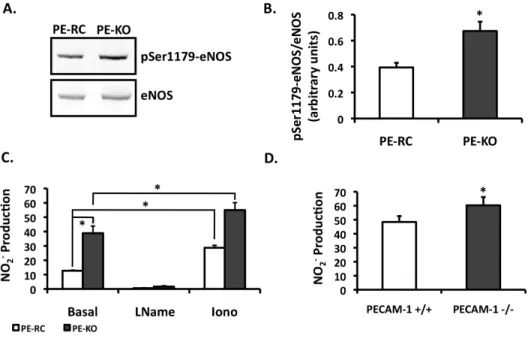
Previous studies suggest a requirement for PECAM-1 in shear-induced eNOS activation 10, 25, 26. Our own results are in agreement with these observations (data not shown). Unexpectedly, our data revealed increased levels of phosphorylated eNOS in PECAM-1 knockout (PE-KO) cells compared to PECAM-1 expressing cells (PE-RC) (Figure 2.1A). PECAM-1 deletion, however, does not affect total eNOS expression 25. To evaluate the functional consequences of increased basal eNOS phosphorylation in the PE-KO cells, we measured NO production. As shown in Figure 2.1B, there is a 3-fold increase in basal NO production in PE-KO cells compared to PE-RC cells. Stimulation with ionomycin increased NO production in both cell types, whereas L-NAME inhibited the production of NO. To determine if these observations are corroborated in whole animals, we measured NO levels from plasma. Indeed, plasma NO levels were higher in PECAM-1-/- compared to
PECAM-1+/+ animals (Figure 2.1C).
To further investigate the role of PECAM-1 in regulation of basal eNOS
phosphorylation, we used siRNA to knockdown PECAM-1 expression in human umbilical vein endothelial cells (HUVECs). The degree of knockdown was assessed by Western blot (Supplemental Figure 2.1A). Interestingly, we observed an increase in both basal eNOS phosphorylation (Supplemental Figure 2.1B) and NO production (Supplemental Figure 2.1C) in PECAM-1-siRNA infected ECs compared to control siRNA cells. Together, these data suggest that absence of PECAM-1 results in higher basal eNOS activity and NO production.
Differential association of eNOS and caveolin-1 in the absence of PECAM-1
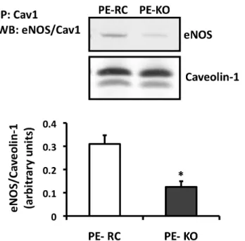
between eNOS and known regulatory proteins we performed co-immunoprecipitation assays. The caveolar scaffolding protein, caveolin-1, has been shown to regulate eNOS activity, primarily as an inhibitor of basal enzyme function 10, 19, 27. Caveolin-1 is able to bind eNOS and block the calmodulin-binding site important for enzyme activation 28. To
determine if PECAM-1 affects the association of eNOS with caveolin-1, we performed co-immunoprecipitation assays. As shown in Figure 2.2, there was a decrease in basal eNOS-caveolin-1 association in the PE-KO cells compared to the PE-RC cells. Additionally, we see a decrease in the colocalization of eNOS and caveolin-1 by confocal microscopy, supporting our immunoprecipitation data (data not shown). These data suggest that PECAM-1 may regulate basal eNOS activity by promoting the association of eNOS with caveolin-1.
Role of PECAM-1 in eNOS localization/trafficking
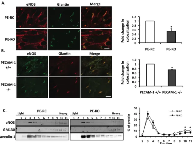
Subcellular localization of eNOS influences its activation 11, 13, 23, 29. To determine if PECAM-1 influences eNOS localization, we performed double-labeling immunofluorescence confocal microscopy in PE-RC and PE-KO cells. Cells were stained for total eNOS and giantin, a membrane-inserted component of the cis- and medial-Golgi complex. In PECAM-1 expressing cells, eNOS is found at both the perinuclear/Golgi complex and the plasma membrane (PM) (Figure 2.3A), consistent with the localization described in blood vessels in
vivo 113. In contrast, in cells that lack PECAM-1, eNOS is redistributed away from the
To further support our in vitro findings, we isolated aortas from PECAM-1+/+ and
-/-mice expressing an eNOS-GFP fusion protein. The aortas were stained en face for giantin and visualized using confocal microscopy (Figure 2.3B). Similar to the PE-KO cells,
quantitative colocalization analysis revealed a significant redistribution of eNOS away from the Golgi complex in the PECAM-1-/- aortas.
As a complementary approach, we utilized sodium carbonate extraction of cells followed by a discontinuous sucrose gradient. In this procedure, cholesterol-rich
microdomains, including lipid rafts and caveolae, float as buoyant membranes at the 5–30% sucrose interface (fractions 3-4), whereas soluble proteins and heavy membranes remain at the bottom of the gradient (fractions 9–11). In all gradients, the distribution of caveolin-1 and GM130, a Golgi marker, were examined to confirm adequate separation of the fractions (Figure 2.3C). In PECAM-1-expressing ECs, eNOS distributed primarily into two distinct pools: light membranes highly enriched in caveolin-1 and heavy membranes enriched in GM130. In PE-KO cells, eNOS is also distributed into two pools. However, we noticed a significant redistribution of eNOS out of the Golgi-enriched fractions (fractions 10-11). These observations are consistent with the immunofluorescence data described above. Notably, PE-KO cells have increased eNOS activity and NO levels despite increased expression levels of the negative regulator caveolin-1 (not shown).
PECAM-1 mediates STAT3-dependent NOSTRIN expression in ECs
distinguish between these possible mechanisms, we first measured ubiquitination of NOSTRIN but did not observe a difference between the PE-RC and PE-KO cells (data not shown). Next, we used quantitative real-time PCR to determine the effect of PECAM-1 deletion on NOSTRIN mRNA levels. Our data demonstrate that NOSTRIN mRNA is significantly decreased in the absence of PECAM-1 (Figure 2.4B).
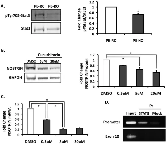
We next addressed the mechanism by which PECAM-1 affects NOSTRIN protein expression. It has previously been shown that the cytoplasmic tail of PECAM-1 is able to function as a scaffold for numerous signaling pathways, including signal transducers and activators of transcription (STAT) protein family members STAT5 and STAT3 30, 31. Additionally, ECs isolated from PECAM-1-/- mice have reduced STAT3 phosphorylation 31.
We asked whether the reduced levels of active STAT3 in the PECAM-1 knockout might result in decreased NOSTRIN expression. To determine if STAT3 phosphorylation is reduced in cells that lack PECAM-1, we probed PE-KO cell extracts for total STAT3 and phospho-STAT3. Importantly, our data are in agreement with previous reports showing that phospho-STAT3 levels are lower in the absence of PECAM-1 (Figure 2.5A). To determine if STAT3 activity affects NOSTRIN expression, we treated the PECAM-1 expressing ECs with cucurbitacin, a selective inhibitor of STAT3/JAK signaling, and assayed for changes in NOSTRIN expression by Western blot and quantitative real-time PCR. Cucurbitacin treatment results in a dose-dependent decrease in both NOSTRIN protein expression (Figure 2.5B) and mRNA levels (Figure 2.5C). These results suggest that PECAM-1
mediates STAT3-induced NOSTRIN expression, which, in turn, regulates eNOS localization.
Binding of STAT3 to the NOSTRIN promoter in endothelial cells
AA duplicates typically separated by 5 bases. In addition to binding TT(N5)AA nanomers, STAT3 is also able to bind octomers and decamers 32, 33. We next investigated the possibility that STAT3 directly binds to the NOSTRIN promoter to modulate mRNA expression by chromatin immunoprecipitation (ChIP) assays with STAT3 antibodies.
Immunoprecipitation of the chromatin lysates was followed by PCR with NOSTRIN promoter primers; NOSTRIN exon 10 primers served as a control. ChIP assays show that STAT3 binds to the NOSTRIN promoter region, but not the NOSTRIN coding region (Figure 2.5D). Together, these data suggest that STAT3 may regulate NOSTRIN mRNA levels by
specifically binding to the NOSTRIN promoter.
Discussion
colocalization between PECAM-1 and caveolin-1 and that these two proteins do not co-migrate on sucrose gels 35. The lack of physical association between eNOS and PECAM-1 led us to investigate differences in eNOS protein interactions and localization as possible mechanisms of regulation.
It is well recognized that correct subcellular targeting of eNOS is critical for proper regulation of its activity and NO bioavailability; thus, tight controlof eNOS targeting to different compartments appears to be essential. In this regard, our data point towards a requirement for tightly regulated levels of NOSTRIN expression within ECs. Overexpression of NOSTRIN can promote the translocation of eNOS from the plasma membrane to
intracellular vesicles, with a concomitant reduction in eNOS enzyme activity 12. Conversely, decreased NOSTRIN expression also influences eNOS subcellular localization and may contribute to the increased NO levels observed in the PECAM-1 knockout. Interestingly, overexpression of the eNOS-binding partner, caveolin-1, leads to accelerated
atherosclerosis formation in mice, partially through reduced NO production 36; while persistent eNOS activation secondary to caveolin-1 deficiency induces pulmonary
hypertension 37. Thus, tight regulation of eNOS regulatory protein levels, including NOSTRIN and caveolin-1 expression, is required for proper eNOS function.
association, however this could be due to other undetermined mechanisms. Of note, it has been reported that eNOS, caveolin-1 and NOSTRIN form a ternary complex to facilitate eNOS translocation 16. Additionally, our data are consistent with the hypothesis that NOSTRIN might serve to stabilize the inhibitory effect of caveolin-1 on eNOS 16.
The signaling pathway identified here relates to the regulation of basal eNOS activity via PECAM-1. However, PECAM-1 is also known to regulate eNOS activation in response to the physiologic stimulus of shear stress 5, 6, 25, 40. PE-KO cells are unable to activate eNOS in response to shear stress, yet they activate eNOS in response to ionomycin, indicating a specific requirement for PECAM-1 in flow-induced eNOS activation (Figure 2.1B and Supplemental Figure 2.2). It is worth noting here that PECAM-1 is also required for flow-induced activation of Akt and Src, two important upstream mediators of eNOS activity 25, 40. The role of shear stress in NOSTRIN-mediated eNOS regulation is currently under
investigation.
Blood flow and the NO signaling pathway are both known modulators of
Figures
Figure 2.1. Basal eNOS phosphorylation and NO production in the PECAM-1 KO.
Figure 2.2. Differential association of the negative eNOS-binding partner, caveolin-1, in the absence of PECAM-1.
PE-RC and PE-KO cells were immunoprecipitated using an antibody against Cav1. Western blots were performed for total eNOS and Cav1. Quantitation is shown on the right (n=6; *P<0.001 vs PE-RC).
Figure 2.3. PECAM-1 regulates eNOS subcellular localization.
Figure 2.4. Reduced NOSTRIN expression in ECs lacking PECAM-1.
Figure 2.5. PECAM-1 regulates NOSTRIN in a STAT3-dependent manner.
Figure 2.6. Model of PECAM-1-mediated NOSTRIN expression and eNOS trafficking.
The cytoplasmic tail of PECAM-1 acts as a scaffold for STAT3 binding. Following activation, STAT3 translocates to the nucleus where it regulates NOSTRIN mRNA expression.
Supplemental Material Cellular Fractionation
Cells were washed twice with cold PBS then scraped into 2 ml of PBS and centrifuged at 1000 rpm for 3 minutes to pellet cells. The PBS was removed and the cells were
resuspended in 750 µl of 500mmol/L sodium carbonate, pH 11. The cells were dounce-homogenized (25 strokes) on ice and sonicated at 50% power with 5 10-second bursts. The homogenate was adjusted to 42.5% sucrose using a 58% sucrose solution prepared in MBS (25mmol/L MES, pH 6.5, 0.15mol/L NaCl). 500 µl of homogenate was placed at the bottom of an ultracentrifuge tube then 1 ml of 30% sucrose was added followed by 600ul of 5% sucrose in MBS. The samples were centrifuged at 48,000 rpm for 3 hours at 4°C in using the SW50 Rotor (Beckman). Aliquots of 200ul were taken starting from the top (for a total of 12 samples) and added to 50ul 10X LSB. The samples (25ul) were run on 4-12% SDS-PAGE gels then transferred to nitrocellulose membrane for Western blot analysis. Membranes were probed with total eNOS, GM130 as Golgi marker and caveolin-1 as the plasma membrane marker.
En Face Staining
RNA Isolation and Quantitative PCR
Total RNA was isolated from a confluent cell monolayer using the TRIzol reagent
(Invitrogen) and DNAse treatment was performed using DNA-free (Ambion). First-strand cDNA was transcribed using random primers and SuperScript II Reverse Transcriptase (Invitrogen). Real-time quantitative PCR was performed using ABsolute SYBR Green ROX mix (Thermo Scientific). Relative levels of gene expression were normalized to mouse 18s expression using the comparative Ct method. Primer sequences are listed in supplemental Table 1.
Chromatin Immunoprecipitation
Supplemental Figures
Supplementary Figure 2.1. PECAM-1 knockdown in HUVECs.
Supplementary Figure 2.2. PECAM-1 is not required for ionomycin-induced eNOS activation.
Supplemental Table 2.1. PCR and ChIP Primers Primers (Mus musculus) Sequence
18s Fwd 5’-CATTCGAACGTCTGCCCTATC-3’
18s Rev 5’-CCTGCTGCCTTCCTTGGA-3’
NOSTRIN Exon 10 Fwd 5’-CCAGGCTCTAATGGAAGAAGAACTG-3’
NOSTRIN Exon 10 Rev 5’-AGACTTCCGTCGCTCTTTGTCC-3’
NOSTRIN Promoter Fwd 5’-GCAGAAGGAAAGGGTTTATTGTGGCCT-3’
Supplemental Table 2.2. Predicted STAT3 binding sites within the NOSTRIN promoter
From JASPAR database 308-317 TGACAGGA 768-777 TTCCAGGCAT 906-915 TTTCAGGAAA From Genomatix
REFERENCES
1. Dudzinski DM, Michel T. Life history of eNOS: partners and pathways. Cardiovasc Res. 2007; 75: 247-260.
2. Huang PL, Huang Z, Mashimo H, Bloch KD, Moskowitz MA, Bevan JA, Fishman MC. Hypertension in mice lacking the gene for endothelial nitric oxide synthase. Nature. 1995; 377: 239-242.
3. Shesely EG, Maeda N, Kim HS, Desai KM, Krege JH, Laubach VE, Sherman PA, Sessa WC, Smithies O. Elevated blood pressures in mice lacking endothelial nitric oxide synthase. Proc Natl Acad Sci U S A. 1996; 93: 13176-13181.
4. Kuhlencordt PJ, Gyurko R, Han F, Scherrer-Crosbie M, Aretz TH, Hajjar R, Picard MH, Huang PL. Accelerated atherosclerosis, aortic aneurysm formation, and ischemic heart disease in apolipoprotein E/endothelial nitric oxide synthase double-knockout mice. Circulation. 2001; 104: 448-454.
5. Dusserre N, L'Heureux N, Bell KS, Stevens HY, Yeh J, Otte LA, Loufrani L, Frangos JA. PECAM-1 interacts with nitric oxide synthase in human endothelial cells: implication for flow-induced nitric oxide synthase activation. Arterioscler Thromb Vasc Biol. 2004; 24: 1796-1802.
6. Fleming I, Fisslthaler B, Dixit M, Busse R. Role of PECAM-1 in the shear-stress-induced activation of Akt and the endothelial nitric oxide synthase (eNOS) in endothelial cells. J Cell Sci. 2005; 118: 4103-4111.
7. Chatzizisis YS, Coskun AU, Jonas M, Edelman ER, Feldman CL, Stone PH. Role of endothelial shear stress in the natural history of coronary atherosclerosis and vascular remodeling: molecular, cellular, and vascular behavior. J Am Coll Cardiol. 2007; 49: 2379-2393.
8. Sessa WC. eNOS at a glance. J Cell Sci. 2004; 117: 2427-2429.
9. Fleming I. Molecular mechanisms underlying the activation of eNOS. Pflugers Arch. 2009; .
10. Garcia-Cardena G, Oh P, Liu J, Schnitzer JE, Sessa WC. Targeting of nitric oxide synthase to endothelial cell caveolae via palmitoylation: implications for nitric oxide signaling. Proc Natl Acad Sci U S A. 1996; 93: 6448-6453.
11. Liu J, Hughes TE, Sessa WC. The first 35 amino acids and fatty acylation sites
determine the molecular targeting of endothelial nitric oxide synthase into the Golgi region of cells: a green fluorescent protein study. J Cell Biol. 1997; 137: 1525-1535.
13. Fulton D, Babbitt R, Zoellner S, Fontana J, Acevedo L, McCabe TJ, Iwakiri Y, Sessa WC. Targeting of endothelial nitric-oxide synthase to the cytoplasmic face of the Golgi complex or plasma membrane regulates Akt- versus calcium-dependent mechanisms for nitric oxide release. J Biol Chem. 2004; 279: 30349-30357.
14. Dedio J, Konig P, Wohlfart P, Schroeder C, Kummer W, Muller-Esterl W. NOSIP, a novel modulator of endothelial nitric oxide synthase activity. FASEB J. 2001; 15: 79-89.
15. Icking A, Matt S, Opitz N, Wiesenthal A, Muller-Esterl W, Schilling K. NOSTRIN functions as a homotrimeric adaptor protein facilitating internalization of eNOS. J Cell Sci. 2005; 118: 5059-5069.
16. Schilling K, Opitz N, Wiesenthal A, Oess S, Tikkanen R, Muller-Esterl W, Icking A. Translocation of endothelial nitric-oxide synthase involves a ternary complex with caveolin-1 and NOSTRIN. Mol Biol Cell. 2006; 17: 3870-3880.
17. Graesser D, Solowiej A, Bruckner M, Osterweil E, Juedes A, Davis S, Ruddle NH, Engelhardt B, Madri JA. Altered vascular permeability and early onset of experimental autoimmune encephalomyelitis in PECAM-1-deficient mice. J Clin Invest. 2002; 109: 383-392.
18. Liu Y, Sweet DT, Irani-Tehrani M, Maeda N, Tzima E. Shc coordinates signals from intercellular junctions and integrins to regulate flow-induced inflammation. J Cell Biol. 2008; 182: 185-196.
19. Feron O, Michel JB, Sase K, Michel T. Dynamic regulation of endothelial nitric oxide synthase: complementary roles of dual acylation and caveolin interactions. Biochemistry. 1998; 37: 193-200.
20. Costes SV, Daelemans D, Cho EH, Dobbin Z, Pavlakis G, Lockett S. Automatic and quantitative measurement of protein-protein colocalization in live cells. Biophys J. 2004; 86: 3993-4003.
21. Chen Z, Peng IC, Sun W, Su MI, Hsu PH, Fu Y, Zhu Y, Defea K, Pan S, Tsai MD, Shyy JY. AMP-Activated Protein Kinase Functionally Phosphorylates Endothelial Nitric Oxide Synthase Ser633. Circ Res. 2009; .
22. Fulton D, Fontana J, Sowa G, Gratton JP, Lin M, Li KX, Michell B, Kemp BE, Rodman D, Sessa WC. Localization of endothelial nitric-oxide synthase phosphorylated on serine 1179 and nitric oxide in Golgi and plasma membrane defines the existence of two pools of active enzyme. J Biol Chem. 2002; 277: 4277-4284.
23. Zhang Q, Church JE, Jagnandan D, Catravas JD, Sessa WC, Fulton D. Functional relevance of Golgi- and plasma membrane-localized endothelial NO synthase in reconstituted endothelial cells. Arterioscler Thromb Vasc Biol. 2006; 26: 1015-1021. 24. Nelson JD, Denisenko O, Bomsztyk K. Protocol for the fast chromatin
25. Liu Y, Bubolz AH, Shi Y, Newman PJ, Newman DK, Gutterman DD. Peroxynitrite reduces the endothelium-derived hyperpolarizing factor component of coronary flow-mediated dilation in PECAM-1-knockout mice. Am J Physiol Regul Integr Comp Physiol. 2006; 290: R57-65.
26. Corson MA, James NL, Latta SE, Nerem RM, Berk BC, Harrison DG. Phosphorylation of endothelial nitric oxide synthase in response to fluid shear stress. Circ Res. 1996; 79: 984-991.
27. Garcia-Cardena G, Fan R, Stern DF, Liu J, Sessa WC. Endothelial nitric oxide synthase is regulated by tyrosine phosphorylation and interacts with caveolin-1. J Biol Chem. 1996; 271: 27237-27240.
28. Michel JB, Feron O, Sacks D, Michel T. Reciprocal regulation of endothelial nitric-oxide synthase by Ca2+-calmodulin and caveolin. J Biol Chem. 1997; 272: 15583-15586.
29. Sessa WC, Garcia-Cardena G, Liu J, Keh A, Pollock JS, Bradley J, Thiru S, Braverman IM, Desai KM. The Golgi association of endothelial nitric oxide synthase is necessary for the efficient synthesis of nitric oxide. J Biol Chem. 1995; 270: 17641-17644.
30. Ilan N, Cheung L, Miller S, Mohsenin A, Tucker A, Madri JA. Pecam-1 is a modulator of stat family member phosphorylation and localization: lessons from a transgenic mouse. Dev Biol. 2001; 232: 219-232.
31. Carrithers M, Tandon S, Canosa S, Michaud M, Graesser D, Madri JA. Enhanced susceptibility to endotoxic shock and impaired STAT3 signaling in CD31-deficient mice. Am J Pathol. 2005; 166: 185-196.
32. Aaronson DS, Horvath CM. A road map for those who don't know JAK-STAT. Science. 2002; 296: 1653-1655.
33. Zhang Q, Wang HY, Marzec M, Raghunath PN, Nagasawa T, Wasik MA. STAT3- and DNA methyltransferase 1-mediated epigenetic silencing of SHP-1 tyrosine phosphatase tumor suppressor gene in malignant T lymphocytes. Proc Natl Acad Sci U S A. 2005; 102: 6948-6953.
34. Shaul PW, Smart EJ, Robinson LJ, German Z, Yuhanna IS, Ying Y, Anderson RG, Michel T. Acylation targets emdothelial nitric-oxide synthase to plasmalemmal caveolae. J Biol Chem. 1996; 271: 6518-6522.
35. Mamdouh Z, Chen X, Pierini LM, Maxfield FR, Muller WA. Targeted recycling of PECAM from endothelial surface-connected compartments during diapedesis. Nature. 2003; 421: 748-753.
37. Zhao YY, Zhao YD, Mirza MK, Huang JH, Potula HH, Vogel SM, Brovkovych V, Yuan JX, Wharton J, Malik AB. Persistent eNOS activation secondary to caveolin-1 deficiency induces pulmonary hypertension in mice and humans through PKG nitration. J Clin Invest. 2009; 119: 2009-2018.
38. Cao X, Tay A, Guy GR, Tan YH. Activation and association of Stat3 with Src in v-Src-transformed cell lines. Mol Cell Biol. 1996; 16: 1595-1603.
39. Fernandez-Hernando C, Yu J, Davalos A, Prendergast J, Sessa WC. Endothelial-specific overexpression of caveolin-1 accelerates atherosclerosis in apolipoprotein E-deficient mice. Am J Pathol. 2010; 177: 998-1003.
40. Bagi Z, Frangos JA, Yeh JC, White CR, Kaley G, Koller A. PECAM-1 mediates NO-dependent dilation of arterioles to high temporal gradients of shear stress. Arterioscler Thromb Vasc Biol. 2005; 25: 1590-1595.
41. Adamo L, Naveiras O, Wenzel PL, McKinney-Freeman S, Mack PJ, Gracia-Sancho J, Suchy-Dicey A, Yoshimoto M, Lensch MW, Yoder MC, Garcia-Cardena G, Daley GQ. Biomechanical forces promote embryonic haematopoiesis. Nature. 2009; 459: 1131-1135. 42. North TE, Goessling W, Peeters M, Li P, Ceol C, Lord AM, Weber GJ, Harris J, Cutting CC, Huang P, Dzierzak E, Zon LI. Hematopoietic stem cell development is dependent on blood flow. Cell. 2009; 137: 736-748.
43. Pardanaud L, Eichmann A. Stem cells: The stress of forming blood cells. Nature. 2009; 459: 1068-1069.
44. Osawa M, Masuda M, Kusano K, Fujiwara K. Evidence for a role of platelet endothelial cell adhesion molecule-1 in endothelial cell mechanosignal transduction: is it a
mechanoresponsive molecule? J Cell Biol. 2002; 158: 773-785.
45. Chen Z, Tzima E. PECAM-1 is necessary for flow-induced vascular remodeling. Arterioscler Thromb Vasc Biol. 2009; 29: 1067-1073.
46. Lin Z, Hamik A, Jain R, Kumar A, Jain MK. Kruppel-like factor 2 inhibits protease activated receptor-1 expression and thrombin-mediated endothelial activation. Arterioscler Thromb Vasc Biol. 2006; 26: 1185-1189.