Evaluation of Potential Inhibitors of Escherichia coli RecA to Attenuate the Rate of Antibiotic Resistance Development and to
Sensitize Escherichia coli to Current Antibiotics by
KERI A.FLANAGAN
A thesis submitted to the faculty of the University of North Carolina at Chapel Hill in partial fulfillment of the requirements for the degree of
Master of Science in the UNC Eshelman School of Pharmacy
CHAPEL HILL 2009
APPROVED BY:
Scott F. Singleton, Ph.D.
Jian Liu, Ph.D.
Qisheng Zhang, Ph.D.
ABSTRACT
Keri A. Flanagan
Evaluation of Potential Inhibitors of Escherichia coli RecA to Attenuate the Rate of Antibiotic Resistance Development and to
Sensitize Escherichia coli to Current Antibiotics Under the direction of Scott F. Singleton, Ph.D.
Antibacterials alike have proven to be an invaluable breakthrough in the treatment of
infectious diseases. Upon their introduction, countless lives were saved. However, bacteria have a profound ability to alter their susceptibility to antibiotics, rendering themselves
resistant to one or more of the current antibiotics available[4,7,26,27]. As in the beginning of the twentieth century, thousands of people are again dying from infectious diseases that were once kept at bay due to the rise in antibiotic resistant bacterial strains. Bacteria become
resistant to newly introduced antibiotics very quickly[5]. Therefore, a new approach to treating bacterial infections is needed and may be accomplished by attenuating bacterial
resistance mechanisms and sensitizing them to current antibiotics. RecA, a recombinase enzyme that is involved in various aspects of DNA repair, horizontal gene transfer and the induction of SOS mutagenesis[19-25], seems to be a promising target whose inhibition would preclude the RecA-dependent activities used by bacteria to reduce their susceptibility to antibiotics. Previous work completed by the Singleton research group has identified various
purified E. coli RecA and demonstrated that such inhibitors can attenuate SOS in live E. coli and potentiate E. coli killing by bactericidal agents. In order to enhance the data validating
RecA as a pharmaceutical target, it was of interest to assess whether such RecA inhibitors could also serve to block its function in horizontal gene transfer[27,27,37]. In this study, we evaluated whether cell-permeable RecA inhibitors could prevent the transfer of genetic material from heat-killed antibiotic-resistant E. coli to live, susceptible E. coli. It was demonstrated that one inhibitor identified from a previous screen (A1) attenuated the rate at
which E. coli developed resistance to chloramphenicol in both the presence and absence of heat-killed chloramphenicol-resistant cells. However, the results also suggest that E. coli may
have a RecA-indepedent pathway for developing resistance. Regardless of this possibility, the hit compound may reveal yet another unique means of impeding the spread of antibiotic resistance genes that could be unraveled in future studies, but does not discredit RecA from
being a novel and promising target for battling antibiotic resistance. Although the latter results may obfuscate the critical path for developing RecA inhibitors as pharmaceutical
agents, it should be emphasized that the results also demonstrate the power of biologically active small molecules to help elucidate complex biological processes. To initiate the process of discovering next-generation RecA inhibitors suited to the task of teasing apart
ACKNOWLEDGMENTS
Despite the fact that I have chosen to leave this amazing and opportunistic program
and school, I am grateful for having the opportunity to study at the University of North Carolina under the direction of many talented and respectable faculty members who have
shared their abundance of knowledge and experience with me. They have always been supportive and wish to see all of their students succeed. I have a strong foundation to enter the work force and am confident they have provided the necessary tools for me to thrive in
life, no matter what endeavors upon which I embark.
Firstly, I would like to thank Dr. Scott Singleton. He has always been extremely kind
to me and concerned about my wellbeing. I will never forget when I was about to come to North Carolina all the way from New Jersey how he personally stopped by an apartment complex I was considering to move to just to ensure its safety and integrity. He accepted me
as a summer rotation student so I could even start the program early. When I needed extra help in my coursework, his door was always open. For him, there were never too many
questions to ask. Lastly, he always trusted and believed in my abilities, even when I did not. I cannot express how much this meant to me.
I need to extend a special thanks to Dr. Tim Wigle, who was my mentor when I first
arrived in the Singleton laboratory. Most of what I learned in my experiences in lab was from Tim. I always appreciated his brotherly concern for me and his need to challenge me to
To the rest of the Singleton lab members, Dr. Anna Gromova, Dr. Daniel Cline, Dr. Malinath Hadamani and Demet Guntas, their support has been invaluable to me. I am glad to
have worked with Anna to learn some chemical synthesis. I am sure I will use it in my future career endeavors at some point. Dan was always there to patiently answer any questions I
had, no matter how rudimentary they may have seemed to him. He is the most patient person I have ever met. Mali was the first person to whom I have ever taught lab techniques, so I was glad to be able to share my knowledge with him. Lastly, Demet was always my pool of
emotional support inside and outside of lab. Not only did she teach me many techniques in the lab, she was always there to give me encouragement and a hug when I was feeling
discouraged. She was more than a co-worker, she was a wonderful friend and I will miss her dearly when I leave. I will miss them all when I leave.
My dissertation committee, Dr. Jian Liu, Dr. Qisheng Zhang, and Dr. Alexander
Tropsha, have all been kind and generous in their advice and desire to aid me in my studies. All I wish is that I utilized their knowledge base more because I know they would have many
more insights into my research to share if I had just asked. Also, Dr. M. Karthikeyan, whom I collaborated with in the Dr. Tropsha laboratory, has been instrumental in furthering the research I wished to accomplish. He ran the molecular simulations and explained all of their
complexities to me. I greatly valued and appreciated his efforts.
Briefly, to Renita Patel and Heidi Clarke, a pharmacy student and undergraduate
To my friends: I have always valued their love, support, advice and companionship. As my family always has (and I thank them for the same reasons), they have always been
there for me and wanted the best for me. They have pushed me through some rough spots and I cannot thank them enough. To Tom Riley especially, who is one of my few friends in the
same field, I am glad to have his knowledge just a phone call away.
And, to everyone whom with I practice kung fu, whether in Hungary or the United States, I thank them for teaching me discipline, self respect, ultimate and divine love, beauty,
patience, strength and for opening my eyes to see what I truly want from life. They have supported and positively changed me in more ways than they will ever know. Hinár Polczer:
he has been more than an instructor, but a most wonderful friend. Encouraging me to go to kung fu camp was the best act he could have ever done for me. It was truly the most wonderful experience of my life and I thank him for that opportunity. And to my master,
Marin Vélin, who is the most spiritual and kind-hearted man I have ever met. I am greatly obliged to be his student and to have had the pleasure of meeting him.
To Mert Sedef, my significant other, who has been an endless supply of emotional support and love, I thank him for being there for me to give me a hug when I needed it, to grocery shop when I lacked the time, and for being my cheerleader on the sideline always
rooting for me.
Last, but not least, to my family: I know deep down they are disappointed that I was
not the first in the family to get a PhD. But I also know they are proud of me nonetheless and will always be proud. They have always wanted me to be happy, no matter how that happiness manifests itself. Thank you for always loving me and caring for me throughout all
TABLE OF CONTENTS
PAGE
LIST OF TABLES ... xi
LIST OF FIGURES ... xii
ABBREVIATIONS ...xv
CHAPTER I INTRODUCTION ...1
Implications of Bacterial Drug Resistance ...1
Targets and Mechanisms of Antibiotic Resistance ...6
Mechanisms of Chloramphenicol Resistance ...9
The Structure and Functions of the RecA Protein ...13
The Involvement of RecA in Recombination ...15
The Role of RecA in the SOS Response to DNA Damage ...18
Responsibility of RecA in the Development of Resistance to Antibiotic Treatment ...21
The Griffith Experiment: Potential Role of RecA in the Incorporation of Exogenous DNA ...25
Observation of Natural Competence in Escherichia coli ...28
Previous Efforts to Develop RecA Inhibitors ...32
Specific Aims of This Project ...36
CHAPTER II MATERIALS AND METHODS ...39
CHAPTER III THE TRANSFER OF CHLORAMPHENICOL RESISTANT GENES MAY BE DETERRED BY THE POTENTIAL INHIBITOR COMPOUND A1 ...48
Bacterial Culture Studies of the Effects of Chloramphenicol
on ∆recA E. coli MG1655 cells ...54
Bacterial Culture Studies of the Effects of A1
on Wild Type E. coli MG1655 cells ...57 Bacterial Culture Studies of the Effects of A1
on ∆recA E. coli MG1655 cells ...59
Conclusions ...60
CHAPTER IV THE APPLICATION OF THE GRIFFITH EXPERIMENT TO
ESCHERICHIA COLI MG1655 CELLS TO OBSERVE THE
POTENTIAL TRANSFER OF ANTIBIOTIC RESISTANT GENES ...64
Bacterial Culture Studies of the Effects of the Addition of Chloramphenicol Resistant Cell Supernatant on
Wild Type E. coli MG1655 cells ...67 Bacterial Culture Studies of the Effects of the Addition of
Chloramphenicol Resistant Cell Supernatant on
∆recA E. coli MG1655 cells ...72
Bacterial Culture Studies of the Effects of the Addition of A1 and Chloramphenicol Resistant Cell Supernatant on
Wild Type E. coli MG1655 cells ...76 Bacterial Culture Studies of the Effects of the Addition of
A1 and Chloramphenicol Resistant Cell Supernatant on
∆recA E. coli MG1655 cells ...80
Conclusions ...83
CHAPTER V UTILIZATION OF MOLECULAR MODELING METHODS OF SCREENING
TO FIND OTHER POTENTIAL INHIBITORS OF RECA ...86 Various Prediction Methods and the Theory
Behind Similarity Searching ...88
Abstraction of Common Scaffolds from Seventy-three
Screening Literature for Similar Compounds
Using SciFinder Scholar ...90
Tanimoto Similarity Coefficients Allow for Clustering in Activity Space ...91
AutoQSAR Analysis Predicts Activity Based on Structure Similarity ...96
Screening for and testing of Commercially Available Compounds ...97
Conclusions ...98
CHAPTER VI CONCLUSIONS ...100
LIST OF TABLES
LIST OF FIGURES
Figure 1.1. Timeline of the introduction of new antibiotics
vs. the reported resistance to the antibiotic ...4
Figure 1.2. Trends in the spread of bacterial resistant strains and approved antibiotics in the United States ...5
Figure 1.3. Targets of antibiotics with representative agents ...7
Figure 1.4. Antibiotic resistance mechanisms ...9
Figure 1.5. Protein Synthesis ...10
Figure 1.6. Signaling cascades responsible for the loss of porins and increased drug efflux ...12
Figure 1.7. Signal-like and motor-like activities of RecA ...14
Figure 1.8. Role of RecA in homologous recombination...16
Figure 1.9. Horizontal gene transfer in bacteria ...17
Figure 1.10. Homologous recombination during horizontal gene transfer ...18
Figure 1.11. Bacterial SOS response ...21
Figure 1.12. Development of chloramphenicol resistance in wild type, super RecA and ∆recA-∆lexA E. coli cells ...24
Figure 1.13 Natural competence and DNA entry into the cell of Gram-positive and gram-negative species ...32
Figure 2.1. General workflow ...45
Figure 3.1. Resistance Development of MG1655 wild type cells in the presence of Cam and A1 ... 53
Figure 3.2. Resistance Development of MG1655 ∆recA cells in the presence of Cam and A1 ...56
Figure 4.2. Resistance Development of MG1655 ∆recA cells
in the presence of Cam and heat-killed Camr cells ... 75 Figure 4.3. Resistance Development of MG1655 wild type cells in the
presence of Cam, heat-killed Camr cells, and A1 ...79 Figure 4.4. Resistance Development of MG1655 ∆recA cells in the
presence of Cam, heat-killed Camr cells, and A1 ... 82 Figure 5.1. Prediction Methods ...89 Figure 5.2. Clustering the molecules in activity space utilizing
LIST OF ABBREVIATIONS
Amp Ampicillin
ATPγS Adenosine 5′-[γ-thio]triphosphate
Cam Chloramphenicol
Camr Chloramphencial resistance/Chloramphenicol resistant cell supernatant CFU Colony-forming units
Cipro Ciprofloxacin
cssDNA circular single-stranded DNA
DHF dihydrofolate
DSB Double-strand break dsDNA double-stranded DNA DTT Dithiothreitol
Em Emission
Ex Excitation
HGT Horizontal Gene Transfer HTS High-throughput screening IRA Inhibitor of RecA
Kan Kanamycin
LB Luria Bertani
MMC Mitomycin C
MOE Molecular Operating Environment
NAD+ β-Nicotinamide adenine dinucleotide, oxidized NADH β-Nicotinamide adenine dinucleotide, reduced
NCI National Cancer Insititute NER Nucleotide excision repair
NMP Nucleotide monophosphate NDP Nucleotide diphosphate NTP Nucleotide triphosphate
NXP Nucleotide mono-, di- or triphosphate NPF Nucleoprotein filament
nts nucleotides (in reference to DNA oligomer concentration)
OD Optical density
ONPG 2-Nitrophenyl-β-D-galactopyranoside
Pi Inorganic phosphate
P-loop Phosphate binding loop
PMB Phosphomolybdate blue
PNP Purine nucleoside phosphorylase PolIV DNA polymerase IV
PolV DNA polymerase V
QSAR Quantitative structure-activity relationship
RDR Recombinational DNA repair
SMILES Simplified Molecular Input Line Entry Specification ssDNA Single-stranded DNA
TCA cycle Tricarboxylic acid cycle THF Tetrahydrofolate
tsDNA Triple-stranded DNA
UV Ultraviolet
VRE Vancomycin-resistant enterococci
CHAPTER I INTRODUCTION
Implications of Bacterial Drug Resistance
Limited organisms in existence have the ability to thrive in every imaginable habit this earth has to offer. Bacteria, being unicellular
microorganisms, are able to survive in water, soil, radioactive waste, acidic hot springs, even in the very living bodies of plants and animals. Millions of bacteria
are present on the skin and within the human body at any given time. Bacteria are perceived as disease-causing, undesirable living organisms, despite the fact that their existence is vital to the cycles of the earth due to their capability to recycle
nutrients. They are even beneficial to humans in that they aid in the health of the digestive tract by performing fermentation of unused energy substrates,
controlling the growth of harmful bacterial species and producing valuable vitamins for their hosts. Most bacteria living in the human body are regulated by the immune system, but pathogenic strains do arise and cause infections diseases,
thus bestowing bacteria with their ominous reputation.
However, before the introduction of antimicrobials, communicable
which included pneumonia (viral and bacterial), tuberculosis and diarrhea. Maintaining personal hygiene, reliance on the immune system and the use of
crude plant extracts were not enough to contend with the microorganisms that caused these diseases. Attempts to discover agents that could have antibacterial
effects were unsuccessfully made in the early 1900’s. Sulfa drugs were introduced in the early 1930’s as the first antimicrobial drugs, but due to adverse effects and toxicity issues, other antimicrobial agents were desired. Sulfa drugs paved the
way for revolutionary agents like penicillin. Also in the 1930’s, Sir Alexander Fleming accidentally stumbled upon a petri dish growing Staphyloccocus
contaminated with mold[1]. However, this plate showed lack of growth of the bacteria near the mold, thus indicating that it released a natural product that was lethal to the bacteria. Upon analysis of the mold, it was determined to be
Penicillium notatum, and thus, the natural product being released from the mold
was coined penicillin. Through collaborative efforts with Howard Florey and
Ernst Chain, penicillin was shown to have bactericidal effects in vivo and proved effective in treating bacterial infections in mice[2]. In the 1940’s, penicillin was finally able to be mass-produced, which enabled it to be extensively used to treat
countless soldiers in World War II, allowing them to treat otherwise fatal and devastating wounds. The Nobel Prize was awarded to Fleming, Florey and Chain
in 1945 in recognition of their discoveries. They paved the road for the introduction of other antibiotics which would lead to a great improvement of the quality of life by combating the infectious diseases that claimed the lives of so
The introduction of antibiotics, which were viewed as “miracle drugs”, was considered to be one of the most imperative medical advancements of the
twentieth century, giving a false sense of hope that humans had triumphed in the war against infectious diseases. Fleming himself foresaw and warned against the
dangers of resistance, stating that underdosage of an antibiotic easily caused microbes to develop resistance in the laboratory and in humans by not exposing them to a lethal dosage of the drug, thus allowing microbes to “educate”
themselves on how to resist antibiotics[3]. Many battles had been victorious with the introduction of new and more effective agents, thus keeping bacterial
resistance from becoming a major medical problem for decades, but resistance to antibacterials escalates today at an alarming rate.
According to the Center for Disease Control and Prevention, two million
people acquire bacterial infections in hospitals each year[4]. Ninety thousand of those people die from these infections due to approximately seventy percent of
those infections being caused by a strain of bacteria that are resistant to one or more of the one-hundred or so available antibiotics[4]. These statistics account only for hospitalized patients. Millions more acquire infections outside of the
A positive trend has been observed when reporting the amount of resistant strains infecting people in the United States. Starting from about 1980, the percent
incidence of resistant strains was very low, less than 5% (Figure 1.2, left panel). However, over the course of twenty years, the percent incidence of resistant strains has drastically increased to about 60% incidence for MRSA and 25-30%
incidence for VRE and FQRP. The need for novel antibacterials is quite apparent, however, an inverse correlation is seen in the number of antibacterials approved
in the United States over the same time span. A total of three new antibiotics were approved for administration between 2003-2007 (Figure 1.2, right panel). How can this be when the need for such treatments is so great? Many large
pharmaceutical companies, including Glaxo Smith-Kline, Wyeth, Aventis and
Figure 1.1. Timeline of the introduction of new antibiotics vs. the reported resistance to the antibiotics. Many antibiotics introduced to the market often acquire resistance quickly
after their introduction. The figure was reproduced from Clatworthy, et al. (2007) Nat. Chem.
Bristol-Myers Squib, have eliminated or downsized their antibacterial research[6]. This is due to the increased cost of drug discovery research, short-comings of
target based drug discovery to unravel novel antibacterials, a shift of interest from short-term acute diseases to long-term chronic diseases and because the rapid
onset of resistance to antibacterial agents gives them a short –lived market time frame. The cost to develop and market the drugs is far greater than the revenue they generate[6].
The moral implications of this biological predicament outweigh the monetary demand. Therefore, the goal of this thesis was to further understand the
implications of the RecA protein in bacterial resistance development and utilize it as a potential novel target in battling resistance onset and therefore battling bacterial infections diseases that are claiming the lives of so many.
Figure 1.2 Trends in the spread of bacterial resistant strains and approved antibiotics in the United States. Left panel: Displays the increase of percent incidence of MRSA, VRE and
FQRP vs. time in years. Right panel: Displays the declining number of approved antibiotics vs time in years. Figures were reproduced from the Infections Disease Society of America, Bad
Targets and Mechanisms of Antibiotic Resistance
Antibiotics are able to function and kill bacteria by inhibiting enzymes and
proteins that are necessary for their survival. Ideal antibiotics have low affinity for host enzymes and proteins and have low toxicity levels within hosts. Four major
categories of targets into which most antibiotics can be classified are inhibitors of cell wall synthesis, nucleic acid synthesis, protein synthesis and agents that impair the cell membrane[7]. Specifically for protein synthesis, antibiotics can bind to the 50S and 30S subunits of ribosomes preventing the translation of RNA to protein. Nucleic acid synthesis can be blocked three ways: antibacterials can inhibit RNA
polymerase, which is needed to initiate transcription; antibacterial agents can act by inhibiting folic acid biosynthesis, which stops the formation of folate that is required to synthesize DNA bases; and lastly can prevent DNA topoisomerases
Since antibiotics function by inhibiting necessary processes within
bacteria, in order to survive, bacteria must find means of bypassing the inhibition capabilities of antibiotics. Therefore, bacteria exhibit three phenotypes in relation to antibiotics: susceptibility, intrinsic resistance and acquired resistance. Any
bacteria species that is susceptible to an antibiotic may develop or acquire resistance to that antibiotic, either through de novo mutations or the inheritance of
DNA encoding resistance either from transformation or mobile genetic elements (including plasmids and transposons)[4]. Intrinsic resistance refers to a bacteria’s natural ability to be resistant to an antibiotic due to the make up of the organism,
meaning the hydrophobic lipopolysaccharide outer membrane of gram-negative bacteria repel hydrophobic antibiotics and large compounds cannot transverse the
Figure 1.3 Targets of antibiotics with representative agents. The various targets of
size-excluding porins in the outer membrane[9]. Basically, no genetic manipulations are needed for intrinsic resistance. On the other hand, acquired
resistance, which is present in only select isolates of certain species, exhibit resistance to antibiotics through four mechanisms, which are alteration of the
antibiotic through enzymatic degradation or modification, acquisition of a drug-insensitive enzyme that functions in place of the drug-sensitive enzyme thus altering the metabolic pathway, decreased accumulation of the antibiotic through
reduced membrane permeability and/or increased efflux and mutation of the target site rendering the antibiotic unable to bind its target[7] (Figure 1.4).
Due to the fact that acquired resistance requires resistant genes from exogenous DNA, either from transformation or mobile genetic elements, or de novo mutations, a mechanism for incorporating these genes or inducing mutations
Mechanisms of Chloramphenicol Resistance
Chloramphenicol was first announced in 1948 by Ehrlich and
coworkers[10]. What made this antibiotic so unique is it was the first natural product to contain a nitro group and to be a derivative of dichloroacetic acid[11]. It is mainly a broad-spectrum bacteriostatic agent with the ability to inhibit all
bacteria tested and organisms that are closely related to bacteria[11]. Chloramphenicol inhibits protein synthesis in concentrations of 10 µM and above,
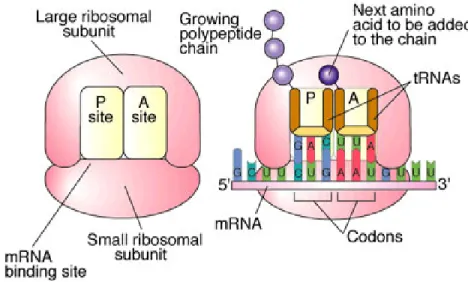
but does not inhibit the activity of proteins[11]. During protein synthesis, aminoacyl tRNA molecules are synthesized in the cytoplasm and are transported
Figure 1.4 Antibiotic resistance mechanisms. The four major mechanisms or resistance
to the ribosome where they bind to associated mRNA at the P site[7]. Another tRNA then binds to the A site that corresponds to the next codon in the mRNA
sequence and a transpeptidation reaction utilizing peptidyl transferase allows for the linkage of the growing peptide chain to the next amino acid to be added to the
chain in the A site[7]. Lastly, the growing chain translocates to the P site, which frees the A site for the next tRNA and this will continue until a stop codon is reached[7] (Figure 1.5). Specifically, chloramphenicol inhibits protein synthesis by binding to the 50S ribosomal subunit of the 70S ribosome and impedes the peptidyl transferase reaction, thus stopping peptide chain formation[7].
Three of the common mechanisms of antibiotic resistance have been
implicated in the development of chloramphenicol resistance: 1) reduced accumulation of chloramphenicol in the cell through reduced membrane permeability and active efflux pumps[9,12-14] 2) altered target[15] and 3) enzymatic degradation of chloramphenicol[7,17]. Chloramphenicol, being a small and
Figure 1.5 Protein synthesis. Image was reproduced from the Biology Active
hydrophilic antibiotic, transverses the outer membrane through porin channels,[9] which span the outer membrane and are water-filled open channels that allow the
passive diffusion of hydrophilic molecules[12]. The three major porins of E. coli are the OmpF, OmpC and PhoE porins and E. coli exhibits modifications of its
porins to become resistant to antibiotics by reducing the amount of OmpF and OmpC porins or mutating OmpC porins[12]. This kind of mutation involves the alteration of the internal loop 3, which in return alters the ability of antibiotics to
transverse the porin channels[12]. Chloramphenicol is believed to pass through the OmpF channel due to its larger size and because of its expression being regulated
by marA, which is de-repressed in response to chemical and antibiotic stress and through downstream signaling, porin synthesis is downregulated while efflux pumps are overexpressed[9,12-13]. Figure 1.6[13] outlines this cascading event. The AcrAB-TolC is a common efflux pump used to export commonly used antibiotics, chloramphenicol included[13,14,15]. The acrA gene encodes a membrane fusion protein, acrB encodes a cytoplasmic membrane efflux pump and tolC encodes for an outer membrane channel[13,14]. MdfA (also known as CmlA and Cmr) is another multi-drug pump that is chromosomally encoded and it provides
resistance to chloramphenicol[13,15].
Also reported, a mutation of guanine to adenine at position 2057 of the
23S RNA gene in the rRNA operon confers resistance to chloramphenicol[16]. This mutation causes an alteration in the sequence in a region of the 23S secondary structure, which is a part of the peptidyl transferase region, and
transferase active site being located near the peptidyl transferase region containing the 23S RNA region[16]. Thus, alterations of the target site of chloramphenicol results in resistance development to chloramphenicol.
Lastly, genes that are responsible for enzymatic degradation of
chloramphenicol are cat genes, which encode for chloramphenicol acetyltransferase[7,18]. This enzyme functions by acetylating the hydroxyl group on
Figure 1.6 Signaling cascades responsible for loss of porins and increased drug efflux.
Activation of marA through de-repression of marR due to antibiotic induced stress activates
micF, which inhibits ompF expression, and activates acrA, acrB and tolC, genes that express
position three on the carbon side chain, utilizing an acetyl group from acetyl-coenzyme A, giving rise to the 3-acetyl derivative; the acetyl group can replace
the hydroxyl group on the carbon on position one on the carbon side chain, thus allowing for a second acetylating reaction to the hydroxyl group on the third
carbon on the side chain[7,17,18]. Some of the cat genes found in E. coli include
catI, catB2, catB3[18], which encode for different types of CATs (types A and B, type A being native to the organism, type B being xenobiotic).
The Functions of the RecA Protein
Genetic preservation and variation are two necessary processes all living
organisms must utilize and balance in order to survive. The genome must be preserved in order for an organism to function properly while it also must be varied in order for organisms to adapt to changing environments. RecA is a key
player in maintaining the balance in bacteria: all bacteria contain RecA and its function is implicated in processes that lead to either genomic preservation or
variation[19, 20]. Metabolic and physiological stresses caused by antibiotic treatment, heat shock, starvation, exposure to UV or harmful chemicals and pressure changes either directly or indirectly lead to DNA damage, which
effectively activates RecA[19,21].
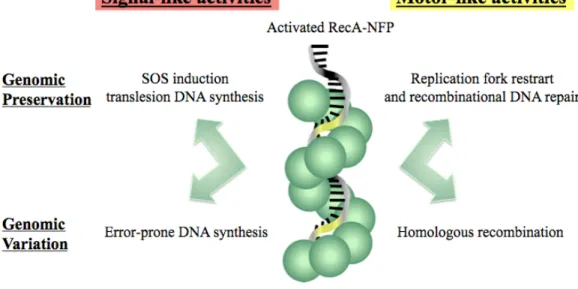
Within the cell, RecA exists as a protein monomer and is biologically
included homologous recombination and replication for restart[23], as well as signaling the initiation of the SOS response[22]. RecA-mediated repair processes, whether they initiate preservation or variation of the genome, aid in the survival of bacteria by providing means of maintaining genetic integrity as well as
adapting to environmental changes. Figure 1.7 summarizes the activities of RecA involved in preservation and adaptation.
Figure 1.7 Signal-like and motor-like activities of RecA. Upon activation of
The Involvement of RecA in Recombination
Recombination in its simplest meaning is the breaking and rejoining of
genetic materials, most often DNA. It is implicated in the genetic diversification of offspring during sexual unions and horizontal gene transfer between
microorganisms. Homologous recombination, or genetic exchange between similar or identical strands of DNA, is needed for recombinational repair in bacteria. However, recombinational repair is only implicated in certain types of
DNA damage due to DNA being double stranded; for example, lesions occurring on one strand can easily be excised and the other strand can be used as a template
to fill in the gap, which does not require recombination[19]. Cross-links, double-strand breaks and lesions in ssDNA would be cases of DNA damage that would require recombination to obtain reliable sequence information from a homologous
strand and RecA is at the heart of recombinational activity[19].
During each of the three repair mechanisms mentioned, ssDNA is either
exposed (stalled replication forks caused by lesions) or generated by the actions of other accessory proteins (UvrA,B, and C and nucleases in cross-link repair and RecBCD and nucleases in double-strand break repair)[19]. Once ssDNA is present, RecA polymerizes on the ssDNA to form the NPF[19,23], conducts a homology search by recruiting a linear dsDNA molecule, then aligns the ssDNA with a
Not only is this process useful for DNA repair, bacteria can utilize
RecA-mediated homologous recombination to incorporate genes into their genome that they acquired through horizontal gene transfer[26]. Horizontal gene transfer can occur in three ways: transformation, transduction and conjugation[27] (Figure 1.9[27]). Transformation is the process in which bacteria uptake exogenous DNA from their environment, usually excreted by a donor bacteria during lysis (usually
upon death of the bacteria). Transduction occurs through a bacteria phage, which carries the genes from the phage-infected donor cell to the recipient cell and injects the DNA into the recipient cell. Conjugation is the process by which donor
bacteria assemble a sex pilus (F factor) and inject DNA into the recipient cell. Once bacteria cells have taken up the exogenous DNA, whether from the same or
Figure 1.8 Role of RecA in homologous recombination. RecA polymerizes onto ssDNA
different species, RecA can pair this DNA, which is heteroduplex DNA, with homologous ssDNA from the host cell (Figure 1.10). Through strand exchange
during a tsDNA intermediate, genes from the donor bacteria can be incorporated into the genome of the host cell. This process has had tremendous effects on the
evolution of bacteria, especially in regards to the spread of antibiotic resistant genes.
Figure 1.9 Horizontal gene transfer in bacteria. This figure displays the
The Role of RecA in the SOS Response to DNA Damage
RecA has proven to be a vital participant in genomic preservation in bacteria by playing a key role in homologous recombination to repair DNA19,23] that has experienced a stalled replication fork, double-strand break or cross-linkage, as well as in the diversification of the bacterial genome through a partial
“mutagenic” role utilizing homologous recombination to incorporate genes acquired during horizontal gene transfer[26,27]. Yet another vital role of RecA in
Figure 1.10 Homologous recombination during horizontal gene transfer. In this case, the
genomic preservation and variation is through induction of the SOS response[21,22].
The expression of SOS genes are regulated by LexA, a repressor protein dimer, which binds to the SOS box of the promoter region of the SOS genes, thus
preventing their expression by preventing RNA polymerase from accessing the promoter[22]. When cells are experiencing normal physiological conditions, the SOS genes are repressed by LexA. However, when DNA damage occurs and
ssDNA accumulates, RecA polymerizes on the ssDNA and is activated, which in return interacts with the LexA repressor protein[28,29,30]. This interaction with LexA facilitates the autoproteolysis of LexA, thus allowing the downstream DNA repair proteins to be turned on[28,29]. It is important to note that the induction of the SOS to genomic damage is a graded response in which different mechanisms
of repair are sequentially activated, starting with excision repair and recombination and eventually leading to mutagenesis if the damage persists
(Figure 1.11). Sequential activation of approximately forty DNA repair proteins depends on the location of the genes to their specific SOS box and the binding affinity of LexA to that SOS box[22]. Generally, proteins can be classified into three main groups: early, middle and late genes.
Early genes consist of uvrA, B, C and D, which encode for endonucleases
that participate in nucleotide excision repair[31]. Small amounts of DNA damage can be repaired this way by simply excising a small length of ssDNA containing the damage and this process allows the cell to attempt to repair the damage
However, if the damage is not repaired after a short amount of time, the amount of de-repressed LexA increases, thus allowing for the expression of the
middle SOS genes, which include recA and recBCD[32,33], the genes required for the expression of recombinase proteins used in double-strand break and
cross-linkage repair. Much larger amounts of DNA can be repaired through recombination as compared to nucleotide excision repair. Because RecA is included in the group of middle SOS genes, the SOS response can be propagated
at a very accelerated rate due to the influx of RecA expression.
DNA damage that still persists even after NER and recombinational repair
attempts will eventually lead to the activation of the late SOS genes, which encode for Polymerase IV, UmuC and D, as well as SulA[22,35]. SulA is responsible for binding FtsZ, an essential protein that initiates cell division[34]. By binding FtsZ, cell division is arrested, thus diverting all of the cell’s efforts to repairing the damage. This includes the atuoproteolysis of UmuD whose
byproducts can bind to UmuC, therefore forming Polymerase V[22]. Both translesion Polymerases IV and V lack the ability to proof read DNA and therefore cannot detect lesions within the DNA, thus allowing the insertion of any
base into gaps across from the site of the lesion[22, 35]. And thus SOS mutagenesis is induced, which allows for the introduction of spontaneous mutations that can
Responsibility of RecA in the Development of Resistance to Antibiotic Treatment
RecA has profound roles in the repair of DNA through homologous recombination and also in inducing the SOS response in bacteria by signaling for the expression of DNA repair proteins, which both processes are imperative in
maintaining bacterial genomic integrity. RecA-mediated homologous recombination is also used by bacteria to incorporate genes acquired through
horizontal gene transfer by pairing the heteroduplex DNA from the donor cell with homologous ssDNA from the host cell, which leads to integration of the donated DNA into its own genome. SOS induction by RecA can also lead to SOS
mutagenesis as a last resort when nucleotide excision repair and recombination
Figure 1.11 Bacterial SOS response. The induction of the SOS to genomic damage is a
fail to repair DNA damage. SOS mutagenesis and homologous recombination of horizontally acquired genes are therefore essential processes for genomic
variation, which provide mechanisms for bacteria to survive in changing environments.
Antibiotics severely alter the environment of bacteria, thus imposing great stress on the bacteria to survive. Antibiotic induced stress has been shown to induce the SOS response through RecA activation[4,26]. As shown in Figure 1.3, there are six major types of antibiotics categorized by their targets: protein synthesis inhibitors, RNA polymerase inhibitors, inhibitors of DNA synthesis and
function, inhibitors of cell wall synthesis, agents that disrupt cell membrane integrity and folic acid biosynthesis inhibitors. Besides DNA damaging agents, other antibiotic classes that have been shown to activate the SOS response are
folic acid biosynthesis inhibitors (since folate is needed for the synthesis of nucleic acids) and inhibitors of cell wall synthesis. It is also pertinent to classify
antibiotics according to their mechanism of action: either bacteriostatic (inhibiting cell growth) or bactericidal (killing > 99.9% of bacteria). Bacteriostatic agents mostly consist of protein synthesis inhibitors.
Recently, it has been demonstrated that all classes examined of bactericidal agents including quinolines, β-lactams and aminoglycosides, produce
hydroxyl radicals in E. coli through the reduction of hydrogen peroxide by ferrous
proposed to occur by bactericidal agents stimulating the depletion of NADH by hyperactivation of the electron transport chain, which in return stimulates
formation of superoxide that damages iron-sulfur clusters. Iron released from these clusters is oxidated, giving hydroxyl radicals as by products that damage
DNA, lipids and proteins, ultimately leading to cell death[36]. As expected, the SOS response was also stimulated via RecA, as demonstrated by a fluorescence assay measure LexA-driven GFP expression and a cellular assay monitoring cell
death in wild type and ∆recA cells. In the fluorescence assay, quinolines and
β-lactams showed a significant increase in the GFP reporter, thus indicating the activation of RecA and the SOS response[36]. Kanomycin, the aminoglycoside tested, did not show an increase in the GFP reporter, but this is expected since kanomycin blocks translation of proteins and therefore the translation of GFP expression. By disabling the SOS response, the killing effect of bactericidals
would be expected to increase. This was demonstrated with all three agents in ∆recA cells, which stresses the importance of the induction of the SOS response
to bypass the killing effects of hydroxyl radicals[36].
The results from the Collins group also suggest that bacteriostatic agents, since they do not produce hydroxyl radicals, may not induce the SOS response.
Experiments to prove this were not done in this study. However, as demonstrated by our laboratory (unpublished data), studies done with chloramphenicol (a bacteriostatic agent) show that at sub-lethal dosages of chloramphenicol, wild
type E. coli cells develop resistance to chloramphenicol at a slower rate then E.
and relativly at the same rate during passaging (Figure 1.12, bottom panel). E. coli cells with RecA knocked out are not able to develop resistance in culture to
chloramphenicol at sub-lethal dosages (Figure 1.12, top panel) and developed resistance much more slowly during passaging (Figure 1.12, bottom panel). These
results imply that the SOS response is needed to confer chloramphenicol resistance.
Figure 1.12 Development of chloramphenicol resistance in wild type, super RecA and ∆∆∆∆
recA-∆ ∆∆
The Griffith Experiment:
Potential Role of RecA in Incorporation of Exogenous DNA
In the 1920’s, pneumonia was a prevalent cause of death, which is an infectious disease affecting the lungs and is caused by a bacterial species
Streptococcus pneumoniae, or pneumococci. Dr. Frederick Griffith, while
studying the distribution of different pneumococcal types obtained from people
infected with pneumonia, noticed four distinct types: Types I, II and III and Group IV. Upon compilation of the data of two two year periods and one three year period, he discerned a decrease in the number of Types I and II pneumococci
infections and a significant increase in the incidents of Group IV infections[37]. However, it was also noted that Group IV pneumococci was always found in
conjunction with another type and was not shown to cause the disease on its own. Therefore, Griffith devised a series of experiments involving the injection of these strains into mice in various combinations of the types in heat-killed virulent, or
smooth (S), versions of the strains with living avirulent, or rough (R), versions of the strains to see if reversion of the R strain to S form could occur. It is also
important to note, what defined the S strain from the R strain was a mucous film around the S strain, which was a polysaccharide capsule, referred to as S antigen by Griffith[37]. The polysaccharide capsule protects the pneumococci from attack by the immune system of its host. What he observed was mice injected with an S strain alone died of pneumonia while mice injected with a R strain alone lived and
also lived and did not produce either S or R cultures when obtained from blood of the mice.
But perhaps the most significant observations of this study when mice were injected with both R and heat-killed S strains were five fold: 1) the mice
died of pneumonia 2) cultures recovered from the blood of the mice were S strains 3) inoculation of the mice of the R strain of on type of pneumococci and the killed S strain of a different type of pneumococci displayed the type of the
heat-killed culture 4) the most successful reversions of the R strain to the S strain were the inoculation with same type of pneumococci (for example, Type II R and
heat-killed Type II S) and 5) reversion is not observed when incubated in vitro, meaning passage through mice is necessary for the reversion[37]. From these results, it can be concluded that the R strain is able to make use of the remnants of
the dead S culture for the synthesis of S antigen. It can also be hypothesized that since this only occurred in mice and not in vitro that the pressure of the immune
system is needed to drive the R strain to survive and seeks the information on how to make S antigen from the killed S cells.
Precisely how transformation occurred from the R strain to the S form was
elucidated by Avery, MacLeod, and McCarty. In their attempts, they used the R36A strain, which was an R Type II pneumococci strain, which was derived
ribonuclease and when these extracts were used to transform the R strain, transformation to the S form was observed[38]. Therefore, it can be concluded that proteins and ribonucleic acid, normally broken down by these substances, are not responsible for transformation. But when the extracts were subjected to treatment
with deoxyribonucleases from various sources, the transformation activity of the R strain to the S form was obliterated[38]. Finally, it was demonstrated that DNA was responsible for the transformation principle.
Since DNA is responsible for transformation, the cell must have a method of uptake and incorporation of virulent genes in the case of pneumococci studies
by Griffith and Avery et al. In the case of this particular study, a method for the incorporation of resistant genes is necessary. Exogenous DNA is taken up by the cell and once ssDNA is present, RecA is activated and forms the nucleoprotein
filament with the ssDNA[19]. From here, RecA will facilitate recombinational repair and integration of the exogenous DNA into the host genome. This process
was necessary for the R strain of the pneomococci to incorporate the DNA encoding for the polysaccharide capsule from the heat-killed S strain, thus making it virulent. This principle can be applied to cultures of E. coli inoculated with
heat-killed antibiotic resistant cells. Theoretically, if E. coli can demonstrate natural competence as S. pneumoniae does (either by a similar or unique
Observation of Natural Competence in Escherichia coli
Competence, the ability of a cell to take up exogenous DNA from its
environment, can be classified as artificial or natural. Artificial induction of competence is a standard laboratory technique in recombinant DNA technology
and is used to incorporate desired genes into host cells[39]. It involves the use of ice-cold CaCl2 or other divalent cations, which is necessary to fluidize the membrane (cold temperature) and aid DNA adsorption and binding to the cell
surface by forming coordination complexes with DNA and the LPS (Ca2+); a brief heat shock that causes the membrane to become rigid, allows for the release of
lipids, which possibly forms pores for DNA to enter and mediates the depolarization of the membrane, therefore reducing its negative charge inside the cell allowing DNA to pass into the cell; and incubation on ice that allows the
pores to close and trap the DNA[39].
Natural competence, on the other hand, is a cell’s natural ability to take up
exogenous DNA from the surrounding medium. Genetic transformation through the utilization of natural competence involves four steps: (1) development of competence through a stimulus; (2) binding of DNA to the cell surface; (3)
processing and uptake of the DNA and (4) integration of the DNA into the chromosome by recombination and expression[40]. Problems that arise from the translocation of DNA are hydrophobic bacterial cytoplasmic membranes act as barriers for DNA, the outer membrane of gram-negative bacteria (like E. coli) is negatively charged due to the LPS content and hinders negative molecules like
may attack DNA during transfer. However, certain strains of gram-positive and gram-negative bacteria have means of uptaking DNA and protecting it while it
transverses. For example, Bacillus subtilis forms a pilin complex with ComGC proteins (competence proteins), allowing the DNA-binding protein complex
ComEA to bind DNA, which delivers the DNA to a nuclease (unidentified) for degradation of one strand of the DNA and the compliment strand is driven into the cytosol by a DNA translocase complex, ComFA, through a channel in the
cytoplasmic membrane constructed from ComEC proteins[41]. S. pneumoniae has a very similar mechanism. An example of a gram-negative bacteria natural
competence pathway is N. gonorrhoeae, which consists of a pilin complex of PilQ proteins that bind DNA and allow it to cross the cell surface[41]. Another pilin complex consisting of PilE allows the DNA to cross the periplasm with the aid of
ComE, a DNA-binding protein, and a nuclease at the cytoplasmic membrane degrades one strand while allowing the other strand to enter the cell through a
channel consisting of ComA proteins[41]. Figure 1.8 demonstrates this[41]. Claverys and Martin also demonstrated that there are some homologous proteins in E. coli to DNA uptake machinery proteins and pore assembly proteins found in other
species[41], which suggests that E. coli may just be naturally competent, contrary to popular believe.
pBluescript KS- plasmid (ampr) were re-grown on solid media harboring all three antibiotics (kan, tet and amp), the cells were able to grow[42]. These results indicated that E. coli colonies on various solid medias can develop moderate competence, as well as that this process was either Ca2+ independent or trace amounts of Ca2+ from the agar or dead cells could act as competence inducing factors. Maeda and collaborators also further demonstrated this possibility by utilizing conjugative deficient strains of E. coli, CAG18439 harboring tetr and DH5a harboring the pHSG299 plasmid with kanr, to co-culture them on LB, water or CaCl2-agar lacking antibiotics then transferring colonies to LB, water or CaCl2 -agar containing both antibiotics[43]. Since colonies grew, it can be concluded that nonconjugative, nonviral horizontal gene transfer is possible in E. coli, which would indicate the need for natural competence.
Baur et al. also have contributed some convincing evidence that E. coli is able to develop natural competence. JM109 cells were incubated in various
natural waters in which the Ca2+ concentrations were known and pUC18 plasmid DNA was added to the culture, which encoded for ampr[44]. After sufficient incubation, samples were plated on LB-amp-agar plates and the transformation
frequencies were determined[44]. The results showed a positive correlation between the calcium concentrations of the waters and the transformation
are sufficient to induce competence in E. coli without the addition of any other competence-inducing factor.
Lastly, Sun et al. observed natural transformation in E. coli without the aid of cations and temperature shifts[45]. In shaking cultures of HB101 E. coli cells, cultures were incubated for 12 hours and statically cultured for up to 12 hours. A plasmid harboring ampr (pDsRED) was added and mixed into the cultures at various incubation times, which were then plated on LB-amp-agar and the
transformation efficiency was determined[45]. The transformation efficiency was shown to be dependent on time in static culture, not on viability, cations or
temperature shifts.
Much more research has been conducted in this area, but significant works were summarized above. This work is important because it demonstrates the
possibility that Griffith’s transformation principle can be applied to cultures of E.
coli. The work presented here demonstrates that antibiotic resistant genes can be
horizontally transferred between E. coli cells and they are capable of taking up exogenous DNA from their environments (plasmids, for example), which implies that DNA released from dead cells, especially heat-killed cells, can be transferred
Previous Efforts to Develop RecA Inhibitors
RecA-mediated activities, including the induction of the SOS response and
homologous recombination through strand exchange, have been implicated in the spread of horizontally transferred antibiotic resistance genes and de novo antibiotic resistance development, therefore making RecA a unique target for
inhibiting the spread of antibiotic resistance. However, few inhibitors of RecA had been revealed as of a few years ago. Since then, other members of the Singleton laboratory have had the opportunity to unearth inhibitors of RecA,
including metal complexes, ATP analogs, peptides and small molecules.
In collaboration with the Kohn laboratory at the UNC Eshelman School of
Pharnamcy, Dr. Andrew Lee had the opportunity to test Zn2+, Hg2+, Cu2+, Ca2+,
Figure 1.13. Natural competence and DNA entry into the cell of gram-positive vs. gram-negative species. a) N. gonorrhea, representative gram-negative bacteria. b) B. subtilis, representative gram-positive bacteria. This figure was reproduced from Claverys
Ba2+, Mn2+, Co2+, Ni2+, Ag+, Cd2+ and Bi3+ metals for inhibition of the RecA protein. The reason for choosing metal complexes was due to the discovery of
Kohn’s laboratory in that the Rho protein was inhibited by Be2+, Cd2+, Ni2+, Zn2+ and Bi3+ metal cations complexed with dithiols[46-48] and due to the fact that Rho is functionally and structurally homologous to RecA[49]. Metal complexes that he was able to show had inhibitory effects on RecA were Zn2+, Hg2+, Cu2+, Ag+, Cd2+ and Bi3+, which in a light-scattering assay[46-48] displayed aggregation of RecA in vitro. After extensive investigation into bismuth-dithiols, Bi3BAL in particular, it was shown that Bi3BAL was able to irreversibly inactivate the RecA protein, but did not do so through competing with ATP or ssDNA (results submitted for publication). Thus, it is a promising lead compound for future inhibitors.
Another means of inhibiting RecA activity explored in the Singleton
laboratory is the use of small peptide inhibitors that are rationally designed to interrupt the monomer-monomer interface of two RecA molecules, which would
essentially disrupt the assembly of the RecA-DNA filaments[50]. Based on the N-terminal domain of RecA, the peptides INPEP and INPEP-SH (INPEP with a salt bridge) were designed to bind more tightly than another RecA monomer[50]. The IC50 value of ATP hydrolysis was lowered to 35 µM with INPEP and 30 µM, a 20-fold decrease as compared to the N-30 peptide modeling the N-terminus[]. By conjugating a cysteine residue on SH with 2-thiopyridine to yield
INPEP-STP, the IC50 was decreased to 3 µM, thus resulting in a very potent 29mer
Since the activity of RecA is dependent upon the formation of the nucleoprotein filament, a process that requires the hydrolysis of ATP to tightly
bind ssDNA[51,53,54], seeking analogs of ATP that could potentially bind the active conformation of RecA to competitively inhibit hydrolysis or bind the inactive
conformation of RecA to promote the dissociation of ssDNA[42] could hinder the recombinational capabilities of RecA. In a study done by Wigle, Lee and Singleton, it was shown that out of twenty-eight potential ligands of RecA
consisting of general nucleotide triphosphates (NTPs) and nineteen synthetic analogs previously untested, six analogs were able to attenuate the
DNA-dependent NTPase activity of RecA[52]. Substitutions that prevented RecA from using the analogs as substrates, and therefore attenuating NTP hydrolysis, were summarized as follows: (1) substitution of groups larger than a hydroxyl group
(OMe, for example) on the C2’ position of the ribose ring prevented ATP and UTP from acting as substrates; (2) adding a methyl or propynyl group on the C5
position of the pyrimidine NTPs; (3) substitution of aromatic groups larger than a benzyl ring on the N6-amino group on the adenine ring[52]. Others were shown to be modest substrates of RecA.
Although nucleotide analogs are able to inhibit ATP hydrolysis of RecA, they were not suitable for cellular assays and therefore, other means of inhibition
were sought out, which included screening of small molecules of a focused set of commercially available compounds[55] and high-throughput screening of a 35,780 compound library at the Biomanufacturing and Research Institiute and
work). From the focused set of commercially available compounds, ATP hydrolysis by RecA was monitored with a fluorescent ATP assay involving the
oxidation of horseradish peroxidase to resorufin as A595[]. Five groups of compounds were tested: (1) vanillin, cinnamaldehyde, curcumin, genistin and
genistein, which were shown to be active in other biological assays;[56-59] (2) adenosine nucleotide-like compounds[60,61]: PMPA, 5’-ASBA and methotrexate; (3) compounds that inhibit the gyrase-Hsp-90-like family of ATPases[62,63]: radicicol, novobiocin and coumermycin; (4) adenine-like inhibitors known to inhibit protein kinases[64,65]: PP2 and PP3; (5) inhibitors of purine nucleotide receptors that are non-nucleotides[66]: PPADS, Bis-ANS, suramin, Congo Red and ANS. The only compounds from this study that were able to inhibit RecA ATPase activity were three suramin-like agents from group 5: Congo Red, suramin and
Bis-ANS[55]. Suramin was also shown to inhibit DNA three strand exchange at 100 µM[55]
.
Lastly, the results of they high-throughput performed at the BRITE center
utilized a PMB ATPase fluorescence assay to measure the hydrolysis of ATP by RecA by measuring the interaction of inorganic phosphate with molybdate to form a phosphomolybdate blue complex, which can be measured at A650 (unpublished data). Of the 35,780 compounds screened, seventy-three were reported as hits and were classified into five compound classes. From the first
compound class, one compound, termed A1, has proven to be a potent inhibitor of
GFP-fluorescence cell based assay in conjunction with ciprofloxacin and inhibited the RecA-mediated DNA three strand reaction (see Wigle dissertation for results).
Specific Aims of This Thesis
As demonstrated by Dr. Tim Wigle, the compound A1 has proven to be a favorable potential inhibitor of RecA, and therefore a means of attenuating the
transfer of antibiotic resistant genes. Therefore, the specific aims of this thesis are as follows: (1) demonstrate that the addition of A1 to cultures grown in sub-lethal dosages of chloramphenicol prevents or slows the development of resistance of
Escherichia coli MG1655 wild type cells; (2) apply the Griffith experiment to
Escherichia coli MG1655 cells to observe the transfer of antibiotic resistant genes
from heat-killed resistant cells to wild type cells; (3) demonstrate that inhibitor compound A1 attenuates the process of transferring antibiotic resistant genes in LB medium from heat-killed resistant cells to wild type Escherichia coli MG1655
cells; (4) utilize molecular modeling methods of screening to find other potential RecA inhibitors based on inhibitory concentration data obtained from various
high throughput screens.
A1 was shown to have a half maximal inhibitory concentration of 8 ±1 µM against the RecA protein in in vitro experiments. Therefore, utilizing a
one-flask resistance assay, monitoring the cultures of MG1655 cells should reveal if
A1 is able to penetrate into the cells and effectively inhibit RecA. This should theoretically attenuate the onset of resistance to antibiotics. However, if such resistance mechanisms of efflux and reduced membrane permeability are utilized,
the resistance development to chloramphenicol, since mutations are not always needed and the SOS response is not normally induced because chloramphenicol
functions by inhibiting protein synthesis. RecA knockout cells will serve as a negative control, which should not develop resistance if RecA is necessary for the
onset of resistance and may develop resistance if RecA is not necessary.
According to the Griffith experiment, DNA can be horizontally transferred from heat killed cells of a virulent strain of Streptococcus pneumoniae to an
avirulent strain of S. pneumoniae, which results in a virulent strain that is able to kill mice. Recent studies have shown E. coli to be naturally competent at a much
lower rate than other species of bacteria, such as S. pneumoniae. Wild type E. coli MG1655 cells, if able to develop competence in liquid medium, would be able to uptake and incorporate antibiotic resistant genes when inoculated with the
supernatant of heat killed resistant cells, therefore resulting in resistant cultures more quickly than cells that are grown without the supernatant of heat-killed
resistant cells in the presence of a sub-lethal dosage of chloramphenicol. RecA knockout cultures would not produce the onset of resistance or a difference in the rates of resistance development would not be observed.
RecA would be required to incorporate the resistant genes into the genome of MG1655 cells. Since A1 has inhibitory effects on RecA, adding it to cultures
deterred if A1 does serve as a RecA inhibitor. Again, DrecA cells will serve as a negative control.
Since A1 has been shown to be a promising candidate for inhibiting RecA and therefore attenuating the spread of antibiotic resistance, finding other
compounds that can serve as inhibitors of RecA is fruitful. Also, utilizing a method that is cost and time effective is favorable. Molecular modeling seemed to be a practical approach to finding potential compounds by minimizing time
spent in the laboratory and the cost of experiments. By utilizing a program entitles Molecular Operating Environment (MOE), common scaffolds can be abstracted
from molecules shown to have inhibitory effects against the RecA protein. The molecules chosen to screen were seventy-three hit compounds obtained from the high-throughput screen performed by Dr. Tim Wigle at the BRITE center where
A1 was discovered. These scaffolds can be input into a search in SciFinder Scholar to obtain similar compounds. Hypothetically, similar compounds will
CHAPTER II
MATERIALS AND METHODS
One-flask resistance assay reagents
LB broth, purchased from Fisher Scientific, was prepared by adding 20 g
of LB per liter of MilliQ de-ionized water. LB was distributed into 250 mL Erlenmeyer flasks, either 30 mL for starter cultures or 50 mL for assay cultures,
and autoclaved for 50 min. LB-agar plates were prepared by adding 20 g of LB and 15 g of agar (Fisher Scientific) per liter of MilliQ de-ionized water, autoclaving for 20-30 min and pouring into culture plates purchased from Fisher
Scientific.
Chloramphenicol was purchased from Sigma-Aldrich (St. Lois, MO) and
was prepared in 34 ng/mL 1 mL stocks in absolute EtOH. Starting materials for A1 were purchased from Sigma-Aldrich and A1 was synthesized by Dr. Anna Gromova or myself according to protocol. Chloramphenicol E-tests strips (0.016
– 250 µg/mL) were purchased from AB-Biodisk (Piscataway, NJ). Dr. Jim
Growing Chloramphenicol resistant cells
Chloramphenicol resistant cells (Camr) were made by growing a 5 mL overnight MG1655 wt culture in LB at 37°C, inoculating 30 mL of LB with 2 mL
of this culture and growing this culture to log phase in the 37°C shaker. Fifty mL
of LB were then inoculated with the MG1655 wt starter culture to an OD600 of
0.05. This culture was then grown in the presence of 5 µg/mL Cam until cells
reached saturation (OD600 close to 2). These cells were streaked and an E-test (AB-Biodisk, Piscataway, NJ) was performed to ensure resistance developed (MIC greater than 32 µg/mL). Cells were stored in 1 mL quantities in the -80°C
freezer. For each assay, one or two 5 mL Camr cultures were grown at 37°C overnight from the freezer stock by stabbing the frozen stock and ejecting the tip
into 5 mL of LB. To obtain heat killed supernatant, the overnight cultures were autoclaved for 15-30 min and were cooled at room temperature until needed.
One-flask resistance assay
Culture plates were prepared by T-streaking cells from a freezer stock and growing them overnight at 37°C. Overnight cultures were grown by picking
individual colonies on the culture plate, harvesting them with a pipette tip and ejecting them into 5 mL of LB broth in glass culture tubes (Fisher Scientific), then placing them in the 37°C shaker. Two mL of the overnight cultures were added