International Consensus Statement on Allergy and Rhinology: Rhinosinusitis
Richard R. Orlandi, MD1, Todd T. Kingdom, MD2, Peter H. Hwang, MD3, Timothy L. Smith, MD, MPH4,
Jeremiah A. Alt, MD, PhD5, Fuad M. Baroody, MD6, Pete S. Batra, MD7, Manuel Bernal-Sprekelsen, MD8,
Neil Bhattacharyya, MD9, Rakesh K. Chandra, MD10, Alexander Chiu, MD11, Martin J. Citardi, MD12,
Noam A. Cohen, MD, PhD13, John DelGaudio, MD14, Martin Desrosiers, MD15, Hun-Jong Dhong, MD16,
Richard Douglas, MD17, Berrylin Ferguson, MD18, Wytske J. Fokkens, MD, PhD19, Christos Georgalas, DLO,
PhD20, Andrew Goldberg, MD, MSCE21, Jan Gosepath, MD, PhD22, Daniel L. Hamilos, MD23,
Joseph K. Han, MD24, Richard Harvey, MD, PhD25, Peter Hellings, MD, PhD26, Claire Hopkins, MD27,
Roger Jankowski, MD, PhD28, Amin R. Javer, MD29, Robert Kern, MD30, Stilianos Kountakis, MD, PhD31,
Marek L. Kowalski, MD, PhD32, Andrew Lane, MD33, Donald C. Lanza, MD, MS34, Richard Lebowitz, MD35,
Heung-Man Lee, MD36, Sandra Y. Lin, MD37, Valerie Lund, CBE, MD, MS38, Amber Luong, MD, PhD39,
Wolf Mann, MD, PhD40, Bradley F. Marple, MD41, Kevin C. McMains, MD42, Ralph Metson, MD43,
Robert Naclerio, MD44, Jayakar V. Nayak, MD, PhD45, Nobuyoshi Otori, MD46, James N. Palmer, MD47,
Sanjay R. Parikh, MD48, Desiderio Passali, MD, PhD49, Anju Peters, MD50, Jay Piccirillo, MD51,
David M. Poetker, MD52, Alkis J. Psaltis, MD, PhD53, Hassan H. Ramadan, MD, MSC54,
Vijay R. Ramakrishnan, MD55, Herbert Riechelmann, MD, PhD56, Hwan-Jung Roh, MD, PhD57,
Luke Rudmik, MD, MSc58, Raymond Sacks, MD59, Rodney J. Schlosser, MD60, Brent A. Senior, MD61,
Raj Sindwani, MD62, James A. Stankiewicz, MD63, Michael Stewart, MD64, Bruce K. Tan, MD65,
Elina Toskala, MD, PhD66, Richard Voegels, MD, PhD67, De Yun Wang, MD, PhD68, Erik K. Weitzel, MD69,
Sarah Wise, MD70, Bradford A. Woodworth, MD71, Peter-John Wormald, MD72, Erin D. Wright, MD73,
Bing Zhou, MD74and David W. Kennedy, MD75
1University of Utah;2University of Colorado;3Stanford University;4Oregon Health Science University;5University of Utah;6University of Chicago; 7Rush University;8Universidad de Barcelona;9Harvard Medical School;10Vanderbilt University;11University of Arizona;12University of Texas Medical School at Houston;13University of Pennsylvania;14Emory University;15Universit ´e de Montr ´eal;16Samsung Medical Center;17University of Auckland; 18University of Pittsburgh;19University of Amsterdam;20University Hospital, Leiden, Netherlands–Hygeia Hospital, Athens, Greece;21University of California, San Francisco;22Dr. Horst Schmidt Kliniken;23Massachusetts General Hospital;24Eastern Virginia Medical School;25University of New South Wales and Macquarie University;26University Hospitals Leuven;27Guy’s Hospital;28University of Lorraine;29University of British Columbia; 30Northwestern University;31Georgia Regents University;32Medical University in Lodz;33Johns Hopkins University;34Sinus & Nasal Institute of Florida; 35Foundation New York University;36Korea University;37Johns Hopkins University;38Royal National Throat Nose and Ear Hospital, London, UK; 39University of Texas Medical School at Houston;40Private Practice;41University of Texas, Southwestern;42Uniformed Services University of Health Sciences;43Harvard Medical School;44University of Chicago;45Stanford University;46Jikei University;47University of Pennsylvania;48University of Washington;49University of Siena;50Northwestern University;51Washington University;52Medical College of Wisconsin;53University of Adelaide; 54West Virginia University;55University of Colorado;56Medical University Innsbruck;57Pusan National University;58University of Calgary;59University of New South Wales;60Medical University of South Carolina;61University of North Carolina;62Cleveland Clinic Foundation;63Loyola University; 64Weill Cornell Medical College;65Northwestern University;66Temple University;67Universidade de S ˜ao Paulo;68National University of Singapore; 69San Antonio Military Medical Center;70Emory University;71University of Alabama at Birmingham;72University of Adelaide;73University of Alberta; 74Capital Medical University;75University of Pennsylvania
Contributing Authors
Isam Alobid, MD, PhD1, Nithin D. Adappa, MD2, Henry P. Barham, MD3, Thiago Bezerra, MD4, Nadieska
Caballero, MD5, Eugene G. Chang, MD6, Gaurav Chawdhary, MD7, Philip Chen, MD8, John P. Dahl, MD,
PhD9, Anthony Del Signore, MD10, Carrie Flanagan, MD11, Daniel N. Frank, PhD12, Kai Fruth, MD, PhD13,
Anne Getz, MD14, Samuel Greig, MD15, Elisa A. Illing, MD16, David W. Jang, MD17, Yong Gi Jung, MD18,
Sammy Khalili, MD, MSc19, Cristobal Langdon, MD20, Kent Lam, MD21, Stella Lee, MD22, Seth Lieberman,
MD23, Patricia Loftus, MD24, Luis Macias-Valle, MD25, R. Peter Manes, MD26, Jill Mazza, MD27, Leandra
Mfuna, MD28, David Morrissey, MD29, Sue Jean Mun, MD30, Jonathan B. Overdevest, MD, PhD31, Jayant M.
Pinto, MD32, Jain Ravi, MD33, Douglas Reh, MD34, Peta L. Sacks, MD35, Michael H. Saste, MD36, John
Schneider, MD, MA37, Ahmad R. Sedaghat, MD, PhD38, Zachary M. Soler, MD39, Neville Teo, MD40, Kota
Wada, MD41, Kevin Welch, MD42, Troy D. Woodard, MD43, Alan Workman44, Yi Chen Zhao, MD45, David
Zopf, MD46
Correspondence to: Richard R. Orlandi, MD, 50 North Medical Drive, Salt Lake City, UT 84132; e-mail: [email protected] Additional Supporting Information may be found in the online version of this article.
Potential conflict of interest: Please see the appendix at the end of this article.
Received: 24 September 2015; Revised: 13 November 2015; Accepted: 16 November 2015 DOI: 10.1002/alr.21695
1Universidad de Barcelona;2University of Pennsylvania;3Louisiana State University Health Sciences Center;4Universidade de S ˜ao Paulo;5ENT Specialists of Illinois;6University of Arizona;7University of Oxford;8University of Texas;9University of Indiana;10Mount Sinai Beth Israel;11Emory University;12University of Colorado;13Wiesbaden, Germany;14University of Colorado;15University of Alberta;16University of Alabama at Birmingham; 17Duke University;18Sungkyunkwan University;19University of Pennsylvania;20Universidad de Barcelona;21Northwestern University;22University of Pittsburgh;23New York University;24Emory University;25University of British Columbia;26Yale University School of Medicine;27Private Practice; 28Department of Otolaryngology, H ˆotel-Dieu Hospital, Centre de Recherche du Centre Hospitalier de l’Universit ´e de Montr ´eal;29University of Adelaide;30Pusan National University;31University of California, San Francisco;32University of Chicago;33University of Auckland;34Johns Hopkins University;35University of New South Wales, Australia;36Stanford University;37Washington University;38Harvard Medical School;39Medical University of South Carolina;40Singapore General Hospital;41Taho University;42Northwestern University;43Cleveland Clinic Foundation;44University of Pennsylvania;45University of Adelaide;46University of Michigan
Background:The body of knowledge regarding rhinosinusi-tis (RS) continues to expand, with rapid growth in number of publications, yet substantial variability in the quality of those presentations. In an effort to both consolidate and critically appraise this information, rhinologic experts from around the world have produced the International Con-sensus Statement on Allergy and Rhinology: Rhinosinusitis (ICAR:RS).
Methods: Evidence-based reviews with recommendations (EBRRs) were developed for scores of topics, using previ-ously reported methodology. Where existing evidence was insufficient for an EBRR, an evidence-based review (EBR) was produced. The sections were then synthesized and the entire manuscript was then reviewed by all authors for con-sensus.
Results:The resulting ICAR:RS document addresses multi-ple topics in RS, including acute RS (ARS), chronic RS (CRS) with and without nasal polyps (CRSwNP and CRSsNP), re-current acute RS (RARS), acute exacerbation of CRS (AE-CRS), and pediatric RS.
Conclusion: As a critical review of the RS literature, ICAR:RS provides a thorough review of pathophysiology and evidence-based recommendations for medical and sur-gical treatment. It also demonstrates the significant gaps in our understanding of the pathophysiology and optimal management of RS. Too oen the foundation upon which these recommendations are based is comprised of lower-level evidence. It is our hope that this summary of the evi-dence in RS will point out where additional research efforts may be directed.C 2016 ARS-AAOA, LLC.
Key Words:
rhinosinusitis; chronic rhinosinusitis; acute rhinosinusitis; recurrent acute rhinosinusitis; evidence-based medicine; systematic review; endoscopic sinus surgery
How to Cite this Article:
Orlandi RR, Kingdom TT, Hwang PH, et al. International Consensus Statement on Allergy and Rhinology: Rhinosi-nusitis.Int Forum Allergy Rhinol. 2016;6:S22-S209.
List of Abbreviations Used
AAOA American Academy of Otolaryngic Allergy AAO-HNS American Academy of Otolaryngology–Head
and Neck Surgery
ABRS acute bacterial rhinosinusitis
AECRS acute exacerbation of chronic rhinosinusitis AERD aspirin-exacerbated respiratory disease
AFRS allergic fungal rhinosinusitis AIFS acute invasive fungal rhinosinusitis
AJC apical junction complex AMCase acidic mammalian chitinase
AMT appropriate medical therapy AOAH acetyl hydroxylase
AR allergic rhinitis ARS acute rhinosinusitis ASA acetyl salicylic acid
AVRS acute viral rhinosinusitis BID twice daily
C3 complement component 3 CBF ciliary beat frequency
CF cystic fibrosis ChT chitotriosidase
CIFS chronic invasive fungal rhinosinusitis CMS Centers for Medicare & Medicaid Services CMT conventional medical treatment
COX cyclooxygenase CRP C-reactive protein
CRSsNP chronic rhinosinusitis without nasal polyps CRSwNP chronic rhinosinusitis with nasal polyps
CT computed tomography
CVID common variable immunodeficiency cysLT cysteinyl leukotriene
DAMP damage-associated molecular pattern DBRCT double-blind randomized controlled trial
DC dendritic cells DSS Dead Sea salt
DTH delayed-type hypersensitivity EBM evidence-based medicine
EBR evidence-based review
EBRR evidence-based review with recommendations EC epithelial cell
ECP eosinophilic cationic protein EDN eosinophil-derived neurotoxin
EER extraesophageal reflux
ELISA enzyme-linked immunosorbent assay EMRS eosinophilic mucin rhinosinusitis
EPOS European Position Paper on Rhinosinusitis and Nasal Polyps
ES endoscopic septoplasty ESR erythrocyte sedimentation rate
ESS endoscopic sinus surgery
FEV1 functional expiratory volume within 1 second FISH fluorescent in situ hybridization
GERD gastroesophageal reflux disease GI gastrointestinal
GIFS granulomatous invasive rhinosinusitis GOSS Global Osteitis Scoring Scale
HBD human beta defensin HLA human leukocyte antigen
HU Hounsfield unit
ICAR:RS International Consensus Statement on Allergy and Rhinology: Rhinosinusitis
IFS invasive fungal rhinosinusitis Ig immunoglobulin
IGS image-guided surgery IHC immunohistochemistry
IL interleukin
ILC innate lymphoid cells
INCS intranasal corticosteroid spray IV intravenous
IVIG intravenous immunoglobulin LL-37 cathelicidin
LM Lund-Mackay score LOE level of evidence
LT leukotriene
MAD mucosal atomization device MBL mannose-binding lectin MCC mucociliary clearance MGO methylglyoxal
MIF migration inhibition factor MMA middle meatal antrostomy
MMP matrix metalloproteinase MMT maximal medical therapy MPRO myeloperoxidase
MRA magnetic resonance angiography MRI magnetic resonance imaging
mRNA messenger RNA MT middle turbinate
NLR nucleotide-binding oligomerization domain-like receptor
NO nitric oxide
NOD nucleotide-binding oligomerization domain NP nasal polyp
NPx nasopharynx NS normal saline
NSAID nonsteroidal anti-inflammatory drug NSD nasal septal deviation
OMC ostiomeatal complex
PAMP pathogen associated molecular pattern PARE pharyngeal acid reflux event
PCD primary ciliary dyskinesia PCR polymerase chain reaction PCRS pediatric chronic rhinosinusitis
P prostaglandin
PID primary immunodeficiency
PLUNC palate, lung, and nasal epithelium clone pro-tein
PND postnasal drip
PNIF peak nasal inspiratory flow PPI proton pump inhibitor
PRISMA Preferred Reporting Items for Systematic Re-views and Meta-Analyses
PRR pattern recognition receptors PSQI Pittsburg Sleep Quality Index
QID 4 times daily QoL quality of life
qRT-PCR quantitative real-time polymerase chain reac-tion
RAGE receptor for advanced glycation end products RARS recurrent acute rhinosinusitis
R-CRS refractory chronic rhinosinusitis RCT randomized controlled trial
RQLQ Rhinoconjunctivitis Quality of Life Question-naire
RS rhinosinusitis
RSDI Rhinosinusitis Disability Index RSI Rhinosinusitis Symptom Inventory RSOM Rhinosinusitis Outcome Measure RT-PCR real-time polymerase chain reaction
SE Staphylococcal enterotoxins SF Short Form Health Survey SNOT Sino-Nasal Outcome Test
SNP single-nucleotide polymorphism SP surface protein
SPECT single proton emission CT TFF trefoil factor family TGF transforming growth factor
TID 3 times daily TLR Toll-like receptor TNF tumor necrosis factor TP-1 thymostimulin
TSST toxic shock syndrome toxin UES upper esophageal sphincter URI upper respiratory infection
FDA U.S. Food and Drug Administration VAS visual analog scale
VD3 vitamin D
Appendix
I. Introduction S25
II. Methods S26
III. Rhinosinusitis Definitions S27
IV. The Burden of Rhinosinusitis S30
V. Acute Rhinosinusitis S33
VI. Recurrent Acute Rhinosinusitis S48
VII. Chronic Rhinosinusitis without Nasal Polyps S53
VIII. Chronic Rhinosinusitis with Nasal Polyps S100
IX. Acute Exacerbation of Chronic Rhinosinusitis S138
X. Surgery for Chronic Rhinosinusitis S139
XI. Pediatric Rhinosinusitis S171
XII. Special Considerations in Rhinosinusitis S178
XIII. Knowledge Gaps and Research Opportunities S180
XIV. References S181
I. Introduction
T
he body of knowledge about rhinosinusitis (RS)con-tinues to expand. A search of the PubMed database us-ing the search terms “sinusitis” or “rhinosinusitis” demon-strates that between 2000 and 2014, 12,847 articles were published on the subject. Further, this search shows that the annual number of publications on RS has continued to grow (Fig. I-1). Besides the daunting number of articles on the topic of RS, there is considerable variation in the quality of these publications.
The practice of evidence-based medicine (EBM) requires a thorough knowledge of the “best external evidence” (Fig. I-2). The sheer number and the varying quality of publications make it increasingly difficult for the clinician to practice EBM in caring for patients with RS.
This International Consensus statement on Allergy and Rhinology: Rhinosinusitis (ICAR:RS) has been developed in order to summarize that best external evidence. The authors’ goal was to assemble and critically appraise all available evidence on the diagnosis, pathophysiology, and management of the various forms of RS. We employed a structured review of the evidence by utilizing over 100 au-thors from around the world, using a stepwise anonymous writing and iterative review process for each of over 140 topics. This methodology has produced a robust review
of current evidence and treatment recommendations based upon the best available evidence.
The methodology we employed seeks to rely principally on higher-level published evidence and to diminish the im-pact of expert opinion such as that which may be seen in a literature review or proceedings of a consensus panel. At the same time, ICAR:RS has limitations. It is neither a meta-analysis nor a clinical practice guideline (CPG). Much of the RS literature does not lend itself well to meta-analysis because there are limited numbers of studies with vari-able methodologies for any given topic. Although ICAR:RS provides evidence-based care recommendations, ICAR:RS should not itself be confused with a CPG. CPGs require strong evidence as well as additional steps of critical review by many stakeholders, including medical specialty societies and patient advocates.
Importantly, although there has been a large and increas-ing number of publications on RS, 2 important caveats should be noted. First, as the number of RS publications has increased, the proportion of all publications annually has held steady at 0.10% throughout the last 15 years (Fig. I-1). Second, review of this ICAR:RS document will reveal that the vast majority of “best external evidence” on RS is relatively weak. As a rhinology community, we should reflect upon this ICAR:RS document for gaps in high-level evidence and, where possible, dedicate ourselves to filling those gaps.
This document as a compendium of recommendations has limitations. This ICAR:RS document does not repre-sent a “cookbook” for providing care for the RS patient. The practice of EBM requires the clinician to have the best available evidence, and then combine that with individual expertise and the patient’s condition, values, and
expec-tations (Fig. I-2).1 RS is a set of highly variable
condi-tions, with different etiologies and a wide breadth of rec-ommended treatments. For example, acute RS differs from acute exacerbation of chronic RS (AECRS), which in turn differs from chronic RS (CRS). Even within CRS, patients with and without nasal polyps (CRSwNP and CRSsNP, re-spectively) have significant differences in pathophysiology and recommended treatments. Applying the diagnostic and treatment recommendations for 1 condition to the others would be erroneous.
In addition to recognizing variability among subsets of RS, the clinician must also recognize the tremendous vari-ability within a subset of RS, especially CRS. CRS patients can be mildly symptomatic or highly symptomatic; they may have limited findings on endoscopy or computed to-mography (CT) or complete involvement of all sinuses; they may be presenting for diagnosis and management for the first time or after many failed treatments or even after multi-ple surgeries. To assume that 1 patient is just like another— and to apply the findings in this document under such an assumption—is not consistent with the practice of EBM.
FIGURE I-1.Results of a PubMed search for the terms “sinusitis” or “rhinosinusitis” by year on the left axis. The right axis shows the sinusitis and rhinosinusitis articles as a percentage of the total number of articles listed for that year.
in this document are based on the best available evidence, they do not define standard of care nor do they define med-ical necessity. Healthcare providers or any others should not assume that a particular treatment is or is not indicated in an individual patient solely based on what is written in this or any other similar document.
Last, the recommendations herein should not be viewed as static. As new and stronger evidence emerges, they will necessarily have to undergo reevaluation and possi-bly change. It is our hope that this summary will guide all who care for RS patients, equipping them to provide our patients with the best possible outcome.
II. Methods
II.A. Topic Development
This document was developed and written so as to have the maximal reliance on published evidence. The authors adapted the method of writing an evidence-based review with recommendations (EBRR), as described by Rudmik
and Smith in 2011.2The subject of RS was initially divided
into 144 topics. Each topic was then assigned to a senior author who is a recognized expert in the field of rhinology, and specifically in RS. Some of the topics had no significant evidence and were assigned as literature reviews. Some had significant evidence but did not lend themselves to providing a recommendation, such as those addressing diagnosis and pathogenesis, and these were assigned as evidence-based reviews (EBRs) without recommendations. Many had evidence to inform recommendations and were assigned as EBRRs.
FIGURE I-2. The practice of EBM. Adapted from: Armstrong EC. Har-nessing new technologies while preserving basic values.Fam Syst Health. 2003;21:351-355. EBM=evidence-based medicine.
To provide the content for each topic, a systematic review
of the literature for each topic using Ovid MEDLINER
(1947 to July 2014), EMBASE (1974 to July 2014), and Cochrane Review databases was performed using the Pre-ferred Reporting Items for Systematic Reviews and
Meta-Analyses (PRISMA) standardized guidelines.3 The search
TABLE II-1. Aggregate grade of evidence
Grade Research quality
A Well-designed RCTs
B RCTs with minor limitations; overwhelming consistent evidence from observational studies
C Observational studies (case control and cohort design)
D Expert opinion; case reports; reasoning from first principles
Because clinical recommendations are best supported by randomized controlled trials (RCTs), the search focused on identifying these studies to provide the strongest level of ev-idence (LOE). Reference lists of all identified studies were examined to ensure all relevant studies were captured. If the authors felt as though a non-English study should be in-cluded in the review, the paper was appropriately translated to minimize the risk of missing important data during the
development of recommendations.3 One major exception
to the search window was made for the Clinical Practice Guidelines of the American Academy of Otolaryngology–
Head and Neck Surgery (AAO-HNS).4 These guidelines
were updated during preparation of the manuscript and were heavily referenced throughout the document. The up-dated 2015 version was therefore used.
To ensure complete transparency of the evidence in EBR and EBRR sections, all included studies were presented in a standardized table format and the quality of each study was evaluated to receive a level based on the Oxford levels
of evidence (from level 1a to 5).5At the completion of the
systematic review and research quality evaluation for each clinical topic, an aggregate grade of evidence was produced for the topic based on the guidelines from the American Academy of Pediatrics (AAP) Steering Committee on
Qual-ity Improvement and Management (Table II-1).6
After providing an aggregate grade of evidence for each EBRR topic (ie, A to D), a recommendation using the AAP guidelines was produced. It is important to note that each evidence-based recommendation took into account the ag-gregate grade of evidence along with the balance of benefit, harm, and costs (Table II-2).
After the development of the initial topic EBRR, the manuscript underwent a 2-stage online iterative review pro-cess using 2 independent reviewers. The purpose of these steps was to evaluate the completeness of the identified lit-erature and ensure the recommendations were appropriate. The topic content was reviewed by another expert on that topic, and all changes were agreed upon by both reviewer and initial authors. The topic content was then reviewed by a second reviewer and changes were agreed upon by the initial authors and the first reviewer. Figures II-1 and II-2 show flowcharts of the topic development and EBRR iterative review processes.
II.B. ICAR:RS Statement Development
After the completion of all topics, the principal edi-tors (R.R.O., T.T.K., and P.H.H.) compiled them into 1
ICAR:RS statement. This draft document was then re-viewed by all contributing authors. The final ICAR:RS manuscript was produced once consensus was reached among the authors regarding the literature and final rec-ommendations.
III. Rhinosinusitis Definitions
III.A. RS Definitions: Acute Rhinosinusitis
Acute rhinosinusitis (ARS) in adults may be defined as sinonasal inflammation lasting less than 4 weeks associated
with the sudden onset of symptoms.4,7–9This definition is
largely based on expert opinion and consensus. Several task forces and consensus groups have all agreed that an acute episode may last up to 4 weeks, though this does not seem to
be based on any objective evidence.4,7–11Adult symptoms
must include nasal blockage/obstruction/congestion or nasal discharge (anterior/posterior) and facial pain/pressure or reduction/loss of smell. ARS in children may be de-fined as sinonasal inflammation associated with the sud-den onset of 2 or more of the following symptoms: nasal blockage/obstruction/congestion, discolored nasal drainage, or a cough that may occur during the day or
night.7
For both adults and children, inquiry should be made about symptoms suggestive of allergy (eg, sneezing, watery rhinorrhea, nasal and ocular pruritus, and watery eyes) in order to help differentiate acute viral or bacterial RS from allergic rhinitis (AR).
The consensus groups have all agreed that in acute viral RS nasal symptoms are generally present for fewer than 10 days. The most recent guidelines from the AAO-HNS included data on the duration of typical viral symptoms in support of the commonly accepted time frames used to
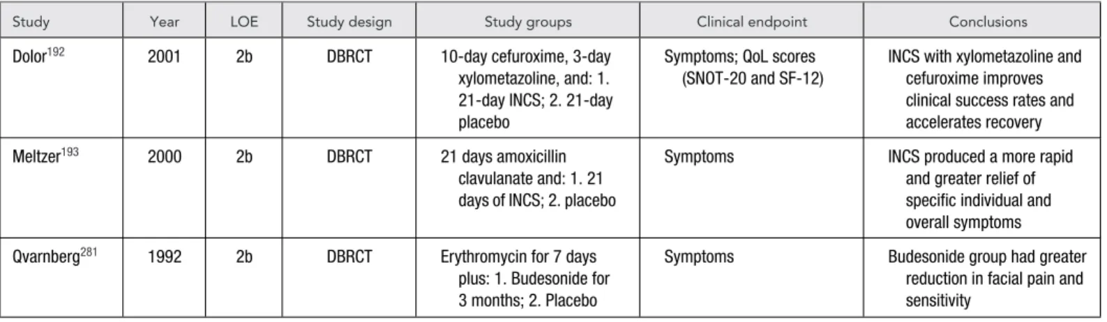
differentiate acute viral RS from acute bacterial RS.4
The EPOS 2012 statement describes a process referred to as “acute postviral rhinosinusitis,” which seems to be based on expert opinion and is defined as a worsening of symptoms after about 5 days, or persistent symptoms after 10 days, but with symptom duration of fewer than
12 weeks.7 This process is not recognized as a separate
entity by the 2015 AAO-HNS guidelines.4
These 2 most recent guidelines also differ slightly on how ARS is diagnosed. Both agree that discolored discharge (with unilateral predominance) and purulent secretions in the nasal passage, moderate to severe local pain, and pro-longed symptoms and/or deterioration of condition after
initial improvement are key for the diagnosis.4, 7The EPOS
statement includes an elevated erythrocyte sedimentation rate and/or C-reactive protein (CRP) and fever as diagnostic
criteria.7The AAO-HNS guidelines cite the low sensitivity
and specificity of fever as a rationale for not including fever
as a diagnostic criterion.4
III.B. RS Definitions: CRS
CRS in adults is defined as sinonasal inflammation
TABLE II-2. AAP-defined strategy for recommendation development6
Preponderance of benefit Balance of benefit Preponderance of harm
Evidence quality over harm and harm over benefit
A. Well-designed RCTs Strong recommendation Option Strong recommendation against
B. RCTs with minor limitations; overwhelmingly consistent evidence from observational studies
Recommendation Option Strong recommendation against
C. Observational studies (case control and cohort design)
Recommendation Option Recommendation against
D. Expert opinion, case reports, reasoning from first principles
Option No recommendation Recommendation against
AAP=American Academy of Pediatrics.
FIGURE II-1.Topic development. PE=principal editor; 10=primary.
based on consensus and has been relatively consistent over the past 25 years. The most recent guidelines agree that CRS in adults is characterized by nasal ob-struction/congestion/blockage, nasal drainage (mucopu-rulent) that may drain anteriorly or posteriorly, facial pain/pressure/fullness, and decreased or loss of sense of
smell.4, 7 Symptoms alone have a high sensitivity but an
unacceptably low specificity, which is why the symptoms must be accompanied by objective findings including tive nasal endoscopy (purulence, polyps, or edema) or posi-tive imaging findings consisting of inflammation or mucosal
changes within the sinuses.4, 7
CRS in children is defined and diagnosed similarly to CRS in adults, with the difference being cough is a much
more significant symptom than is decreased sense of smell.7
Interestingly, this definition too is essentially based on con-sensus but does have some data supporting headache, nasal obstruction, postnasal drainage/rhinorrhea, and cough as the 4 most common symptoms identified in children with
sinusitis.12
For both adults and children, CRS with nasal polyps (CRSwNP) is diagnosed when nasal polyps (NPs) can be visualized in the nose and/or middle meati, in the
FIGURE II-2. Topic EBRR iterative review process. 10=primary; 20=secondary; 30=tertiary; EBRR=evidence-based review with recommendations; PE=
principal editor.
may require further investigation to exclude neoplastic pathologies.
III.B.1. CRS Definition: Disease or Syndrome?
In view of the different clinical phenotypes and inflamma-tory endotypes of CRS, it can be considered an umbrella term covering several inflammatory disease states of the sinonasal cavities. On the basis of clinical and/or radio-logic examination, CRS is generally divided into CRSsNP and CRSwNP. Apart from these 2 major clinical pheno-types, other phenotypes relate to the variety of presenting symptoms in CRS patients and the presence or absence of
concomitant bronchial disease.4, 7It is not surprising to find
different phenotypes of the disease, given the multitude of underlying etiologic factors.
A wide range of inflammatory patterns may act together with mucociliary and/or structural abnormalities to give rise to the development of CRS. The multifactorial etiology of CRS, involving genetic factors, environmental influences, occupational factors, infection, allergy, immune dysfunc-tion, and systemic diseases, has led to the recent attempt to define endotypes of disease. CRS has been classified into different inflammatory clusters, such as T helper 1 (Th1)-driven or neutrophilic inflammation, and T helper 2
(Th2)-driven or eosinophilic inflammation.13Several specific
in-flammatory mediators have been associated with CRS, and the beneficial effects of novel treatments with biologics like anti–interleukin 5 (IL-5), anti–immunoglobulin E (IgE), and others support their importance. We should, however,
re-alize that mixed inflammatory clusters are likely common and important in these patients; this would help explain the limited beneficial effects of targeting 1 inflammatory mediator such as IgE or IL-5 in isolation.
Taken together, CRS represents a condition with differ-ent phenotypes and endotypes, which we are only starting to better understand. In a single CRS patient, pinpointing the different etiologic factors responsible for the develop-ment of the disease remains the challenge for the future.
III.C. RS Definitions: Recurrent
Acute Rhinosinusitis
Recurrent acute rhinosinusitis (RARS) has been defined as 4 episodes per year of ARS with distinct symptom-free
in-tervals between episodes.4, 7, 8, 14 Each episode must meet
the criteria listed in Section III.A for ARS. The number of episodes required for the diagnosis of RARS has var-ied between consensus statements, but the 2007 guidelines
in Rosenfeld et al.15 addressed this number. Citing the
published literature, they reported that the average adult gets between 1.4 and 2.3 viral upper respiratory infections (URIs) per year. They felt that setting the number of acute bacterial rhinosinusitis (ABRS) episodes at 4 for RARS de-creased the chances of misdiagnosis.
Only a few cohort studies have examined the clinical course of this group of patients. Patients with RARS tend not to be treated surgically, and when they are, they
un-dergo less extensive surgery than CRS patients.16, 17
may possibly be due to a relatively higher number of
sinonasal anatomic variants in this patient group.18
III.D. RS Definitions: AECRS
AECRS is defined in a patient in whom a previous diagnosis of CRS exists, and a sudden worsening of symptoms occurs,
with a return to baseline symptoms following treatment.8, 11
A more stringent definition has not been proposed to this point nor have more precise diagnostic criteria been put forward. The concept for AECRS was based on a similar disease pattern in otitis media (personal communication with Donald Lanza, MD). Although the literature on this condition is limited, potential diagnostic criteria are pro-posed in Section IX.C.
III.E. RS Definitions: Subacute RS
Although absent from recent guidelines, subacute RS has been a term used to describe clinical presentations that fall between the time frames of ARS and CRS (symptoms of 4 to 12 weeks’ duration). To date, there have been very few clinical reports on which to base the delineation of these patients as a distinct clinical entity; reports that do make that distinction define the process based on consensus. The term has been used at least since 1975, and Lanza and
Kennedy11recommended reintroducing the term although
there was no formal definition provided by the U.S. Food and Drug Administration (FDA) for disease lasting 4 to
12 weeks.12It is likely that patients who fall into this group
either have slow-to-resolve ARS or an early presentation of evolving CRS. Use of this definition or classification should be limited until a better understanding of this condition is achieved.
III.F. RS Definitions: Sinusitis or RS?
In 1996, the Task Force on Rhinosinusitis sponsored by the AAO-HNS reached a consensus regarding specific
di-agnostic criteria and working definitions to describe RS.19
The Task Force agreed to replace the word “sinusitis” with the more descriptive term “rhinosinusitis” to emphasize the close relationship between the nose and paranasal si-nuses, the similarities in both morphology and physiology between the mucosal lining of the 2, and the fact that the 2 conditions often exist simultaneously in the same pa-tient, even when 1 might be the predominant feature of the presentation.
One of the key arguments used to support the devel-opment of this terminology is that rhinitis not only occurs
concomitantly with “sinusitis” but often heralds its onset.19
Furthermore, nasal obstruction and drainage, which are 2 of the key features of sinusitis, are closely related to
symptoms of rhinitis.20 Since the Task Force reached a
consensus regarding the use of the term “rhinosinusitis,” several multidisciplinary position papers have supported the notion that “sinusitis” is almost always accompanied
by rhinitis.9,19–22However, there are very few studies that
have examined objective evidence to support the use of this terminology.
A key study providing evidence regarding this
relation-ship was conducted by Gwaltney et al.23The authors
eval-uated computed tomography (CT) data from 31 patients with an acute rhinovirus infection and detected simultane-ous involvement of both the nasal cavity and the paranasal sinuses. Specifically, thickening of the walls of the nasal passages, engorgement of the inferior turbinates, obstruc-tion of the ethmoid infundibulum, and abnormalities of the maxillary, sphenoid, and frontal sinuses were found in 77%, 87%, 39%, and 32%, respectively. The radiographic abnormalities resolved after 2 weeks without any antibiotic therapy in 79% of the patients.
Two studies have evaluated for the presence of in-flammation in the nasal cavity in patients with CRS.
Bhattacharyya24 identified inflammatory cells in the nasal
septal mucosa in patients with CRS. A more recent study
conducted by Van Crombruggen et al.25evaluated the
pres-ence of inflammatory mediators in the ethmoidal mucosa or polyp tissue from patients with CRSsNP and those with CRSwNP, respectively, and compared them to infe-rior turbinate nasal mucosal tissue from the same patients and to inferior turbinates from healthy controls. In both CRSwNP and CRSsNP, inflammatory mediator levels were increased in both sinus and nasal mucosa, compared to healthy control tissues.
Although limited published experimental evidence ex-ists supporting the term “rhinosinusitis,” it has clearly been accepted by several multidisciplinary, international expert groups, and it is physiologically logical and clinically evident.
IV. The Burden of RS
IV.A. Societal Burden of RS
IV.A.1. RS Societal Burden: Direct Costs
RS (both acute and chronic forms) affects approximately 12% to 15.2% of the adult population in the United States,
annually.26, 27This annual prevalence exceeds that of other
common respiratory conditions such as hay fever (8.9%),
acute asthma (3.8%), and chronic bronchitis (4.8%).26
The direct costs of managing ARS and CRS exceed US$11
billion per year.4These figures, however, do not distinguish
between ARS and CRS; further stratification is presented in the next sections.
IV.A.1.a. Societal Direct Costs: ARS.
Direct cost estimates attributable to the diagnosis and treatment of ARS are sparse in the literature. The disease burden of ARS has been primarily assessed using utilization mea-sures such as office visits and antibiotic prescription rates. For example, there are approximately 5.1 million ambula-tory office visits per year with a coded diagnosis of ARS and approximately 86% of these visits result in an oraldiagnosis associated with antibiotic therapy.4 In 1 of the few prospective, observational studies on ARS, Scandina-vian researchers determined the direct costs of 1 episode of
ARS at€266 (about US$300).29
IV.A.1.b. Societal Direct Costs: CRS.
The direct costs of CRS include the costs for both RARS and the tra-ditional form of CRS. The direct costs of CRS have been ascertained on multiple levels based on single-institutional cohorts, analyses of claims databases, and analyses of na-tionally representative healthcare cost data sets. For exam-ple, individual patient cohorts, most commonly from aca-demic medical centers, have quantified the direct medicalcosts at US$921 to US$1220 per patient-year.30, 31 These
data may, however, represent a bias toward more severely diseased patient populations and also rely on some extrap-olation of costs.
Recent claims-based studies have provided more refined and generalized cost data for CRS. In a study of 4.4
mil-lion patients, Bhattacharyya et al.32identified 4460 patients
undergoing endoscopic sinus surgery (ESS). The healthcare costs for CRS in the year leading up to ESS (therefore, the medically refractory group) were US$2449, US$1789 of which were attributable to facility and physicians’ charges. Costs related to the management of CRS are not limited to medical management only; economic studies have demon-strated the cost of ESS to range from US$3500 per case (in
Canada) to US$7700 per case (in the United States).32, 33
The presence or absence of polyps also influences direct medical costs in CRS. Patients with recurrent polyps af-ter prior surgery demonstrated higher direct medical costs per year (US$866) than non-polyp patients (US$570) or
primary polyp patients (US$565).34
Finally, population-based assessments have determined incremental costs of CRS relative to those without CRS.
Bhattacharyya35 determined significantly increased
incre-mental healthcare utilization costs of US$772 for total healthcare expenses, US$346 for office-based expenditures, and US$397 in prescription expenditures for CRS in a
na-tionally representative healthcare economics database (p
0.01 vs those adults without CRS). From an international perspective, also utilizing a national healthcare insurance
database, Chung et al.,36 found that Taiwanese patients
with CRS diagnoses incurred significantly higher outpatient
costs (US$953 vs US$665;p<0.001) and total healthcare
costs (US$1318 vs US$946; p< 0.001) than comparison
subjects without CRS. Although less commonly studied, recent claims-based data indicate an annual direct cost of treatment attributable to RARS of US$1091 per
patient-year.37The overall direct cost burden of CRS in the United
States has been estimated at US$8.6 billion per year.35
IV.A.2. RS Societal Burden: Indirect Costs
In contrast to direct healthcare costs, the indirect healthcare costs of RS include societal costs related to absence from work (absenteeism), decreased work productivity while at
work (presenteeism) and other forms of lost productivity (e.g. home life). In a nationally based household study, among the 15.2% of those reporting acute or chronic RS annually, 5.7 workdays were missed vs 3.7 for those
with-out (p<0.001).26This translates into 61.2 million
poten-tial workdays missed per year among adults in the United States and an estimated work productivity loss of US$3.79
billion per year.26, 38Data for presenteeism and other forms
of lost productivity due to RS as a whole are currently lack-ing, but data for subtypes of RS are available.
IV.A.2.a. Societal Indirect Costs: ARS.
Data for the indirect costs of ARS are somewhat limited, with most data coming from control arms of interventional stud-ies for ARS. Recently, Spanish investigators found theindirect cost of an ARS episode to range from €224 to
€439 (about US$250 to US$490) depending on treatment
intervention.39 If patients are assumed to be absent from
work during the symptomatic days of an ARS episode, the indirect costs increase to US$747 to US$820, depending on
whether antibiotic treatment is offered.38
IV.A.2.b.
Societal Indirect Costs: CRS.
The overall indirect cost burden of CRS is substantial and relates to the underlying severity of the CRS. A recent national health-care expenditure database investigation found that patientswith CRS experienced 1.0±0.4 incremental workdays lost
per year due to CRS.40This figure includes both
nonrefrac-tory and refracnonrefrac-tory patients and directly compares those with and without CRS diagnoses. European investigators
found 57% of CRS patients (n=207) reported absenteeism
from work due to CRS.41In patients with relatively limited
CRS but planning balloon dilatation (n=56), Stankiewicz
et al.42found substantial proportions of patients reporting
absenteeism (6.5%), presenteeism (36.2%), and productiv-ity loss (38.3%) via a validated work-specific survey. In a multi-institutional study from tertiary-level rhinology clin-ics, likely representing a cohort of the most severely
dis-eased refractory patients (n=55), Rudmik et al.43 found
mean annual rates of absenteeism of 24.6 days per year and presenteeism of 38.8 days per year, with an overall annual productivity cost of US$10,077 per patient. Although cau-tion must be exercised when extrapolating these figures to the general CRS population, it is clear that CRS imparts significant indirect costs.
The indirect costs of CRS are not only work-related.
Stankiewicz et al.42identified a 40.0% rate of impairment
of activity with CRS. In a nationally representative sample,
Bhattacharyya40determined activity limitations of 13.3%,
work limitations of 12.0%, social limitations of 9.0%, and cognitive limitations 6.0% with CRS.
episode of RS. Although relatively limited data are available for indirect costs for this RS subtype, investigators found an average of 4.4 workdays missed per year specifically due
to RARS.44Economic studies of RARS have identified
ab-senteeism and preab-senteeism rates of 1.7 and 0.66 days per
acute episode, respectively.45
IV.B. Individual Burden of RS
By definition, patients with CRS will suffer with some com-bination of cardinal sinonasal symptoms, including nasal congestion, nasal drainage, facial pressure/pain, and loss of smell. Description of individual sinonasal symptoms and overall burden of disease is often done using individual symptoms scales or sinus-specific quality-of-life (QoL) in-struments. However, the impact of CRS often extends be-yond the sinonasal region and can have profound effects on functional well-being and general health-related QoL. Sev-eral studies have explored the burden of CRS using either general health-related QoL or health-state utility scores and compared these findings to scores from patients with other chronic disease states. Health-state utility scores are partic-ularly useful for comparing the burden of different diseases because these instruments measure disease impacts using a single, common metric. Using transformations of the Short Form 6D instrument (SF-6D), health states of 230 patients
with CRS were found to average 0.65 (0=dead, 1=perfect
health), a valuation that was worse than what has been re-ported for congestive heart failure, chronic obstructive
pul-monary disorder, and Parkinson’s disease.46Similar studies
have been performed showing severe impairment in general QoL and well-being using the Short-Form 36 (SF-36) and
Euroqol 5 Dimension (EQD-5) questionnaires.47–49When
responses of CRS patients are examined in detail, the most common extrasinus disease manifestations include fatigue and bodily pain, sleep dysfunction, and depression.
Severe fatigue is commonly reported by patients with CRS. A systematic review with meta-analysis, including data on 3427 patients from 28 studies, examined fatigue
in patients with CRS.50 The baseline median prevalence
of fatigue was 54%, ranging from 11% to 73% across studies. Another systemic review with meta-analysis
exam-ined bodily pain in 11 studies with 1019 patients.51Using
primarily the SF-36 instrument, pooled mean bodily pain scores were 0.89 standard deviations below national or
lo-cal population norms (p <0.001), exceeding bodily pain
scores reported in patient populations aged 25 years older. Both fatigue and bodily pain were shown to significantly improve after sinus surgery, with combined effects sizes of 0.77 (95% confidence interval [CI], 0.59 to 0.95) for fa-tigue and 0.55 (95% CI, 0.45 to 0.64) for bodily pain.
Poor sleep quality is a frequent complaint of patients with CRS but this dysfunction has only recently been ex-plored in depth. Using the Pittsburgh Sleep Quality Index (PSQI), subjective sleep quality was assessed in a
multi-institutional cohort of 268 patients with CRS.52The PSQI
is a self-reported questionnaire (range, 0 to 21, with higher
scores indicating worse sleep) measuring sleep quality and disturbance over the preceding 1-month period, with high internal consistency, reliability, and construct validity. The mean PSQI score in this group was 9.4, with 75% report-ing “poor” sleep based on accepted cutoffs (ie, abnormal
is>5). In this group, PSQI scores significantly correlated
with sinus-specific QoL scores on both the 22-item Sino-Nasal Outcome Test (SNOT-22) and Rhinosinusitis
Dis-ability Index (RSDI) instruments (r=0.55 andr=0.53,
respectively).53, 54A recent contemporary review examined
potential mechanisms of sleep dysfunction in CRS, includ-ing alterations in nasal airflow and direct effects of anti-somnogenic cytokines, but these hypotheses remain spec-ulative, and further research is required to understand the
association between CRS and sleep.55
Another prominent factor that impacts overall QoL and well-being in patients with CRS is the presence of depres-sion. Studies have reported prevalence rates for depression
in CRS ranging from 9% to 26%.56–61 The wide range
likely reflects differences in patient populations and diag-nostic accuracy for depression (ie, patient-report, physi-cian diagnosis, validated questionnaire). Regardless, the frequency of depression in patients with CRS is above
pop-ulation norms of between 5% and 10%.62 The
comor-bid presence of depression is associated with worse sinus-specific and general QoL compared to CRS patients who
are not depressed.58, 59, 61 Not surprisingly, those CRS
pa-tients with depression have higher healthcare utilization, including increased antibiotic usage and physician visits, as well as more missed workdays than CRS patients
with-out this comorbidity.60 A number of studies have
exam-ined the impact of depression on outcomes after sinus
surgery.56, 58, 59, 61 Universally, patients with comorbid
de-pression and CRS have worse sinus-specific QoL at both baseline and postoperative time points compared to those without depression even after controlling for other factors. However, patients with depression do appear to have a similar degree of overall improvement compared to those without depression; they just start and end with worse QoL. Further studies are required to understand whether depres-sion is simply a comorbid disease commonly found along-side CRS or whether the presence of CRS contributes to depression.
IV.C. Measurement of Disease Severity
tailor treatment, neither endoscopic nor radiographic find-ings have been shown to correlate strongly with preinter-vention and postinterpreinter-vention QoL outcomes. However, re-finements of current measures and potential development of novel measures of CRS may someday improve outcome prediction and clinical decision-making.
Endoscopic exams can be performed in both a preoper-ative and postoperpreoper-ative setting, and multiple efforts have
been undertaken to standardize these exams.63–68 A
vari-ety of scoring systems exist, but fundamentally each sys-tem comprises a weighted composite of some combination of the available endoscopic variables: extent and location of mucosal inflammation, presence and character of dis-charge, presence of scar, presence of crust, and middle turbinate (MT) position. Investigation of the test-retest re-liability and content validity of several of these scales has been investigated. Unfortunately, at best, endoscopic
scor-ing systems only weakly correlate with current QoL64, 67
and postoperative gains in QoL.69Although there is room
for refinement of endoscopic scoring, endoscopic exams likely provide only a portion of the data required to accu-rately predict symptomatology.
CT of the sinuses has been a mainstay of clinical
out-comes research,70yet several studies have shown that this
method of scoring does not correlate well with
contem-poraneous measures of QoL.71–74 However, there is
con-flicting evidence on the ability of CT severity to predict QoL gains postintervention, with some studies showing
correlations71, 75 but more studies showing that CT stage
is not an independent predictor of outcome.71, 76, 77Among
currently available radiographic scoring systems, there is simply binary data; eg, presence or absence of inflamma-tion. However, there is likely missing information in this scheme. CT scans do provide important information on anatomy, location of disease, extent of disease, and pres-ence of osteoneogenesis. Some have tried to identify pre-dictive data from CT scans, and have noted that presence of osteoneogenesis on CT is associated with diminished
QoL gains after surgery,78 and also that intrasinus
quan-tity and density of opacification may correlate with QoL
measures.79
Although both endoscopic and radiographic measures may be further refined in the future to better correlate with QoL measures and outcomes, it seems clear that they are not measuring exactly the same constructs, and failure to identify a high level of correlation with symptoms does not imply that the measure is not useful. Future research might identify other factors that predict outcome, such as mea-sures of immune response and regulation and the status of the microbiome.
V. ARS
V.A. ARS: Incidence/Prevalence
The reported incidence of ARS is significantly affected by how ARS is defined. For this consensus statement, ARS is defined as the symptomatic inflammation of the paranasal
sinuses and nasal cavity lasting less than 4 weeks. Although viral, bacterial, or fungal pathogens can cause ARS, the majority of cases begin when a viral URI involves the nasal cavity and paranasal sinuses.
It is estimated that adults will experience between 1 and 3 episodes of viral ARS per year and 12% of the adult
popu-lation will be diagnosed with RS.27,80–83Furthermore, ARS
accounts for 2% to 10% of primary care and
otolaryn-gology visits.84, 85Data from the United States often fail to
differentiate between the various types of RS to a degree that renders it challenging to provide an accurate estimate
of ARS prevalence.86 However, 1 prospective study
esti-mated ARS incidence at 9%.87
The current literature shows that a broad range of epi-demiologic methods have been used to assess the preva-lence of RS. Estimation of disease prevapreva-lence based on review of medical records only covers patients who sought and received medical attention, and may thus reflect a biased selection strategy. These methods almost certainly result in underestimation because they do not capture episodes of disease for which patients did not seek care. The use of household visits sought to eliminate sampling bias by including patients who may not have had access to medical care, thus encompassing a truly representative
population.27 These data represent the best available
esti-mates of ARS prevalence currently available.
V.B. ARS: Pathophysiology
Important in the defense of the sinonasal tract are sneezing to remove large particles, mucus to trap smaller particles, and ciliary transport to propel mucus to the gut for degra-dation. Sinus health also involves identification of foreign particles and mounting the appropriate response through
the innate and adaptive immune systems.88It is the
down-stream effects of these defensive responses that we perceive as the symptoms of ARS.
The immune response within the sinonasal cavity is mul-tifaceted, complex, and interrelated. Allergic, viral, bacte-rial, and fungal insults as well as environmental irritants are implicated in causing ARS. Resulting mucosal swelling causes sinus ostial obstruction, exacerbating ARS. Also, nose blowing has been hypothesized to seed pathogen-bearing mucus into the sinuses, and thereby serves as
an-other mechanism of sinus infection.89 As traditionally
de-fined, allergy involves a systemic IgE-mediated response to local antigen exposure. Although data correlating al-lergy and ARS are weak, changes at the cellular receptor level suggest a relationship. For example, intercellular ad-hesion molecule 1 (ICAM-1) is a receptor for rhinovirus
and is upregulated in patients with AR.90This suggests a
possible mutually-reinforcing relationship among allergic and infectious immune insults. Environmental exposure to
smoking91, 92and air pollution86, 93, 94have also been linked
to ARS, though mechanisms remain unclear.
TABLE V-1. Anatomic variants as risk factors for RS*
ARS Mixed group CRS
Pathology Effect No effect Effect No effect Effect No effect Effect size
Concha bullosa S16 S17(+trend) S99,S100,M101 S102,M103 S104,M105,S106,S107 Mild effect if large
Intralamellar cell S99 None
Paradoxical middle turbinate S16 S102,S99,S100 S106, S107 None
Infraorbital ethmoid S17 S16 M97 S99 S106, S107 >3 mm may have effect
Septal deviation S17(+trend) R108,S100,M101 S102 R109,S106,S107 Small effect with increasing angle
Accessory ostium S16 Effect noted
Infundibulum stenosis S17 S16 Possible effect
Uncinate bullosa S99 S106 None
*When the classification of RS is separated into acute, chronic, or a mixed distribution, anatomical variants have greater impact on ARS than CRS. Superscripted numbers are reference citations.
S=study with confirmed symptoms of RS in addition to CT evidence of inflammation; M=study that only identified mucosal thickening and did not confirm cases had symptoms of RS; R=well-performed systematic review.
phenomenon, conchae bullosa, and nasal septal deviations
(NSDs).95 Periapical infections of maxillary molars can
lead to direct inoculation of the overlying sinus cavity, although dental causes of ARS are rare and more
com-monly seen in CRS.96The evidence addressing the relative
contribution of several factors is explored in greater detail in the next sections.
V.B.1.a. ARS Pathophysiology Contributing
Fac-tors: Anatomic Variants.
Because radiographic imagingis not indicated for uncomplicated cases of ARS,15there is
no direct research to determine anatomic causes of ARS. Instead, inferences are made from studies of complex cases including RARS, complications of ARS, and patients with AECRS.
Anatomical anomalies that have the potential to cause sinusitis include stenosis of the infundibulum, recirculation phenomenon, anomalies of the uncinate and MT,
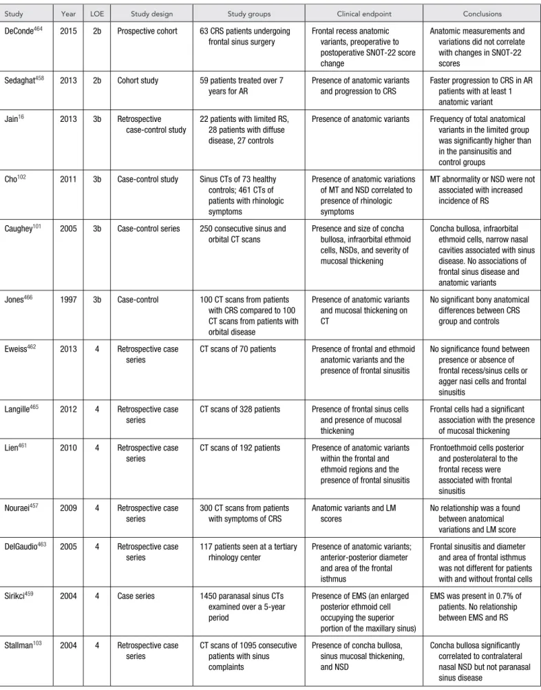
infraor-bital ethmoid cells (Haller cells), and NSDs.95A 97-patient
case-control study by Jain et al.16examined patients with
clinical symptoms of CRS and either CT evidence of pansi-nusitis or isolated maxillary/ostiomeatal complex (OMC) disease (classified as “limited sinusitis”). Both groups were compared to normal controls. The premise of that study was that, if anatomical anomalies truly caused obstructive RS, patients with limited sinusitis should have a higher in-cidence of pathologic anatomic variants relative to patients with pansinusitis or controls. The authors searched for con-cha bullosa, infraorbital ethmoid cells, lateralized uncinate processes, accessory ostium, and paradoxical MTs in both groups. They found that only the presence of a concha bullosa and accessory ostium were significantly related to those cases associated with obstructive pathology (“limited disease”).
Another study, by Alkire and Bhattacharyya,17
com-pared 36 patients meeting strict criteria for RARS to 42
contemporaneous control patients, searching for causative anatomical anomalies. The presence of infraorbital ethmoid
cells (39.9% vs 11.9%, p = 0.006) and a narrowed
in-fundibulum (0.591 mm vs 0.823 mm, p < 0.001) were
identified as potential causative factors. Additionally, the authors noted a trend that did not achieve statistical signif-icance for increased presence of concha bullosa and septal spurs in cases of RARS.
When the literature is collectively considered by sorting studies according to ARS vs CRS, a few findings emerge (Table V-1). CRS appears unrelated to anatomic variation and is more likely inflammatory in nature. ARS appears to be related to several anatomical anomalies: concha
bul-losa, infraorbital ethmoid cells greater than 3 mm in size,97
accessory ostia in the common drainage pathway,98 and
stenosis of the infundibulum.16
Non–OMC-related causes of ARS include oroantral fis-tula and dental infections. One retrospective case series showed that a periapical abscess of a maxillary tooth
has a 9.75 odds ratio (p < 0.001) of causing
substan-tial reactive mucosal thickening on cone beam CT.110
Additionally, another study showed that periodontal dis-ease with tooth roots emerging into the antrum and oroantral fistulas can cause the symptoms and signs of
ARS.96
In summary, the evidence for an association between ARS and anatomic variants is weak and largely inferred from studies on RARS, CRS, and mixed groups of RS (Table V-2).
r
Aggregate Grade of Evidence: C (Level 4: 16 studies).TABLE V-2. Evidence for anatomic variants and ARS
Study Year LOE Study design Study groups Clinical endpoint Conclusions
Jain16 2013 4 Diagnostic case-control 1. Maxillary/OMC
inflammation (limited); 2. Pansinusitis; 3. Normal (control)
CT evidence of sinus disease
Limited disease showed increased concha bullosa and accessory ostium
Shanbhag110 2013 4 Retrospective case series CT with maxillary
sinusitis
1. Fluid filling sinus (by thirds); 2. Mucosal thickening
Oroantral fistula, periodontal disease and projected root or abscess predict maxillary sinusitis
Azila106 2011 4 Diagnostic case-control 1. CRS symptoms; 2.
Normal
CT evidence of sinus disease
No effect of concha bullosa, paradoxical MT, Infraorbital ethmoid cell, NSD, or uncinate bullosa
Cho102 2011 4 Diagnostic case-control 1. CRS symptoms; 2.
Normal
CT evidence of sinus disease
No effect of NSD, concha bullosa, or paradoxical MT
Alkire17 2010 4 Diagnostic case-control 1. RARS symptoms; 2.
Normal
CT evidence of sinus disease
RARS associated with Infraorbital ethmoid cell and smaller infundibular width
Bomeli96 2009 4 Retrospective case series CT with mucosal
thickening
1. Periapical tooth lucencies; 2. Periodontal disease
Periapical lucencies increase presence of sinus inflammation by 9.75 times (odds ratio)
Caughey101 2005 4 Diagnostic case-control 1. CT evidence of
mucosal thickening; 2. Normal CT
CT evidence of sinus disease
Concha bullosa, NSD, and infraorbital ethmoid cell increases risk of sinus disease
Kieff107 2004 4 Diagnostic case-control 1. Ipsilateral maxillary
CRS; 2. Contralateral normal side
CT evidence of sinus disease
No effect from concha bullosa, Infraorbital ethmoid cell, NSD or paradoxical middle turbinate
Stallman103 2004 4 Diagnostic case-control 1. CT with mucosal
disease with concha bullosa; 2. CT with mucosal disease without concha bullosa
CT evidence of sinus disease
In cases of mucosal thickening, no increased chance of concha bullosa
Stackpole97 1997 4 Diagnostic case-control CT evidence of
mucosal thickening and Infraorbital ethmoid cells
CT evidence of sinus disease
Infraorbital ethmoid cell size predicts mucosal thickening on CTs
Lam104 1996 4 Diagnostic case-control CRS with concha
bullosa
CT evidence of sinus disease
No evidence that concha bullosa has effect on CRS
Nadas105 1995 4 Diagnostic case-control Concha bullosa:
absent, small, medium, and large
CT evidence of sinus disease
Concha bullosa appears unlikely to have an effect on CRS
Bolger99 1991 4 Diagnostic case-control 1. CRS symptoms; 2.
Normal
CT evidence of sinus disease
Concha bullosa showed association with CRS; infraorbital ethmoid cell showed no association
Calhoun100 1991 4 Diagnostic case-control 1. Any sinus
symptoms; 2. No sinus symptoms
CT evidence of sinus disease
TABLE V-3. Evidence for allergy and ARS
Study Year LOE Study design Study groups Clinical endpoint Conclusions
Baroody115 2008 1b DB crossover (n=20) Allergic subjects who
underwent nasal challenge; controls
Eosinophils in maxillary sinus Nasal challenge with allergen causes increased eosinophils in the maxillary sinus
Rantala120 2013 2a Cross-sectional
(n=1008)
Atopic and nonatopic adults age 21–63 years
Upper and lower respiratory tract infections
Individuals with atopic disease had higher risk of developing URIs including RS
Chen114 2001 2a Questionnaire (n=8723) Children in Taiwan Rhinosinusitis Children reporting allergic more
likely to have RS
Holzmann113 2001 2b Retrospective review
(n=102)
Children with orbital complications of ARS
Prevalence of AR Orbital complications more common in allergy season
Frerichs121 2014 3a Systematic review Allergic and nonallergic
patients
Prolonged course (>4 weeks) of RS
No significant increase in prolonged RS
Naclerio116 1997 3a Observational (n=10) Allergic subjects at peak
of season
Sinus CT abnormality 60% had CT abnormalities
Savolainen112 1989 3b Case control (n=224) Acute maxillary sinusitis
with and without allergy
ARS Prevalence of AR 25% and 16.5% in non-AR patients
DB=double-blind.
evidence, plausible mechanisms that may explain their in-teraction, relevant human and animal studies, and whether treatment of AR changes disease expression in ARS.
The estimated prevalence of AR is about 20%, whereas over 50% of the U.S. population has evidence of IgE sen-sitization, demonstrating that a positive skin test does not
necessarily indicate nasal allergic disease.111Thus, positive
skin tests in ARS patients are not proof of a relationship between AR and ARS because they only indicate sensiti-zation. Conversely, local nasal allergic reactions can occur without evidence of systemic IgE sensitization. Thus, even the absence of a positive allergy test does not rule out a role for AR in ARS.
Many, but not all, studies support an association
be-tween AR and ARS. Savolainen112 found the incidence of
allergy to be 25% in a group of 224 patients with acute maxillary sinusitis, which was significantly greater than the
16% incidence in a control group. Holzmann et al.113
re-ported an increased prevalence of AR in children who had orbital complications of ARS, and also reported that these complications occurred more commonly during pollinating
seasons. In a study involving 8723 children, Chen et al.114
found the prevalence of RS to be significantly higher in children with AR than in children without allergies. Impor-tantly, having AR did not predict a prolonged course of ARS.
Studies suggest that allergic inflammation may lead to inflammation in the sinuses. On a pathologic basis, nasal challenge with allergens in allergic individuals leads to an
influx of eosinophils into the maxillary sinus.115 Similarly,
the majority of subjects with ragweed-sensitive AR (60%) had sinus mucosal abnormalities on CT imaging during the peak of ragweed season, yet resolution of symptoms after
treatment did not correlate with radiologic imaging.116
Fur-thermore, individuals with ragweed AR had significantly more eosinophils in the maxillary sinus during the ragweed
season compared to outside the ragweed season.117 These
studies suggest that AR could affect the inflammation in the sinuses.
Because of the challenges of human studies, a mouse model was developed to address the influence of AR on ARS.118, 119 Mice with ongoing nasal allergic reaction (but not mice with lower airway reaction or sensitization alone) had a worsened episode of ARS, and this effect could be transferred by Th cells. These studies suggest that local al-lergic inflammation plays an important role in the expres-sion of ARS.
Currently, there are no definitive data that show that medical treatment or immunotherapy prevents the devel-opment of ARS. There are no studies demonstrating that treatment for seasonal or perennial rhinitis reduces the inci-dence of ARS during allergen-exposed periods. The limited frequency of ARS occurring during seasonal AR makes the prospect of a definitive immunotherapy study whose out-come is the development of ARS highly unlikely.
In summary, observational studies provide a modest LOE supporting a relationship between AR and ARS. This is fur-ther supported by evidence from a mouse model of ARS in-duction in allergen-sensitized, allergen-exposed mice. There is some evidence that AR increases the likelihood of orbital complications of ARS but no evidence that AR prolongs the duration of ARS (Table V-3).