INTERNATIONAL JOURNAL OF APPLIED
SCIENCE AND RESEARCH
ISSN IS :
2581-7876
www.ijasr.org
2019
V0L.-2
ISSU.-1
Copyright © 2019 IJASR All rights reserved Page 1
Radiological Evaluation of Missing or Root canal treated of First permanent molar
tooth in the Patient age group between 12 to14 years, Al Rass General Hospital.
Daij AL Daiji, Hisham Al Owaimer.Mashael Alhebs, Monirah Alfawaz, Asma AlHazza.
AlRass General Hospital Dental Department
Abstract
The first permanent molars usually erupt between ages (6-7) years and the important role of these teeth in the correct development of the adult dentition, Objectives: to evaluate the condition of FPM`s in patients aged 12,13 &14 years old .Method: 256 patients between 12 to14 years old panoramic radiographs was evaluated for missing or endodontically treated, the Result show alarming poor condition of PFM`s despite the effort done to control dental caries in the region.
Keywords: First molar, dentistry, radiology.
1.
Introduction
“We know that good oral health is an integral component of good general health. Although enjoying good oral health includes more than just having healthy teeth".
Our target is to save it for life time use, and nothing gives better replacement. It is a real problem if adolescents will lose their teeth in such young age.
In this research will evaluate the first posterior molar if it’s missing, or endodontically treated. The first permanent molars usually erupt between ages (6-7) years and the important role of these teeth in the correct development of the adult dentition can be compromised by their vulnerability to dental caries and developmental structural defects which, without treatment, can lead to their progressive destruction, so loss of permanent molars is always associated with serious consequences .
The main causes of tooth loss are dental caries, dental trauma, periodontal disease.
dental caries is the single most common chronic childhood disease, all experts on dental caries generally agree that it is an infectious and communicable disease and that multiple factors influence the initiation and progression of the disease
Many reasons are correlated with tooth loss such socio-economic status (children and adolescents living in poverty suffer twice as much tooth decay), unhealthy diets (studies have shown a relationship between diet and dental caries, evidence confirm a relationship between-meal snacking and the frequency of eating and drinking are related to dental caries incidence.
Preventive Dental Programs:
Preventive dental programs and awareness programs are implemented in the region education and the whole Kingdome of Saudi Arabia through ministry of health and ministry of health to reduce the caries which is the main reason for destruction of teeth, the 133000-population city has 17 primary health centers and a referral Hospital. Al Rass General Hospital.
INTERNATIONAL JOURNAL OF APPLIED
SCIENCE AND RESEARCH
ISSN IS :
2581-7876
www.ijasr.org
2019
V0L.-2
ISSU.-1
Copyright © 2019 IJASR All rights reserved Page 2
Diagnostic campaigns for school students and campaigns in big malls also done to retrieve data about the condition of the dental oral hygiene of the community. But it’s limited to visual diagnostic alone which cannot detect impacted or endodontics treated teeth.
Primary health centers providing service to the patients starting from making proper treatment planning, implement caries detection methods, restore cavitated teeth and applying pit & fissure sealants
PIT AND FISSURE SEALANTS, the cariostatic properties of sealants are attributed to the physical obstruction of the pits and grooves. This prevents colonization of the pits and fissures with new bacteria and also prevents the penetration of fermentable carbohydrates to any bacteria remaining in the pits and
fissures, so that the remaining bacteria cannot produce acid in cariogenic concentration,
Many clinical studies have reported on the success of pit and fissure sealants with respect to caries reduction. As the longevity of the sealant increases, the retention rate becomes a determinant of its effectiveness as a caries preventive measure .Studies have shown that the placement of pit and fissure sealants is an effective caries preventive measure.
Another role of the primary care performing a routine recall for patients to make sure they are brushing their teeth correctly to reduce the plaque accumulation, diet analysis, CRT and fluoride application should be ready protocol to keep teeth minimally exposed to caries process.
Why this, why now?
While working as a team in ALRass General hospital it was noticed that many patients of adolescent are referred to oral surgery clinic or endodontics away more than orthodontics or prosthodontics. Questions was put on the table and the first was, what is the common status of these category? In this study the whole sample of patients ages 12 – 13 – 14 years visiting the referral hospital of Al Rass was taken to reflect a common picture of the status of FPM, which is the first tooth to erupt and exposes to caries process.
2.
2. Materials and Methods:
3.
2.1. Study setting:
Retrospective, Hospital based study to examine the prevalence of tooth loss or endodontically treated in Saudi adolescents aged between 12-13-14 years old.
2.1.1. Population of the Study:
The sample was recruited from patients visiting and treated in AL Rass Al Qassim Region SAUDI ARABIA.
2.1.3. Study Instruments:
The data will be collected in the predesigned and pretested perform, the following variables will be collected like Age, sex, oral hygiene, and panoramic showing healthy, extraction, root canal treated of tooth related to first permanent molar.
A program that reviews and stores information (Digiview© program),
INTERNATIONAL JOURNAL OF APPLIED
SCIENCE AND RESEARCH
ISSN IS :
2581-7876
www.ijasr.org
2019
V0L.-2
ISSU.-1
Copyright © 2019 IJASR All rights reserved Page 3
No patient will examined physically, only historical radiographic images (Panoramic) that have detailed information about patient date of birth, name, and time panoramic image was done.
Radiographic examination:
Panoramic radiographs are the most widely used radiological diagnostic technique in dental practice, offering full vision of the maxilla and mandible and adjacent regions.,
A standardized step radiographic protocol to take panoramic radiographs as a basic tool examination as Al Rass General Hospital Dental department is a Referral hospital for 17 primary health care centers.
Radiographic assessment:
The PAN images were exported from their dedicated software and viewed in general software (DigiView™ picture archiving and communication system) with possibilities to use zoom function and image enhancement, including brightness, contrast and gamma curve functions.
Data treatment
Data were analyzed using SPSS® 13.0 (SPSS Inc., Chicago, IL). Descriptive statistics calculated the number of operated first molars based on each of the radiographic methods.
2.2. Methodology
A Cross sectional study, descriptive analytic method by using a (Digiview© program) and test the hypothesis was followed.
Statistical method followed was based on using Microsoft Excel 2010 in analysis and Fisher's Exact Test to measure the validity and reliability.
2.2.1. The Sample
256 patients aged between 12-14years old panoramic image studied for detecting of missing or endodontic treated. Sample included all patients reported through 2004-2005-2006.
Inclusion criteria
Patient group between 12, 13 and 14 years old. Starting from 1/04/2018. Patient visiting & treated at dental department at Al Rass general hospital. Male patient and female patients.
Saudi and non-Saudi patients.
Exclusion criteria
Patients above 14 years old. Starting from 15/04/2018.
INTERNATIONAL JOURNAL OF APPLIED
SCIENCE AND RESEARCH
ISSN IS :
2581-7876
www.ijasr.org
2019
V0L.-2
ISSU.-1
Copyright © 2019 IJASR All rights reserved Page 4
2.2.2. Ethical Approval
The ethical approval was attained from Saudi Arabia ministry of health, through the local committee of ethical approval, higher studies relations office, medical education and training affairs, Buraidah city, ALQassim Region, Saudi Arabia. Feb15.2018.
3. Results and Observations
3.1. Section 1: General Information
3.1.1. Distribution of Population
In the present study, a total of 256 patients visited or treated in the Dental Department of AlRass General Hospital.
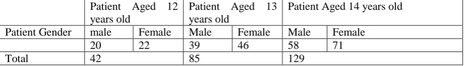
3.1.2. Sample Characteristics
Table 1 showing sample characteristics:
Patient Aged 14 years old Patient Aged 13
years old Patient Aged 12
years old
Female Male
Female Male
Female male
Patient Gender
71 58
46 39
22 20
129 85
42 Total
3.2. Section 2: Final Radiographic Result
Data collected from this section and based on the results displayed in this table the results are summarized below.
Table 2. The number of permanent mandibular first & second molars found missed in patients aged 12 years old.
left permanent mandibular second molar left permanent
mandibular first molar
Right permanent
mandibular second molar
Right permanent
mandibular first molar
Tooth number
Female Male
Female Male
Female Male
Female male
Patient Gender
0 0
2 1
0 0
1 1
Number of
extracted teeth detected
0 3
0 2
Total
INTERNATIONAL JOURNAL OF APPLIED
SCIENCE AND RESEARCH
ISSN IS :
2581-7876
www.ijasr.org
2019
V0L.-2
ISSU.-1
Copyright © 2019 IJASR All rights reserved Page 5
left permanent
mandibular second molar
left permanent mandibular first molar
Right permanent mandibular second molar
Right permanent mandibular first molar Tooth number Female Male Female Male Female Male Female male Patient Gender 0 11 9 0 0 11 10
Number of
extracted teeth detected 0 20 0 21 Total
Table 4. The number of permanent maxillary first & second molars found missed in patients aged 12 years old.
left permanent maxillary second molar left permanent
maxillary first molar
Right permanent maxillary second molar
Right permanent maxillary first molar Tooth number Female Male Female Male Female Male Female male Patient Gender 0 0 0 0 0 0 0 0
Number of
extracted teeth detected 0 0 0 0 Total
Table 5. The number of left permanent maxillary first & second molars found with Root canal Treated in patients aged 12 years old.
left permanent maxillary second molar left permanent
maxillary first molar
Right permanent maxillary second molar
Right permanent maxillary first molar Tooth number Female Male Female Male Female Male Female male Patient Gender 0 0 1 1 0 0 5 2
Number of
extracted teeth detected 0 2 0 7 Total
Table 6. The number of permanent mandibular first & second molars found missed in patients aged 13 years old.
left permanent mandibular second molar left permanent
mandibular first molar
Right permanent mandibular second molar
Right permanent mandibular first molar Tooth number Female Male Female Male Female Male Female male Patient Gender 0 1 4 4 0 0 3 6
Number of
INTERNATIONAL JOURNAL OF APPLIED
SCIENCE AND RESEARCH
ISSN IS :
2581-7876
www.ijasr.org
2019
V0L.-2
ISSU.-1
Copyright © 2019 IJASR All rights reserved Page 6
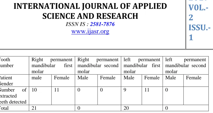
Table 7. The number of permanent mandibular first& second molars found with Root canal Treatment in patients aged 13 years old.
left permanent mandibular second molar left permanent
mandibular first molar
Right permanent mandibular second molar
Right permanent mandibular first molar Tooth number Female Male Female Male Female Male Female male Patient Gender 0 0 12 10 0 0 12 8
Number of
extracted teeth detected 0 22 0 20 Total
Table 8. The number of permanent maxillary first & second molars found missed in patients aged 13 years old.
left permanent maxillary second molar
left permanent maxillary first molar
Right permanent maxillary second molar
Right permanent maxillary first molar Tooth number Female Male Female Male Female Male Female male Patient Gender 0 0 3 3 0 0 1 1
Number of
extracted teeth detected 0 6 0 2 Total
Table 9. The number of left permanent maxillary first & second molars found with Root canal Treated in patients aged 13 years old.
left permanent maxillary second molar
left permanent maxillary first molar
Right permanent maxillary second molar
Right permanent maxillary first molar Tooth number Female Male Female Male Female Male Female male Patient Gender 0 0 4 2 0 0 7 4
Number of
extracted teeth detected 0 6 0 11 Total
Table 10. The number of permanent mandibular first & second molars found missed in patients aged 14 years old.
left permanent mandibular second molar left permanent
mandibular first molar
Right permanent mandibular second molar
Right permanent mandibular first molar Tooth number Female Male Female Male Female Male Female male Patient Gender 1 0 11 6 0 1 9 8
Number of
INTERNATIONAL JOURNAL OF APPLIED
SCIENCE AND RESEARCH
ISSN IS :
2581-7876
www.ijasr.org
2019
V0L.-2
ISSU.-1
Copyright © 2019 IJASR All rights reserved Page 7
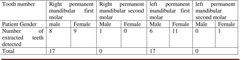
Table 11. The number of permanent mandibular first& second molars found with Root canal Treatment in patients aged 14 years old.
left permanent mandibular second molar left permanent
mandibular first molar
Right permanent mandibular second molar
Right permanent mandibular first molar Tooth number Female Male Female Male Female Male Female male Patient Gender 1 0 18 17 2 1 12 13
Number of
extracted teeth detected 1 35 3 25 Total
Table 12. The number of permanent maxillary first & second molars found missed in patients aged 14 years old.
left permanent maxillary second molar
left permanent maxillary first molar
Right permanent maxillary second molar
Right permanent maxillary first molar Tooth number Female Male Female Male Female Male Female male Patient Gender 0 1 4 3 0 1 4 2
Number of
extracted teeth detected 1 7 1 6 Total
Table 13. The number of permanent maxillary first & second molars found with Root canal Treated in patients aged 14 years old.
left permanent maxillary second molar
left permanent maxillary first molar
Right permanent maxillary second molar
Right permanent maxillary first molar Tooth number Female Male Female Male Female Male Female male Patient Gender 0 0 4 2 0 0 5 3
Number of
extracted teeth detected 0 6 0 8 Total Discussion
INTERNATIONAL JOURNAL OF APPLIED
SCIENCE AND RESEARCH
ISSN IS :
2581-7876
www.ijasr.org
2019
V0L.-2
ISSU.-1
Copyright © 2019 IJASR All rights reserved Page 8
The ministry of health in Saudi Arabia provide oral health care free of charge, starts from primary health care centers then to referral hospitals if needed. 17 PHC’s referring to AL Rass General Hospital (the site for this study).
The result of this study shows that patients visited the dental department at age of 12 years old is only 42 patients, the number doubled by age of 13 (85) patients and increased up to (129) patients at age of 14 years old.
Among patients aged 12 years old root canal treatment of lower mandibular first molar was significant.
The missing mandibular first molar increased in patients aged 13 years old while 17 maxillary first molars out of 85 patients were shown with root canal treatment.
At age of 14 years old the patients presented with root canal filled in first mandibular molars were proximally 50% of the sample.
Bar chart showing number of patients aged 12-13-14 years old having root canal treatment.
INTERNATIONAL JOURNAL OF APPLIED
SCIENCE AND RESEARCH
ISSN IS :
2581-7876
www.ijasr.org
2019
V0L.-2
ISSU.-1
Copyright © 2019 IJASR All rights reserved Page 9
Conclusion
4.
This study answered the question of the common status of FPM which was not promising,
the study can be a basic for more studies in regarding accomplishing best oral health care
to patients in primary care.
There is a need for guidelines advising primary care dentists in how to be more effective to control the high-risk caries process.
It highlights the need for extensive prevention public programs targeted the population through mass media and one to one interview in how to brush effectively, visit the dentist for routine check.
References:
[1] Eichenberger M1, Erb J1, Zwahlen M2, Schätzle M1.. The timing of extraction of non-restorable first permanent molars: a systematic review. Send to Eur J Paediatr Dent. 2015 Dec;16(4):272-8. .2013. [PubMed]
[2] DC-V Ong,*JE Bleakley. Compromised first permanent molars: an orthodontic perspective. Australian Dental Journal2010; 55: 2–14.
[3] D.S. GILL, R.T. LEEAND C.J. TREDWIN . Treatment Planning for the Loss of First Permanent Molars. Dental Update – July/August 2001.
[4] S. Albadri,1 H. Zaitoun, Extraction of first permanent molar teeth: results from three dental hospitals. BRITISH DENTAL JOURNAL 2007.
[5] L H Matzen,corresponding author1 S Schou,2 J Christensen,1,3 H Hintze,1 and A Wenzel1. Audit of a 5-year radiographic protocol for assessment of mandibular third molars before surgical intervention. Proceedings of the IADMFR; 22–27 June; Bergen, Norway, 2013.
[6] Wenzel A, , Gotfredsen E. Audit for extraoral radiographic examinations in a digital department. Dentomaxillofac Radiol 2005; 34: 228–30. [PubMed]
[7] Williams JK, Gowans AJ. Hypomineralised first permanent molarsand the orthodontist. Eur J Paediatr Dent 2003;4:129–132.