Automated Classification and Detection of
Macular hole by using OCT image
M.Saya Nandini Devi1, S.Santhi2
Research Scholar, Dept. of Electronics &Instrumentation Engg., Annamalai University, Chidambaram,
Tamil Nadu, India1
Professor, Dept. of Electronics & Instrumentation Engg., Annamalai University, Chidambaram, Tamil Nadu, India.2
ABSTRACT:The major cause of blindness in India and worldwide is recognized as Diabetic Retinopathy [DR], which is an eye related disorder caused by diabetes. Diabetic retinopathy is a severe problem which occurs in retinal part of the inner eye. It causes blur or distorted vision leading to vision loss. Optical Coherence Tomography (OCT) provides important additional information about the retina. It produces dependable, reproducible and objective retinal images especially in diabetic macular hole and provides information that can clearly only be detected with OCT. Macular hole is a small break in the macula located in the centre of the eye light sensitive tissue called retina. The macula provides the sharp, central vision we need for reading, driving and seeing fine detail. In this paper, we investigate an automated detection of macular hole by using BPNN (Back Propagation Neural Network) classifier and implemented by using MATLAB. These processes can automatically detect the problem of macular hole.
KEYWORDS: Diabetic Retinopathy; Optical Coherence Tomography; Macular hole; BPNN classifier; MATLAB software.
I.INTRODUCTION
Diabetic retinopathy is a difficulty of diabetes and the leading cause of vision impairment and blindness among working-age adults. It occurs when diabetes damages the tiny blood vessels in the retina, which is the light-sensitive tissue at the backside of the eye. Diabetic retinopathy may lead to diabetic macular edema (DME), which is a swelling in an area of the retina called the macular [National Eye Institute]. Thus significant reduction in morbidity can be achieved.
The various methods of assessment of the macular hole are
Slit-lamp bio- microscopy
Binocular indirect ophthalmoscopy
Ultrasound B scan
Fundus fluoresce in angiography
Optical coherence tomography
Each method has its advantages and disadvantages.
Slit-lamp bio- microscopy
Binocular indirect ophthalmoscopy
Binocular indirect ophthalmoscope has become an indispensable tool to diagnose and manage a variety of vitreoretinal disorders. The instrument has a rich history evolving through many generations to yield the current diagnostic marvel. Sophisticated additions to the basic technology include high magnification lenses built in to the ophthalmoscope, video adapters that facilitate patient and student education as well as open up an array of telemedical possibilities and laser photocoagulation systems mounted on to the indirect ophthalmoscope to treat peripheral tears through a 20 diopter lens [7].
FFA (Fundus fluorescein angiography)
Fluorescein Angiography as an important tool is understanding the diagnosis and treatment of retinal disorders. The diagnostic procedure utilizes a specialized fundus camera to capture rapid sequence of photographs of retina following an intravenous injection of fluorescein sodium. FFA facilitates in vivo study of retinal circulation and it is particularly useful in Diabetic Retinopathy and macular degeneration, the two leading causes of retinal blindness [6].
.
OCT (optical coherence tomography)
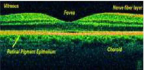
OCT is a non-invasive method by which the layers of the retina can be imaged in a very short time. It has the highest resolution of 3 microns, even though presently the OCT machines are very expensive and not available at every center in our country. OCT gives a quick evaluation of the retinal anatomy and functional status both in quality and quantity Fig. 1 shows an OCT scan of retina.
OCT can be particularly helpful in diagnosing:
Macular hole
Macular pucker
Vitreomacular traction
Macular edema
Macular Degeneration
In diabetics, OCT is most useful to measure and quantify macular hole, and in the era of anti-VEGF therapy, it has become the most important imaging tool in managing patients with diabetic macular hole.
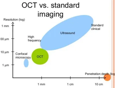
An OCT (Optical Coherence Tomography) camera can instantly obtain cross-section images of sub-surface layers of transparent or opaque materials such as the structure of the retina. Fig. 2 shows the merits of OCT in comparison with normal imaging standard.
Fig. 2 Comparison of OCT and standard imaging
In OCT light reflected from the tissue in a similar way as in Ultrasonography. Hence OCT light scans the tissue rapidly several times and generating multiple images very rapidly. This is combined to form a linear image like a B-scan ultrasound image Light travels too rapidly to be detected directly hence the reflected light is measured indirectly using a method called low coherence interferometry, the axial resolution is determined by the band width of the light source. The transverse resolution is reduced to 10-15 micrometer due to natural aberration of the human eye.
Time Domain and Spectral Domain technology
From its inception, OCT images were acquired in a time domain fashion. Time domain systems acquire approximately 400 A-scans per second using 6 radial slices leaning 30 degree apart. Because the slices are 30 degree apart, care must be taken to avoid missing pathology between the slices.
Spectral domain technology, on the other hand, scans around 20,000-40,000 scans per second. This increased scan rate and number diminishes the likelihood of motion artefact, enhances the resolution and decreases the chance of absent lesions. Most time domain OCTs are precise to 10-15 microns but newer spectral domain machines may approach 3 micron resolution. Whereas most time domain OCTs image 6 radial slices, spectral domain systems continuously image a 6mm area.
A reference mirror was continuously moved to detect signal from the various depth of tissue being analysed and an image is created, this image created is called time domain OCT.
But with newer method the signal is detected by a stationary arm allowing for much faster scan speed( imaging speed) and is termed as spectral domain OCT Analysis of the data obtained by the software is the most important function of the OCT device. These algorithms are used to calculate the retinal thickness by automatically delineating the inner and outer retinal boundaries
Advantages of OCT:
Non-contact and noninvasive Method
The retinal and intra retinal measurements can be done
Rapid scan can capture high resolution images
Very short time to capture images
OCT can be done in undilated pupil particularly in angle closure glaucoma.
Disadvantages:
Equipment cost is high
Patient with poor fixation
Inherent limitation of the automated software producing image artifacts.
Needs operator training and skill to get good images
II.REVIEWOFLITERATURE
In this paper [1], compare the retinal thickness by evaluation of stereo fundus photography with that assessed objectively by OCT. The degree of agreement between subjectively and objectively assessed retinal thicknesses was very good implying that changes can be accurately and prospectively measured with OCT. In next paper [2], Analysis of the segmentation layers of intra retinal layers which are important for disease detection present in SDOCT images is presented. Graph based segmentation is employed which solely based on pixel intensity variation and distance between neighbour pixels. This segmentation method is less prone to noise and the pre-processing step can be considered as optional. In another paper [3], author used to localise the intra retinal boundaries. It uses near infra red light ranging from 800-1300mm. It is widely used because of the clinically appreciable features like non-invasive, non contact, no radiation, painless and fast procedure. The technique is sensitive up to 10 microns. OCT can help in diagnosing conditions like macular hole, macular edema, macular degeneration, DR and Glaucoma. OCT image suffer from speckle noise. In another paper [4], Speckle noise is caused due to constructive or destructive interference of the light waves from the object. Initially the image is converted into gray scale as processing of 2D image is easier. The literatures have suggested the use of filter like median, mean and Gaussian filter. In another paper [5], Automatic screening will help the doctor to fast identify the situation of the patient in a more accurate way. The macular abnormalities caused due to DR can be detected by applying morphological operation, filters and threshold on the fundus images of the patient. Early detection of RNFL layer thickness from the OCT images of the patient. The RNFL thickness estimation involves the use of active countor based deformable snake algorithm for segmentation of the anterior and posterior boundaries of the RNFL. This method produces accurate, reliable, and robust detection. DR can be diagnosis using fundus image and Glaucoma can be diagnosed using OCT image.
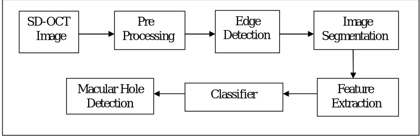
III.PROPOSED ALGORITHM
A. DESIGN CONSIDERATIONS:
This section introduces method for detecting retinal diseases by classifying SD-OCT images.
Fig. 3 Block Diagram of Proposed Method
B. DESCRIPTION OF THE PROPOSED METHOD:
1. Gray Conversion
The intensity plays a major role in the detection of hole in retina OCT images represented with low numbers of pixel intensity values. Each pixel in image having intensity value ranging from minimum 0 (darkest pixel) to maximum value
SD-OCT
Image
Pre
Processing
Edge
Detection
Macular Hole
Detection
Classifier
is 255 (lighter pixel). The region has high and low intensities vary based on image intensity which is taken as image objects. The first step of in pre-processing is mainly to converting RGB color image into a gray scale image.
2. Median Filter
The ‘speckle’ noise can be reduced by Median filter, and comparing to convolution is better. It also preserve from blur edges.
3. Image Enhancement
Contrast-limited adaptive histogram equalization (CLAHE) is used to enhance contrast of all the small regions in an image. CLAHE operates on small regions in the image generally called as tiles. It not takes the entire image during this process. To enhances the contrast of each tile’s. Then the neighboring tiles are combined using bilinear interpolation for eliminate the artificially induced boundaries. In homogeneous areas, it can be used to limit for avoiding amplifying of any noise which prevent an image.
4. Morphological Operation
Morphological process is mainly suitable for analyzing the shapes in images. Two main processes are dilation and erosion which is come under the category of morphological process. In dilation, each and every point in an image is super imposed by the means kernel along with its surrounding pixels. The dilation process is mainly for increasing the size of an original object. Erosion is an inverse procedure of dilation. It is mainly to thin an image through that these are subtracted under a structuring element or kernel. Opening consists of erosion which is followed by dilation and then leads to smooth an image for breaking narrow joints and also removing thin protrusions areas in images. Closing consists of dilation which is followed by erosion mainly to smooth a images, and also eliminate the small holes. These algorithms are used to detect the edges and also by removing noise which occurs in an image for find the specific shapes in images.
4. Edge Detection
Edge detection is mainly performed on the green component image by applying canny edge filter along with suitable threshold. Logical AND operation is used on two images, one is being a threshold image and another one is being an edge detected image. The thresholding is done based on an adaptive histogram equalized image for increase the contrast of an image. Edge can be detected by using canny edge detector.
5. Image Segmentation
To generate the coefficient from the canny edge detector mainly to detect the retinal layer by using wavelet filter. Then once again the detected retinal layer are eroded for find out that OCT image having hole disease having or not based on feature extraction.
6. Feature Extraction
Gray level co-occurrence matrix used to detect the features values of the OCT images. To create a GLCM, use the gray co matrix function based on the pixel intensity in gray level. Hence we find out GLCM for find out the intensity pixels of images.
7. Classifier
above-stated problems are tried with different number of hidden layers; that start ranging from 1 up to 4 layers. Based on these results, a neural network with one hidden layer can be found out and also eight neurons gave the best classification result. A learning constant in presence case is g = 0.9 is chosen by trial and error.
IV.SIMULATIONRESULTS
The simulation study involves with OCT images. The images are taken from OCT, an image which is implemented in MATLAB software. It removes speckle noise from images by using Median filter. Edge can be deducted from OCT images using the Canny edge detector. Wavelet Filters used to segment this process to detect and recover the false edges. Features can be extracted from OCT images based on the shape and size appearance of the images. The values of the noise and de-noise can be vary based on shape and size which are segmented. BPNN classifier used to classify the segmented images. The classifier specifies the sensitivity of 90%, specificity of 90% and performance can be evolved with 80% accuracy. The overall specification can be evolved with the OCT images..
V.CONCLUSION
In this investigated and proposed algorithm, a method is used based on anatomical structural details and retinal image information. This system intends to help the ophthalmologists not only in DR screening process but any other eye related abnormality which is based on retinal photography. It is not a final result application but it can be a preliminary diagnosis tool or a decision support system for ophthalmologists. Human ophthalmologists are still needed for the cases where detection results are not very obvious. This type of presentation will enable clinicians to identify retinal landmarks more quickly and will also help to take decision while treating the abnormality, particularly maculopathy.
REFERENCES
[1] Charlotte Strøm, Birgit Sander, Nicolai Larsen, Michael Larsenv and Henrik Lund-Anders, et al (2002)Diabetic Macular Edema Assessed with Optical Coherence Tomography and Stereo Fundus Photography Investigative Ophthalmology & Visual Science, January 2002,Vol. 43, No. 1
[2] Appaji M. Abhishek, IEEE Member, Tos T. J. M. Berendschot, IEEE Member, Shyam Vasudeva Rao,IEEE Member and Supriya Dabir, Non IEEE member, et al” Segmentation and Analysis of Retinal Layers (ILM & RPE) inOptical Coherence Tomography Images with Edema’’, IEEE Conference on Biomedical Engineering and Sciences, 8 - 10 December 2014, Miri, Sarawak, Malaysia
[3] Hortensia Sa´nchez-Tocino, Aurora Alvarez-Vidal, Miguel J. Maldonado,Javier Moreno-Montan˜e´s, andAlfredo Garcı´a-Layana , et al “Retinal Thickness Study with Optical Coherence Tomography in Patients with Diabetes”, Investigative Ophthalmology & Visual Science, May 2002, Vol. 43, No. 5 Copyright © Association for Research in Vision and Ophthalmology
[4] Kuo A. N., McNabb R. P., Chiu S. J., El-Dairi M. A., Farsiu S., Toth C. A., Izatt J. A., “Correction of Ocular Shape in Retinal Optical Coherence Tomography and Effect on Current Clinical Measures,” Am. J. Ophthalmol. 156(2), 304–311 (2013).10.1016/j.ajo.2013.03.012 [PMC free article] [PubMed]
[5] Arulmozhivarman Pachiyappan, Undurti N Das, Tatavarti VSP Murthy and Rao Tatavarti, et al “Automated diagnosis of diabetic retinopathy and glaucoma using fundus and OCT images”,Lipids inHealth and Disease 2012 11:73.
[6] Mr. John, Retina Clinic, Aravind Eye Hospital,” Fundamental of Fundus Fluorescein Angigography”, Vol. IV, No.3, July - September 2004
[7] Dr. Sonia Rani John DNB, Dr. Meena Chakrabarti MS DO DNB, Dr. Arup Chakrabarti MS DO, et al “Binocular Indirect Ophthalmoscope“,Kerala Journal of Ophthalmology Vol. XXI, No. 4
[8] Funatsu H, Yamashita 9H, Shimizu E, Mimurac T, Nakamura S, et al. (2004)Quantitative measurement of retinal thickness in patients with diabetic macularedema is useful for evaluation of therapeutic agents. Diabetes Res ClinPract66: 219–227
[9] Davis MD, Bressler SB, Aiello LP, Bressler NM, Browning DJ, et al. (2008) Browning and associates. Comparison of Time-DomOCT and Fundus Photographic Assessments of Retinal Thickening in Eyes with Diabetic Macular Edema. Invest Ophthalmol Vis Sci 49: 1745–1752.
[10] Afef Maalej*, Wathek Cheima, Khallouli Asma, Rannen Riadh and Gabsi SalemMilitary Department of Ophthalmology (Tunis), Tunisia,“Optical Coherence Tomography for Diabetic Macular Edema: Early Diagnosis, Classification and Quantitative Assessment” Maalej et al., J Clinic Experiment Ophthalmol 2012, S:2
[11] Massin P, Girach A, Erginay A, Gaudric A (2006)” Optical coherence tomography a key to the future management of patients with diabetic macular oedema”. ActaOphthalmolScand 84: 466-474.
[12] Pratul P. Srinivasan, Leo A. Kim,Priyatham S. Mettu, Scott W. Cousins,Grant M. Comer, Joseph A. Izatt,1,4 and Sina Farsiu1“Fully automated detection of diabetic macular edema and dry age-related macular degeneration from optical coherence tomography images” published 12 Sep 2014 (C) 2014 OSA 1 October 2014 | Vol. 5, No. 10 | DOI:10.1364/BOE.5.003568 | BIOMEDICAL OPTICS EXPRESS 3568
[13] Burt P. J., Adelson E. H., “The Laplacian Pyramid as a Compact Image Code,” IEEE Trans. Commun. 31(4), 532–540 (1983).10.1109/TCOM.1983.1095851
[14] Massin P, Vicaut E, Haouchine B, Erginay A, Paques M, et al. (2001) Reproducibility of retinal mapping using Optical Coherence Tomography. ArchOphthalmol 119: 1135-1142.