Copyright1999 by the Genetics Society of America
Centromere Mapping Functions for Aneuploid Meiotic Products: Analysis of
rec8,
rec10
and
rec11
Mutants of the Fission Yeast
Schizosaccharomyces pombe
Michelle D. Krawchuk and Wayne P. Wahls
Department of Biochemistry, Vanderbilt University School of Medicine, Nashville, Tennessee 37232-0146 Manuscript received January 14, 1999
Accepted for publication May 27, 1999
ABSTRACT
Recent evidence suggests that the position of reciprocal recombination events (crossovers) is important for the segregation of homologous chromosomes during meiosis I and sister chromatids during meiosis II. We developed genetic mapping functions that permit the simultaneous analysis of centromere-proximal crossover recombination and the type of segregation error leading to aneuploidy. The mapping functions were tested in a study of the rec8, rec10, and rec11 mutants of fission yeast. In each mutant we monitored each of the three chromosome pairs. Between 38 and 100% of the chromosome segregation errors in the rec8 mutants were due to meiosis I nondisjunction of homologous chromosomes. The remaining segregation errors were likely the result of precocious separation of sister chromatids, a previously described defect in the rec8 mutants. Between 47 and 100% of segregation errors in the rec10 and rec11 mutants were due to nondisjunction of sister chromatids during meiosis II. In addition, centromere-proximal recombination was reduced as much as 14-fold or more on chromosomes that had experienced nondisjunction. These results demonstrate the utility of the new mapping functions and support models in which sister chromatid cohesion and crossover position are important determinants for proper chromosome segregation in each meiotic division.
D
URING meiosis each chromosome undergoes one mutations that decrease meiotic recombination ( Ponti-celliandSmith1989;Linet al. 1992;LinandSmith round of DNA replication; homologouschromo-somes then pair and undergo recombination. Subse- 1995;Liet al. 1997). In each mutant, crossover recombi-nation is preferentially reduced toward the centers of quently, homologous chromosomes segregate from one
another during meiosis I (MI) and then sister chromatids each of the three chromosomes and, intriguingly, the mutations differentially affect chromosome segregation segregate from one another during meiosis II (MII). In
most organisms, reciprocal recombination events (cross- during MI and MII (Molnaret al. 1995;Krawchuket al. 1999; Parisi et al. 1999). The rec8 mutants suffer overs) help ensure proper MI segregation (Roeder1997).
Recent evidence suggests that the position of cross- aberrant segregation of homologous chromosomes dur-overs has an important role in chromosome segregation ing MI, at least part of which is attributable to the preco-during both MI and MII. In several organisms, chromo- cious separation of sister chromatids (PSS). The rec10 somes that suffer spontaneous MI nondisjunction (MI and rec11 mutants suffer aberrant segregation of sister NDJ) exhibit a preferential reduction of crossovers near chromatids during MII. The three genes are proposed the centromeres (Gethmann1984;Rasoolyet al. 1991; to define a pathway of “meiotic chromatid cohesion” Rosset al. 1992;MacDonaldet al. 1994;Mooreet al. important for proper chromosome segregation in both 1994; Sherman et al. 1994; Hassold et al. 1995). In meiotic divisions (Krawchuket al. 1999). We therefore contrast, chromosomes that suffer spontaneous MII developed mapping functions to simultaneously mea-nondisjunction (MII NDJ) exhibit an elevated fre- sure centromere-proximal recombination and to deter-quency of crossovers near their centromeres (MacDon- mine the segregation patterns of each of the three aldet al. 1994;Fisheret al. 1995;Koehleret al. 1996a,b; S. pombe chromosomes in aneuploid meiotic products Lambet al. 1996). Models invoking a synergistic interac- from each of the rec8, rec10, and rec11 mutants. tion between crossover position and sister chromatid
cohesion can best explain these observations (Moore
andOrr-Weaver1998;Krawchuket al. 1999). MATERIALS AND METHODS The rec81, rec101, and rec111genes of the fission yeast
S. pombe strains and culture media: The S. pombe strains
Schizosaccharomyces pombe were identified in a screen for
used for this study are listed in Table 1. Rich medium was yeast extract agar (YEA) or liquid (YEL); sporulation media was synthetic sporulation agar (SPA); minimal media was nitro-gen base agar (NBA) or liquid (NBL). All media formulations
Corresponding author: Wayne P. Wahls, Department of Biochemistry,
and culture techniques were as described (Gutzet al. 1974).
Vanderbilt University School of Medicine, 621 Light Hall, Nashville,
TN 37232-0146. E-mail: [email protected] YEA-B was YEA containing 2.5mg/ml of Phloxin-B (Sigma,
Development of mapping functions:In a meiosis that gener-TABLE 1
ates four haploid products the centromeres undergo first
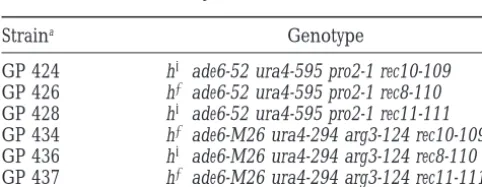
divi-S. pombestrains sion segregation (FDS). Loci unlinked to the centromere
un-dergo second division segregation (SDS) 67% of the time.
Straina Genotype This is due to the statistical probability of the various patterns
by which the unlinked alleles can enter the ascus. By extension, GP 424 h2ade6-52 ura4-595 pro2-1 rec10-109 disomics or diploids arising from MI NDJ will be 100% hetero-GP 426 h1ade6-52 ura4-595 pro2-1 rec8-110 zygous for the centromere and 67% heterozygous for unlinked GP 428 h2ade6-52 ura4-595 pro2-1 rec11-111 loci. Likewise, disomics or diploids arising from MII NDJ will GP 434 h1ade6-M26 ura4-294 arg3-124 rec10-109 be 0% heterozygous for the centromere and 67% heterozy-GP 436 h2ade6-M26 ura4-294 arg3-124 rec8-110 gous for unlinked loci. TheHaldane(1919) mapping func-GP 437 h1ade6-M26 ura4-294 arg3-124 rec11-111 tion describes the relationship between the frequency of re-combinants and genetic distance in haploid meiotic products,
aStrains were as described (DeVeauxet al. 1992).
RF51⁄
2(12e22M), (1)
where M is the genetic distance in Morgans (1M51 exchange) St. Louis) and 100mg/ml of adenine. SPA, NBA, and NBL
between two markers and RF is the fraction of recombinants. were supplemented with required amino acids, adenine, and
In the Haldane mapping function the RF reaches a theoretical uracil at 100mg/ml. The growth factor requirement of pro2
maximum of 1⁄
2 because of multiple recombination events: mutants is partially satisfied by arginine. Therefore, citrulline
the second exchange event in a genetic interval restores the (50mg/ml), which does not satisfy the growth factor
require-parental linkage of markers. As the genetic distance becomes ment of the pro2 mutants, was used to satisfy the growth factor
large the probability of even numbers of crossovers (that re-requirement of the arg3 mutants.
move recombinants) equals the probability of odd numbers Mating and meiosis:Strains to be crossed were grown at 328
of crossovers (that create recombinants). Hence, for two un-in 5 ml of YEL to a density of 13107cells/ml. The cultures
linked markers and a sufficiently large sample size the in-were combined, the cells in-were harvested by centrifugation,
termarker RF will reach a plateau of (1⁄
2), which is the limiting washed with water, resuspended in 100ml of water, and spotted
multiplier in the Haldane equation. on SPA. The mating mixtures were incubated at room
temper-In a similar fashion, the following equation relates diploid ature for 3–5 days. Asci and vegetative cells were harvested
heterozygosity to genetic distance in those diploids arising into 0.5 ml of 0.5% glusulase and incubated at room
tempera-from MII NDJ: ture for 8 hr to liberate spores and kill vegetative cells. The
suspensions were then treated with an equal volume of 60% RF
heterozygous52⁄3(12e22m). (2) ethanol for 30 min to kill any remaining vegetative cells. Spore
suspensions were washed once with water, diluted, and plated In this case, the RFheterozygousis the fraction of diploids that are
on the appropriate media. heterozygous for the locus being studied and m is the genetic
Diploid spore isolation and haploidization analysis:Spores distance between the marker being studied and the centro-were plated on YEA-B and incubated for 3 days at 328 to mere. (We apply a lowercase m for the genetic distance be-distinguish the diploid spore colonies from the haploid spore tween a marker and the centromere to distinguish from the colonies. On Phloxin-B-containing plates, haploid cells pro- uppercase M used for the genetic distance between two mark-duce light pink colonies whereas diploid cells promark-duce dark ers.) Because of the way that alleles assort into the diploids, pink colonies (Gutzet al. 1974). The ploidy was confirmed the heterozygosity resulting from MII NDJ can range from 0 by microscopic examination of the cells. Diploid cells are to 67%, so the Haldane multiplier of1⁄
2becomes2⁄3, but the both longer and wider than haploid cells and can be readily remainder of the equation is unchanged. By solving for m we distinguished under the microscope. can determine the genetic map distance of the locus from the The parental haploid strains that were crossed had different centromere as a function of the frequency of heterozygosity in alleles at several loci on all three chromosomes. The genotypes the MII NDJ diploids:
of the resulting diploid spore colonies were determined by
m5 21⁄
2[Ln(121.5RFheterozygous)]. (3) analysis of haploidized colonies on diagnostic plates. Diploid
spore colonies were haploidized with m-fluorophenylalanine A similar equation describes the relationship between fre-(m-FPA;Kohliet al. 1977). After 2 days incubation at 328in quency of heterozygosity and genetic distance in those diploids supplemented NBL containing 0.4–0.8% m-FPA, the liquid arising from MI NDJ:
cultures were diluted and plated on YEA-B to determine
hap-12RFheterozygous51⁄3(12e22m). (4) loidization efficiency, which ranged from 40 to 95%. Fifty
haploidized colonies derived from each individual diploid
In this case, 12RFheterozygousis the fraction of diploids that are spore colony were patched onto YEA to score for the two
homozygous for the locus being studied. Because of the way different ade6 alleles. On media with limiting amounts of
ade-that alleles assort into the diploids, the homozygosity values nine, such as YEA, S. pombe strains carrying the ade6-M26 allele
for MI NDJ diploids can range from 0% for the centromere form dark red colonies while strains carrying the ade6-52 allele
to 33% for an unlinked marker. Thus, the Haldane multiplier form pink colonies (Gutz1971;Gutzet al. 1974). After
geno-of 1⁄
2 becomes 1⁄3. Solving for m gives the MI NDJ diploid typing the ade6 locus, the patches were replica plated to various
mapping function differentially supplemented minimal media to genotype the
pro2 and arg3 loci. The mating-type locus on chromosome II m5 21⁄
2[Ln(3RFheterozygous22)]. (5) was scored for allele type by analysis of the diploid spore
For PSS during MI, followed by random assortment of all colonies. Diploid spore colonies were patched onto SPA and
the chromatids in both meiotic divisions, 67% of the diploid incubated at room temperature for 2–3 days. Iodine vapor
meiotic products are expected to be heterozygous for the staining was used to distinguish the heterozygous diploids
centromere. This is because the centromeres become essen-(h1/h2, sporulate and stain with iodine) from the homozygous
tially nonfunctional for segregation and thus partition into diploids (h1/h1 or h2/h2, do not sporulate and stain with
Figure 1.—Types of chromosome segregation in meiosis based upon inheritance of centromere-linked markers. Only a single homologous chromosome pair is shown for the sake of clarity. The lines represent individual chromatids within homologous chromosomes. (A) Normal meiosis results in four haploid products. (B) Meiosis I nondisjunction: homologous chromosomes fail to segregate during the reductional division, resulting in meiotic products that contain two heterozygous chromosomes. (C) Meiosis II nondisjunction: sister chroma-tids fail to segregate during the equational division, resulting in meiotic products that contain two homozygous chromo-somes. (D) Precocious separation of sister chromatids: if both sets of sister chromatids separate precociously, and individual chromatids distribute at random during both MI and MII,
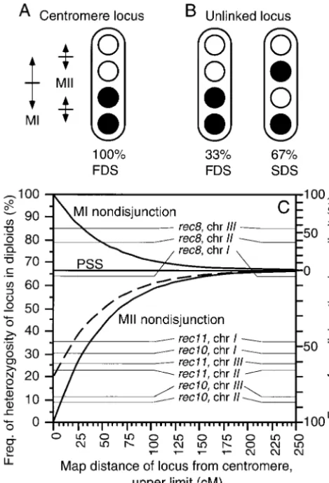
67% of disomic or diploid spores will be heterozygous (mate- Figure2.—Centromere mapping principles and practice. rials and methods). (E) Fates of meiotic products. Loci can be mapped relative to the centromere based upon the frequencies of first division segregation (FDS; markers segregate during MI) and second division segregation (SDS; markers segregate during MII). (A) The centromere exhibits patterns by which the unlinked alleles can enter the ascus.
100% FDS. (B) Recombination between the centromere and Therefore, no diploid mapping function can be derived for
an unlinked locus generates 33% FDS and 67% SDS of alleles PSS events, because markers linked to the centromere and
at that locus. Any deviation from 67% SDS of alleles indicates markers unlinked to the centromere will each yield the same
that the locus is linked to the centromere and the genetic fraction (67%) of heterozygous diploids.
distance can be calculated with a mathematical equation de-rived from the mapping function of Haldane(1919). This principle can be applied to the meiotic diploid (or disomic)
RESULTS AND DISCUSSION spores to reveal both the segregational and recombinational
history of loci relative to the centromere. (C) Mapping
func-Centromere mapping functions for aberrantly
segre-tions showing the relasegre-tionship between the frequency of
het-gated chromosomes:In a normal (wild-type) meiosis, erozygous diploids, the type of chromosome segregation error, two rounds of chromosome segregation produce hap- and genetic distance. By definition, MI NDJ generates diploids that are all heterozygous at the centromere, while MII NDJ loid meiotic products (Figure 1A). MI NDJ (Figure 1B),
generates diploids that are all homozygous at the centromere. MII NDJ (Figure 1C), and PSS (Figure 1D) can be
in-Departures from these diploid heterozygosity values are due ferred from the segregation patterns of
centromere-to the genetic distance of the locus from the centromere (i.e., linked genetic markers. In S. pombe, most disomic aneu- recombination, thick lines), or NDJ events of the opposite ploids are unstable and cannot be readily studied (Fig- type (e.g., dashed line), or the presence of PSS, or some combi-nation of the three. The diploid heterozygosity mapping func-ure 1E;NiwaandYanagida1985). However, because
tion thus sets the upper limit for genetic distance and the lower fission yeast contains only three pairs of chromosomes,
limit for the type of NDJ. Diploid heterozygosity values from aberrant chromosome segregation leads to a relatively
Table 2 are superimposed upon the mapping functions (thin high frequency of stable diploid meiotic products, thus lines)—these are further described in the text.
permitting segregation analyses of all three pairs of chromosomes within individual meioses (Krawchuket
al. 1999). Haldane(1919), allowed us to develop mathematical
functions that describe the relationship between the The principle of centromere mapping (Figure 2, A
het-erozygosity of any given locus in the meiotic diploids observed (Table 2). In other words, a 2.5-fold increase in recombination relative to wild-type cells (Table 3) (or disomics), and the genetic distance of that locus
from the centromere (Figure 2C). For alleles of loci and 100% MI NDJ can explain the data. Recombination cannot have been increased by.2.5-fold, because this unlinked to the centromere, 67% of the diploids will
be heterozygous regardless of the type of missegregation would further reduce the genetic linkage between cen-tromere III and ade6, thereby producing a frequency of that occurs. PSS will also produce 67% heterozygous
diploids regardless of whether or not the marker is heterozygosity,85% (Figure 2C). However, 30 cM only defines the upper limit to the amount of recombination linked to the centromere. In contrast, any significant
departure from 67% heterozygosity demonstrates that because some fraction of the homozygous diploids could have been generated by PSS and/or MII NDJ the locus in question is genetically linked to its
centro-mere. Furthermore, heterozygosity values of.67% re- (Figure 1).
Model 2, normal recombination and 70–90% MI NDJ: veal MI NDJ events, while heterozygosity values of,67%
reveal MII NDJ events (Figure 2C). In wild-type cells, ade6 is 12 cM from centromere III. Examination of the mapping functions (Figure 2C) or
The assay system:As a test of our mathematical
func-tions, we examined chromosome segregation patterns Equations 2 and 4 (materials and methods) can con-vert this into the frequency of heterozygosity at ade6 in diploid meiotic products from one mutant (rec8) with
a known defect in MI segregation and two mutants (rec10 expected for each type of segregation error. Exclusively MI NDJ would produce 93% heterozygosity, exclusively and rec11) with known defects in MII segregation (
Mol-naret al. 1995;Krawchuket al. 1999). We used strains MII NDJ would produce 14% heterozygosity, and exclu-sively PSS would produce 67% heterozygosity. One can that allowed us to monitor simultaneously the
segrega-tion of markers on all three chromosomes. As an inter- determine algebraically what proportions of each would generate the observed 85% heterozygosity. Segregation nal control we used alleles of the ade6 locus, which is
linked (12 cM) to the centromere of chromosome III. errors of 70% MI NDJ and 30% PSS, or of 90% MI NDJ and 10% MII NDJ, would fit the data if recombination For chromosome II, we selected the mat1 locus, which
maps z100 cM from centromere II in wild-type cells. in the rec8 mutants occurred at wild-type levels. If both PSS and MII NDJ contribute to the segregation errors, We also monitored the segregation of alleles of the pro2
and arg3 genes, which map close together z300 cM then, depending upon their relative contributions, the frequency of MI NDJ would be somewhere between 70 from the centromere of chromosome I. If
centromere-proximal recombination on chromosomes I and II were and 90%.
Model 3, reduced recombination and 55–85% MI NDJ: In normal in the rec8, rec10, and rec11 mutants, we would
expect the chromosome I and II marker alleles to assort the absence of recombination, ade6 would be genetically inseparable from centromere III. Exclusively MI NDJ independently of their centromeres. In other words, the
markers on chromosomes I and II would be heterozy- would produce 100% heterozygosity, exclusively MII NDJ would produce 0% heterozygosity, and exclusively gous in z67% of the diploid meiotic products.
Con-versely, if centromere-proximal recombination were suf- PSS would produce 67% heterozygosity. Thus, segrega-tion errors of 55% MI NDJ and 45% PSS, or of 85% MI ficiently reduced in the rec mutants, the markers would
become linked to their respective centromeres and NDJ and 15% MII NDJ, would fit the data if centromere III-proximal recombination were absent in the rec8 mu-would therefore exhibit missegregation patterns
charac-teristic of centromere-linked markers (Molnar et al. tants.
In summary, the mapping functions demonstrate that 1995;Krawchuket al. 1999).
MI NDJ inrec8mutants:In the rec8 mutants, the ade6 between 55 and 100% of the rec8 mutant meioses suf-fered MI NDJ of chromosome III. While the mapping alleles on chromosome III were heterozygous in 85%
of the meiotic diploids (Table 2). This value falls on functions cannot provide the precise frequency, we fa-vor a combination composed predominantly of MI NDJ the MI NDJ curve (Figure 2C) and is significantly higher
than the frequency (67%) of heterozygosity expected and PSS. Centromere-proximal recombination is reduced in the rec8 mutant meioses (Krawchuket al. 1999;Parisi as a result of PSS. Thus, the chromosome III segregation
defect is unlikely the result of PSS alone, and some MI et al. 1999), and 4.4% (genetic assay)–20% (cytological assay) of rec8 mutant meioses exhibit unambiguous PSS NDJ must have occurred. But how much? It is not
possi-ble to determine the precise value, but the mapping (Molnaret al. 1995), thus supporting model 3.
Similar results were obtained for the markers on chro-functions allow us to set the absolute limits. Three
possi-ble models are considered. mosome II. In the rec8 mutants, the mat1 locus was
heterozygous in 79% of the meiotic diploids (Table 2). Model 1, elevated recombination and 100% MI NDJ:
Ex-clusively MI NDJ accompanied by some level of recombi- This value falls on the MI NDJ curve (Figure 2C) and is significantly higher than the frequency (67%) of het-nation might account for the data. Examihet-nation of the
mapping functions (Figure 2C) or Equation 5 (materi- erozygosity expected as a result of PSS. Exclusively MI NDJ accompanied by wild-type levels of recombination als and methods) reveals that a genetic distance of
TABLE 2
Segregation patterns of markers in aneuploid meiotic products
Heterozygosity of marker in diploids (%)a
Relevant Diploids pro2-arg3 mat1 ade6
genotype genotyped Chromosome I, 300 cM Chromosome II, 100 cM Chromosome III, 12 cM
rec82 92 63 (53–73) 79 (71–87) 85 (78–92)
rec102 35 31 (16–46) 9 (0–18) 12 (1–23)
rec112 45 36 (22–50) 23 (11–35) 26 (13–39)
aMeiotic crosses between pairs of haploid strains were heterozygous for the indicated marker loci; the genetic
distances of those markers from their respective centromeres in wild-type cells (Munz1994) are also shown. Diploid (aberrant chromosome segregation) spore colonies were identified and a clonal population of each was haploidized. Fifty haploid derivatives from each diploid spore colony were genotyped for each locus. Data are the frequency of diploid spore colonies heterozygous for the indicated markers on each of the chromosomes. Values in parentheses indicate the 95% confidence interval for each frequency.
confidence interval). However, the mapping functions diploids (Table 2). This value falls on the MII NDJ curve (Figure 2C) and is significantly lower than the frequency (Figure 2C) indicate that recombination is reduced
two-fold or more between centromere II and mat1 (Table (67%) of heterozygosity expected as a result of PSS. Thus, the rec10 mutants suffer some level of MII NDJ 3). Thus, the rec8 mutants suffer missegregation of
chro-mosome II as a result of MI NDJ events accompanied of centromere III. Exclusively MII NDJ accompanied by normal levels of recombination would produce 14% by some level of PSS, MII NDJ, recombination, or some
combination of the three. By the same rationale and heterozygosity at ade6 (Figure 2C; materials and methods, Equation 2), close to the observed value. The types of calculations applied to the chromosome I data,
the frequency of MI NDJ for chromosome II in the rec8 mapping functions also revealed an upper limit to the crossover frequency near that of wild-type meioses (Ta-mutants must fall between 38 and 100%.
The chromosome I marker loci were heterozygous in ble 3), consistent with such a model. However, centro-mere-proximal recombination might be reduced much 63% of the rec8 meiotic diploids (Table 2). The 95%
confidence interval of the data encompasses an uninfor- further, perhaps even eliminated, in the rec10 mutants. In that case, the observed 12% heterozygosity would be mative area at which the MI NDJ, PSS, and MII NDJ
mapping functions converge. Exclusively MI NDJ ac- due to a low level of MI NDJ and/or PSS. Segregation errors of 82% MII NDJ and 18% PSS, or of 88% MII companied by recombination, PSS, and exclusively MII
NDJ accompanied by recombination would each pro- NDJ and 12% MI NDJ, would fit the data if centromere III-proximal recombination were absent. Therefore, the ducez67% heterozygosity (Figure 2C).
MII NDJ in rec10 mutants:In the rec10 mutants the mapping functions demonstrate that between 82 and 100% of the rec10 mutant meioses suffer MII NDJ of ade6 markers were heterozygous in 12% of the meiotic
TABLE 3
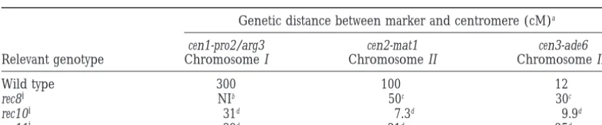
Genetic map distances in meioses with aberrant chromosome segregation
Genetic distance between marker and centromere (cM)a
cen1-pro2/arg3 cen2-mat1 cen3-ade6
Relevant genotype Chromosome I Chromosome II Chromosome III
Wild type 300 100 12
rec82 NIb #50c #30c
rec102 #31d #7.3d #9.9d
rec112 #39d #21d #25d
aWild-type genetic distances were as reported (Munz1994). The maximum possible genetic distances in
meioses suffering aberrant segregation were calculated from the heterozygosity values in Table 2 using the mapping functions (Figure 2C;materials and methods, Equations 3 and 5).
bNot informative. The heterozygosity value (63%) is too close to the value (67%) at which the various
mapping functions converge (Figure 2C) to permit meaningful calculation of the genetic distance.
cBased on the assumption of 100% MI NDJ of centromeres. The genetic distances would be smaller if any
of the segregation errors resulted from MII NDJ or PSS.
dBased on the assumption of 100% MII NDJ of centromeres. The genetic distances would be smaller if any
chromosome III, and the precise value is dependent of the centromere-proximal recombination defects. If, for example, some fraction of rec10 or rec11 mutant upon the relative additional contributions from
recom-bination, MI NDJ, and PSS. meioses suffered MI NDJ, then the amount of centro-mere-proximal recombination that occurred could actu-Nine percent of the rec10 mutant meiotic diploids
were heterozygous for the mat1 locus on chromosome II ally be considerably lower than calculated based upon the assumption of 100% MII NDJ at the centromeres (Table 2). Thus, the rec10 mutants suffer predominantly
MII NDJ of chromosome II (Figure 2C). Interestingly, (e.g., dashed line in Figure 2C). A similar rationale ap-plies to the rec8 mutant meioses, which have predomi-the maximum possible genetic distance in those meioses
leading to aberrant segregation was 7.3 cM, which is 14- nantly MI NDJ errors.
Cohesion, crossovers, and segregation: It has been fold (or more) below that of wild-type cells (Table 3). In
other words, centromere-proximal crossover recombi- reported that the rec81, rec101, and rec111genes encode chromosome III-specific activators of recombination nation on chromosome II was reduced in those meioses
with aberrant segregation. As discussed previously, the (DeVeauxandSmith1994;FoxandSmith1998). That claim seems indefensible in light of the results of this heterozygous diploids (9%) could be due to this residual
recombination, to a low level of MI NDJ, to a low level study and others (Molnaret al. 1995;Krawchuket al. 1999;Parisiet al. 1999). The rec81gene actually encodes of PSS, or to some combination of the three. By any of
the models, between 87 and 100% of the rec10 mutant a sister chromatid cohesion (cohesin) protein required for normal sister chromatid cohesion, homologous meioses suffer MII NDJ of chromosome II.
Similar observations and conclusions apply to the seg- chromosome pairing, and MI chromosome segregation. The rec8-110 mutation is epistatic to rec10-109 and to regation patterns of chromosome I in the rec10 mutants
(Tables 2 and 3; Figure 2). A reduction of 10-fold (or rec11-111, thereby defining a genetic pathway. And the rec101and rec111genes are required for proper segrega-more) in centromere-proximal recombination was
ob-served in those meioses with aberrant segregation, and tion of sister chromatids during MII. These genes are proposed to encode components of a pathway required between 54 and 100% of the segregation errors are due
to MII NDJ. to establish, maintain, and appropriately release meiotic
interactions between homologous chromosomes (
Kraw-MII NDJ inrec11mutants:The results and conclusions
for the rec11 mutants parallel those of the rec10 mutants chuket al. 1999). Hypotheses of how sister chromatid cohesion and crossover position work synergistically to (Tables 2 and 3; Figure 2C). Each of the three
chromo-somes suffered MII NDJ of sister chromatids and centro- ensure the fidelity of chromosome segregation in each of the two meiotic divisions can be found in that study. mere-proximal recombination was reduced at least
eightfold and at least fivefold on chromosomes I and II, The utility of the mapping functions:The new map-ping functions permit the simultaneous analysis of cen-respectively, in those meioses with aberrant segregation.
Between 47 and 100% (chromosome I), 66 and 100% tromere-proximal recombination and chromosome seg-regation defects. There are three principal advantages (chromosome II), and 61 and 100% (chromosome III)
of the segregation errors are due to MII NDJ. of these functions. First, it is not necessary for the marker to be tightly linked to the centromere in
wild-Reduced centromere-proximal recombination in the
rec8, rec10, and rec11 mutants: Reduced centromere- type cells, nor is it even necessary to know the map distance between the marker and its centromere in wild-proximal recombination was demonstrated for rec8
(chromosome II), rec10 (chromosomes I, II, and III), type cells. Any significant deviation from 67% heterozy-gosity in the meiotic aneuploids proves that the locus and rec11 (chromosomes I and II) mutant meioses with
aberrant chromosome segregation (Table 3). Two stud- is linked to its centromere. Thus, one can readily map loci relative to the centromeres in mutant meioses or ies of crossing over between pairs of markers have shown
that recombination is reduced markedly near the cen- even in otherwise wild-type meioses with spontaneous nondisjunction events. Second, the frequency of hetero-tromeres of each of the three chromosomes in each of
the rec8, rec10, and rec11 mutants, but recombination zygosity provides a direct readout of the type of meiotic missegregation. Any significant deviation from 67% het-toward the telomeres occurs at nearly wild-type levels
(Krawchuk et al. 1999; Parisi et al. 1999). Because erozygosity must be due to either MI NDJ or MII NDJ; the type of error and the lower limit to its magnitude those studies looked at the majority of meioses, and
this study examined only those meioses in which the can be read directly from the mapping functions (Figure 2C). And third, these functions may be applied to any chromosome missegregation could be scored
geneti-cally, it is not possible to make a direct comparison. organism in which diploid or disomic meiotic products can be studied. While it is easiest to visualize the deriva-However, centromere-proximal recombination may
ac-tually be reduced on each chromosome in each of those tion of the mapping functions for organisms with or-dered tetrads (Figure 2), the functions will work equally rec8, rec10, and rec11 mutant meioses leading to diploidy.
The mapping functions reveal the maximum possible well for organisms without ordered tetrads.
There are also three limitations to the new mapping genetic distance between a marker and its centromere
and meiotic analysis and induced haploidization. Genetics 87:
genetic distance between any given marker and its
cen-471–489.
tromere (Figure 2C). Second, they only reveal the lower Krawchuk, M. D., L. C. DeVeauxandW. P. Wahls,1999 Meiotic
chromosome dynamics dependent upon the rec81, rec101and
limit to the type of NDJ (Figure 2C). And third, it is
rec111genes of the fission yeast Schizosaccharomyces pombe. Genetics
not possible to map the distance between a marker and
153:57–68.
the centromere in those meioses that undergo predomi- Lamb, N., S. B. Freeman, A. Savage-Austin, D. Pettay, L. Taftet
al., 1996 Susceptible chiasmate configurations of chromosome
nantly PSS. Nevertheless, these functions can provide a
21 predispose to non-disjunction in both maternal meiosis I and
better understanding of the relationship between
mei-meiosis II. Nat. Genet. 14: 400–405.
otic recombination and chromosome segregation. Li, Y. F., M. Numata, W. P. WahlsandG. R. Smith,1997
Region-specific meiotic recombination in Schizosaccharomyces pombe : the We thank Aaron Graff, Ju¨ rg Kohli, Ning Kon, Michael Lichten,
rec11 gene. Mol. Microbiol. 23: 869–878.
Gisela Mosig, and Wallace Sharif for helpful suggestions; Calley
Har-Lin, Y.,andG. R. Smith,1995 Molecular cloning of the meiosis-din and Steve Lindsey for laboratory assistance; and Gerry Smith for induced rec10 gene of Schizosaccharomyces pombe. Curr. Genet. 27: providing yeast strains. This work was supported by a grant from the 440–446.
National Institutes of Health (GM54671) and by pilot project funds Lin, Y., K. L. Larson, R. DorerandG. R. Smith,1992 Meiotically from the Vanderbilt University Research Council. M.D.K. was sup- induced rec7 and rec8 genes of Schizosaccharomyces pombe. Genetics
132:75–85. ported in part by a training grant from the National Institutes of
MacDonald, M., T. Hassold, J. Harvey, L. H. Wang, N. E. Morton
Health (CA09582) and W.P.W. was a Leukemia Society of America
et al., 1994 The origin of 47,XXY and 47,XXX aneuploidy: het-Special Fellow (3021-94) for a portion of this research.
erogeneous mechanisms and role of aberrant recombination. Hum. Mol. Genet. 3: 1365–1371.
Molnar, M., J. Bahler, M. SipiczkiandJ. Kohli,1995 The rec8 gene of Schizosaccharomyces pombe is involved in linear element
LITERATURE CITED
formation, chromosome pairing and sister-chromatid cohesion
DeVeaux, L. C.,andG. R. Smith,1994 Region-specific activators during meiosis. Genetics 141: 61–73.
of meiotic recombination in Schizosaccharomyces pombe. Genes Dev. Moore, D. P.,andT. L. Orr-Weaver,1998 Chromosome
segrega-8:203–210. tion during meiosis: building an unambivalent bivalent. Curr.
DeVeaux, L. C., N. A. HoaglandandG. R. Smith,1992 Seventeen Top. Dev. Biol. 37: 263–299.
complementation groups of mutations decreasing meiotic recom- Moore, D. P., W. Y. Miyazaki, J. E. TomkielandT. L. Orr-Weaver, bination in Schizosaccharomyces pombe. Genetics 130: 251–262. 1994 Double or nothing: a Drosophila mutation affecting meiotic
Fisher, J. M., J. F. Harvey, N. E. MortonandP. A. Jacobs,1995 chromosome segregation in both females and males. Genetics Trisomy 18: studies of the parent and cell division of origin and 136:953–964.
the effect of aberrant recombination on nondisjunction. Am. J. Munz, P.,1994 An analysis of interference in the fission yeast Schizo-Hum. Genet. 56: 669–675. saccharomyces pombe. Genetics 137: 701–707.
Fox, M. E.,andG. R. Smith,1998 Control of meiotic recombination Niwa, O.,andM. Yanagida,1985 Triploid meiosis and aneuploidy in Schizosaccharomyces pombe. Prog. Nucleic Acid Res. Mol. Biol. in Schizosaccharomyces pombe: an unstable aneuploid disomic for
61:345–378. chromosome III. Curr. Genet. 9: 463–470.
Gethmann, R. C.,1984 The genetic analysis of a chromosome-spe- Parisi, S., M. J. McKay, M. Molnar, M. A. Thompson, P. J. van der cific meiotic mutant that permits a premature separation of sister Spek et al., 1999 Rec8p, a meiotic recombination and sister chromatids in Drosophila melanogaster. Genetics 107: 65–77. chromatid cohesion phosphoprotein of the Rad21p family,
con-Gutz, H.,1971 Site specific induction of gene conversion in Schizo- served from fission yeast to humans. Mol. Cell. Biol. 19: 3515–
saccharomyces pombe. Genetics 69: 331–337. 3528.
Gutz, H., H. Heslot, U. LeupoldandN. Loprieno,1974 Schizosac- Ponticelli, A. S.,andG. R. Smith,1989 Meiotic recombination-charomyces pombe, pp. 395–446 in Handbook of Genetics, edited by deficient mutants of Schizosaccharomyces pombe. Genetics 123:
R. C. King.Plenum Press, New York. 45–54.
Haldane, J. B. S.,1919 The combination of linkage values, and the Rasooly, R. S., C. M. New, P. Zhang, R. S. HawleyandB. S. Baker, calculation of distances between loci of linked factors. J. Genet. 1991 The lethal(1)TW-6cs mutation of Drosophila melanogaster is
8:299–309. a dominant antimorphic allele of nod and is associated with a
Hassold, T., M. Merrill, K. Adkins, S. FreemanandS. Sherman, single base change in the putative ATP-binding domain. Genetics
1995 Recombination and maternal age-dependent nondisjunc- 129:409–422.
tion: molecular studies of trisomy 16. Am. J. Hum. Genet. 57: Roeder, G. S.,1997 Meiotic chromosomes: it takes two to tango.
867–874. Genes Dev. 11: 2600–2621.
Koehler, K. E., C. L. Boulton, H. E. Collins, R. L. French, K. C. Ross, L. O., D. Treco, A. Nicolas, J. W. SzostakandD. Dawson, Hermanet al., 1996a Spontaneous X chromosome MI and MII 1992 Meiotic recombination on artificial chromosomes in yeast. nondisjunction events in Drosophila melanogaster oocytes have dif- Genetics 131: 541–550.
ferent recombinational histories. Nat. Genet. 14: 406–414. Sherman, S. L., M. B. Petersen, S. B. Freeman, J. Hersey, D. Pettay
Koehler, K. E., R. S. Hawley, S. ShermanandT. Hassold,1996b et al., 1994 Non-disjunction of chromosome 21 in maternal mei-Recombination and nondisjunction in humans and flies. Hum. osis I: evidence for a maternal age-dependent mechanism involv-Mol. Genet. 5: 1495–1504. ing reduced recombination. Hum. Mol. Genet. 3: 1529–1535.
Kohli, J., H. Hottinger, P. Munz, A. StraussandP. Thuriaux,