en
t
re
v
ie
w
s
re
ports
de
p
o
si
te
d r
e
se
a
rch
refer
e
e
d
re
sear
ch
interacti
o
ns
inf
o
rmation
developing medaka forebrain
Ivan Conte and Paola Bovolenta
Address: Departamento de Neurobiología Celular, Molecular y del Desarrollo, Instituto Cajal, CSIC, Dr Arce, Madrid 28002, Spain.
Correspondence: Paola Bovolenta. Email: [email protected]
© 2007 Conte and Bovolenta.; licensee BioMed Central Ltd.
This is an open access article distributed under the terms of the Creative Commons Attribution License (http://creativecommons.org/licenses/by/2.0), which permits unrestricted use, distribution, and reproduction in any medium, provided the original work is properly cited.
Six3 transcriptional regulation
<p>A cluster of highly conserved non-coding sequences surrounding the Six3 gene were identified in fish genomes, and transgenesis in medaka fish demonstrates that these sequences have enhancer, silencer and silencer blocker activities that are differentially combined to control the distribution of Six3.</p>
Abstract
Background: Embryonic development is coordinated by sets of cis-regulatory elements that are collectively responsible for the precise spatio-temporal organization of regulatory gene networks. There is little information on how these elements, which are often associated with highly conserved noncoding sequences, are combined to generate precise gene expression patterns in vertebrates. To address this issue, we have focused on Six3, an important regulator of vertebrate forebrain development.
Results: Using computational analysis and exploiting the diversity of teleost genomes, we identified a cluster of highly conserved noncoding sequences surrounding the Six3 gene. Transgenesis in medaka fish demonstrates that these sequences have enhancer, silencer, and silencer blocker activities that are differentially combined to control the entire distribution of Six3.
Conclusion: This report provides the first example of the precise regulatory code necessary for the expression of a vertebrate gene, and offers a unique framework for defining the interplay of
trans-acting factors that control the evolutionary conserved use of Six3.
Background
Embryonic development is coordinated by networks of evolu-tionary conserved regulatory genes that encode transcription factors and components of cell signaling pathways, which in many instances are repetitively exploited in space and time to generate appropriate outcomes in target cells.
Progressive specification of the vertebrate prosencephalon indeed follows this rule [1,2] and requires, among other fac-tors, recurrent use of Six3, which is a member of the Six/sine oculis family of homeobox transcription factors [3]. In all
ver-tebrates, Six3 is expressed from the neurula stage in the
ante-riormost neural plate and then in its derivatives: the developing eyes and olfactory placodes, the hypothalamic pituitary regions, and the ventral telencephalon. In mouse and chick, this distribution overlaps with that of its closely
related homolog, namely Six6 [3]. However, with time Six3
and Six6 expressions progressively segregate to different
brain regions, and Six3 - but not Six6 - is additionally
expressed in the olfactory bulb, cerebral cortex, hippocam-pus, midbrain, and cerebellum [4]. Consistent with this
expression, Six3-null mice die at birth, lacking most of the
head structures anterior to the midbrain, including eyes [5],
and mutations in SIX3 have been found in humans affected
Published: 6 July 2007
Genome Biology 2007, 8:R137 (doi:10.1186/gb-2007-8-7-r137)
Received: 23 February 2007 Revised: 5 June 2007 Accepted: 6 July 2007 The electronic version of this article is the complete one and can be
by holoprosencephaly and aprosencephaly/atelencephaly
[6,7]. During mammalian lens induction, Six3 is essential in
the presumptive lens ectoderm to activate Pax6 and possibly
Sox2 expression [8]. In addition, morpholino-based
knock-down of the medaka fish Six3 demonstrates the
concentra-tion-dependent need for the function of this transcription factor for proximo-distal patterning of the optic vesicles [9]. Biochemical and functional studies have also shown that
Six3, as well as Six6, can induce ectopic retinal tissues and
control retinal neuroblast proliferation, acting as transcrip-tional repressors through the interaction with members of the
groucho family of transcriptional co-repressors [10-15]. Fur-thermore, Six3, but not Six6, functionally interacts with the DNA replication inhibitor Geminin, controlling the balance between cell proliferation and differentiation with a mecha-nism that is independent of transcriptional regulation [16].
How the activity of Six3 - or that of any other gene with
mul-tiple functions during embryo development - is diversified remains to be elucidated. This could be facilitated by defining the precise gene regulatory network that controls its spatio-temporal expression. It is now well established that control of
gene expression is executed through sets of cis-regulatory
regions within the noncoding DNA of animal genomes. These
cis-regulatory modules have variable length and contain
clus-ters of DNA-binding sites for different transcription factors. These modules work as promoter enhancers or silencers and collectively constitute a unique code for the switching on and off of gene activity [17-19].
The experimental definition of the organization of these
spe-cific cis-regulatory elements has progressed substantially in
both Drosophila and sea urchin [17]. In contrast, our
under-standing of how these modules are combined to generate pre-cise gene expression patterns in vertebrates is still rather limited. Possible causes of this are the increased genome complexity and the slow and laborious process of testing the functional significance of identified elements in mammals [20]. Recently, however, computational approaches based on multispecies genomic sequence alignments, combining both closely related and highly divergent organisms, have facili-tated identification of highly conserved noncoding sequences, which in many cases appear to coincide with the regulatory modules of genes that play critical roles in development.
Analyses of the complex regulation of genes such as Sox2,
Sox9, Otx2, Shh, and Irx provide some illustrative examples
[21-27]. Functional testing of 'enhancer' activity has also pro-gressed, thanks to the use of alternative and relatively faster 'transgenic' approaches based on the use of nonmammalian vertebrate model systems [20,25].
Here, we have taken advantage of both the power of compu-tational analysis and the particular compact genome and high
transgenesis efficiency of the medaka fish (Oryzia latipes)
[28] to dissect the regulatory control of one of the two Six3
medaka homologs, olSix3.2, that we identified during the
course of this study. olSix3.2 is more closely related to the
mammalian Six3 than the previously described medaka
homolog [29] (hereafter referred to as 'olSix3.1'). Similar to
other related studies [23-25], we identified and functionally
characterized sets of cis-regulatory modules that control the
olSix3.2 promoter, showing that at least some of these cis -regulatory elements are conserved in other vertebrates, although they are dispersed over a greater stretch of DNA. Going a step further, we have also used combinations and
deletions of the identified cis-regulatory modules to elucidate
the regulatory code of olSix3.2, which is composed of two
enhancers, two silencers, and two 'silencer blockers' used in a combinatorial manner. This comprehensive description of the olSix3.2 cis-regulatory code provides a unique framework
for defining the network of trans-acting factors that control
the evolutionary conserved activity of Six3 during forebrain
development.
Results
Isolation, characterization, and expression of olSix3.2
In order to identify the elements that regulate Six3 expression
using the medaka fish (Oryzia latipes) as a model, we used
the available olSix3.1 coding sequence (AJ000937) as a query
to search public databases (see Materials and methods, below) for the ortholog genomic loci of the closely related
spe-cies Fugu rubripes, Tetraodon nigroviridis, and Danio rerio
(zebrafish). This search retrieved four different loci, one for
the fugu and the tetraodon, and two for the zebrafish (six3a
and six3b). Alignment of about 20 kilobases (kb) of the
retrieved sequences upstream of the Six3 translational start
sites identified a cluster of conserved noncoding blocks roughly contained within the first 4.5 kb (data not shown). In the case of the zebrafish, alignment of the six3a or six3b loci yielded comparable results. This information was used to
amplify from genomic DNA a fragment of the medaka Six3
locus that contains the corresponding conserved noncoding blocks and the entire first exon.
Interestingly, nucleotide and amino acid sequence alignment
of the partially amplified olSix3 coding region did not
com-pletely overlap with that reported for the previously identified
olSix3.1 [29] but identified - as in zebrafish and Xenopus
[30,31] - a second Six3-related gene in the medaka genome,
namely olSix3.2 (AM494407).
Cloning and sequencing of the entire olSix3.2 coding region
revealed a two-exon structure, similar to that of olSix3.1 and
the mouse Six3, in which the first exon encodes the Six and
homeobox domains. olSix3.1 and olSix3.2 exhibited 76% and
63% identity at the nucleotide and amino acid levels, respec-tively. Interestingly, comparison of the amino acid sequence (81% versus 59%; Additional data file 1) and genomic organi-zation, together with phylogenetic analysis (Additional data
file 2), demonstrated that olSix3.2 was more closely related to
comm
en
t
re
v
ie
w
s
re
ports
refer
e
e
d
re
sear
ch
de
p
o
si
te
d r
e
se
a
rch
interacti
o
ns
inf
o
rmation
the family (Additional data file 2).
olSix3.1 is expressed in the anterior embryonic shield and the developing eye [29]. To determine whether the newly identi-fied gene and the initially identiidenti-fied homolog had similar
dis-tributions, we compared the expression domain of olSix3.2
with those of olSix3.1 and the related olSix6 [13] using whole-mount in situ hybridization. As for olSix3.1, olSix3.2 was first detected in the anterior neural plate at late gastrula stages but was additionally expressed in the anterior axial mesoderm at
St16 (Figure 1a-c). At the optic vesicle stage, both olSix3.2 and
olSix3.1, but not olSix6, were expressed in the forebrain.
However, although olSix3.2 was more abundant in the
pre-sumptive telencephalon (Figure 1e,h), olSix3.1 was
predomi-nant in the optic area (Figure 1d,g). This distribution was more evident at later stages of development, when both
olSix3.1 and olSix6, which first appears at the optic cup stage (Figure 1l) [13]), were strongly expressed in the developing neural retina, optic stalk, and preoptic and hypothalamic
areas (Figure 1j,l,m,o,p,r). In contrast, olSix3.2 mRNA was
distributed in the developing lens, olfactory pits, telen-cephalon, neural retina, anterior hypothalamus, and anterior and posterior thalamus (Figure 1k,n,q). During retinal
neuro-genesis, olSix3.1 was mostly confined to the inner nuclear
layer (Figure 1s), and olSix3.2 and olSix6 to the retinal
gan-glion and amacrine cells (Figure 1t,u).
In conclusion, the distribution of olSix3.2 appeared closely
related to that reported for the chick and mouse Six3
[4,32,33], whereas the combined expression patterns of
olSix3.1 and olSix6 resembled that reported for Six6 [34,35]. The cis-regulatory elements responsible for olSix3.2 expression are contained in a 4.5 kb genomic region ending with a distal 'silencer'
On the basis of this expression pattern, we next searched for the elements that could be involved in the regulation of
olSix3.2 expression. Alignment of the amplified olSix3.2
genomic sequence with the corresponding sequences from
fugu, tetraodon, and zebrafish (analyses involving six3a and
six3b yielded similar results) identified ten conserved non-coding blocks within the 4.5 kb upstream of the translational start site olSix3.2 (Figure 2a).
Owing to selective pressure, functional elements in genomes evolve at a slower pace than nonfunctional regions [36-39]. A number of recent studies have functionally demonstrated that a proportion of the highly conserved noncoding regions present in vertebrate genomes correspond to regulatory ele-ments with enhancer activity [21,39]. We therefore asked whether the region containing the cluster of ten highly con-served noncoding elements was necessary and sufficient to
control the entire expression of olSix3.2.
[image:3.612.313.553.82.540.2]Comparative analysis of olSix3.1, olSix3.2, and olSix6 expression pattern during embryonic development
Figure 1
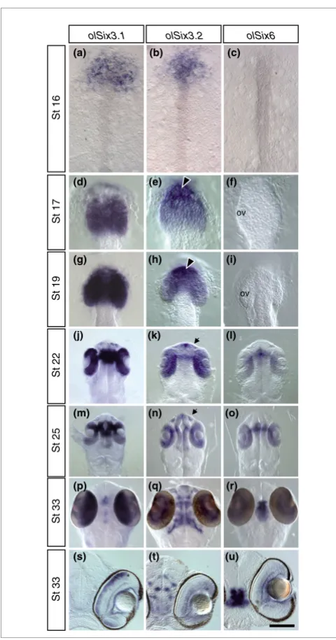
Comparative analysis of olSix3.1, olSix3.2, and olSix6 expression pattern during embryonic development. Medaka embryos at different
developmental stages (as indicated in the panels) were hybridized in toto with specific probes, as indicated on the top of each column. (a to r) Anterior dorsal views; (s to u) frontal vibratome sections through the eye. From St16 to St19, only olSix3.1 and olSix3.2 are expressed in the anterior neural plate (panels a to c) and then in the presumptive telencephalon and optic vesicles (panels d to i), although olSix3.1 is more abundant in the optic vesicles (panels d and g) and olSix3.2 in the telencephalic region (arrowheads in panels e and h). From St22 onward, when olSix6 mRNA also becomes detectable, the three genes are co-expressed, albeit at different levels, in the developing neural retina, optic stalk, and pre-optic and hypothalamic area (panels j to r). In addition, olSix3.2 is distributed in the developing lens, olfactory pits (panels k and n; arrow), telencephalon, and anterior and posterior thalamus (panels k, n, and q). During retinal neurogenesis, olSix3.2 and olSix6 are restricted to the retinal ganglion and amacrine cells (panels t and u), whereas olSix3.1 is restricted to the inner nuclear layer (panel s).
St 17
St 22
St 25
St 33
(a)
olSix3.1
(b) (c)
olSix3.2
St 19
(g) (h) (i)
(l)
(m) (n)
olSix6
(o)
(p) (q) (r) (d) (e) (f)
St 16
(j) (k)
St 33
(t) (u)
(s)
The cis-regulatory elements responsible for the olSix3.2 expression are contained in a 4.5 kb genomic region
Figure 2
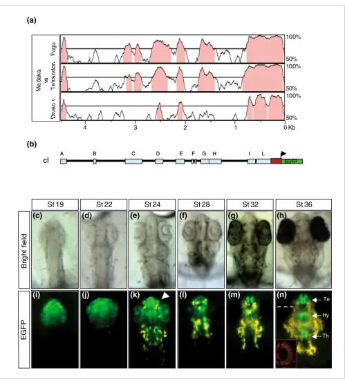
The cis-regulatory elements responsible for the olSix3.2 expression are contained in a 4.5 kb genomic region. (a) VISTA comparison of the 5' olSix3 genomic region plotted against those from Fugu rubripes, Tetraodon nigroviridis, and Danio rerio. The blocks of sequences (75% identity over 100 base pairs) conserved among the four species are indicated in pink. (b) Schematic structure of the 5' olSix3.2 genomic region/enhanced green fluorescent protein (EGFP) reporter construct (cI) containing ten highly conserved noncoding regions represented as light blue rectangles A to L. The red rectangle represents the 5'-untranslated region and the first nine nucleotides of the olSix3.2 coding sequence in frame with a nuclear EGFP reporter (green). (c to h) Bright field images; and (i to n) epi-fluorescence dorsal views of cI transgenic embryos at different stages of development (as indicated). Note that the cI construct drives EGFP reporter expression to the same olSix3.2 expression domain, recapitulating its entire pattern (compare with Figure 1). The arrowhead in panel k points to the olfactory pits. The inset in panel n shows a frontal section through the eye (dotted line), where EGFP is expressed in the amacrine cells. The section was counter-stained with propidium iodine (red). Hy, hypothalamus; Te, telencephalon; Th, thalamus.
(a)
Me
da
k
a
vs
D
n
a
io
r.
T
e
tr
a
o
do
n
F
ug
u
r.
100%
50% 100%
50% 100%
50%
St 32
St 36
Br
ig
h
t f
ie
ld
EG
F
P
St 22
St 24
St 28
St 19
(c)
(d)
(e)
(f)
(h)
(
(g)
(i)
(j)
(k)
(l)
(m)
(n)
0 Kb 1
2
4 3
(b)
A B C D E F G H I L
EGFP
cI
A B C D E F G H I L
EGFP
cI
Te
Hy
comm
en
t
re
v
ie
w
s
re
ports
refer
e
e
d
re
sear
ch
de
p
o
si
te
d r
e
se
a
rch
interacti
o
ns
inf
o
rmation
[image:5.612.55.558.82.643.2]The most distal conserved module, A, is a silencer that restrains olSix3.2 expression to the anterior neural plate
Figure 3
The most distal conserved module, A, is a silencer that restrains olSix3.2 expression to the anterior neural plate. (a) Drawings to the left of the panel are schematic representations of the different constructs (cI to cV) used to study the potential regulatory activity of modules A to C, whereas the tables to the right summarizes the presence (+) or absence (-) of enhanced green fluorescent protein (EGFP) reporter expression observed with each construct and corresponding to the endogenous olSix3.2 expression domain (NE) or with an ectopic posterior expansion (EPE). The A module with silencer activity is depicted in purple. (b to d) Bright field images, and (e to g) epi-fluorescence dorsal views of cII transgenic embryos at different stages of development (as indicated). Note that the domain of EGFP expression is progressively expanded in the caudal direction (arrows in panels e and f), invading the spinal cord at St36 (panel g). Equivalent patterns were observed with the cIII and cIV transgenic lines. Dotted lines in panels e to g indicate the caudal limit of endogenous olSix3.2 expression.
(a)
A D E G
G
A B C D E F G H I L
EGFP
B
C D E F G H I L
D E F H I L
F H I L
D E F G H I L
EGFP
EGFP
EGFP
EGFP
+
-+
+
+
+
+
+
-+
EPE
NE
Bright field
EG
FP
St 19
St 22
(e)
(f)
(c)
(b)
cI
cII
cIII
cIV
cV
St 36
(d)
To this end we fused this 4.5 kb genomic region, including the first nine nucleotides of the coding sequence, in frame with a
nuclear EGFP (enhanced green fluorescent protein) reporter
(Figure 2b). This construct, containing the ten conserved noncoding blocks (termed A-L; Figure 2b), was used to gen-erate three independent stable transgenic medaka lines, which all exhibited a spatio-temporal distribution of the reporter virtually identical to that observed for the
endog-enous olSix3.2 both at embryonic (compare Figure 1 with
Fig-ure 2c-n) and adult stages (not shown). We thus concluded that this region was sufficient to control the entire expression of olSix3.2.
In addition to regulatory elements, sequence conservation could reflect the existence of natural anti-sense mRNAs [40]
or of alternative and yet uncharacterized exons of Six3.
How-ever, reverse transcription polymerase chain reaction
(RT-PCR) analysis and in situ hybridization studies excluded
these possibilities (data not shown). We thus assumed that the ten modules, identified on the basis of their conservation among teleosts (the precise nucleotide sequence of each mod-ule is provided in Additional data file 3), could all potentially contain elements that are involved in the regulation of
olSix3.2. To test whether this assumption was correct, we generated a series of constructs (named cI to cXXVII) carry-ing different combinations of the A-L modules, which were then functionally assayed by generating and analyzing three independent stable transgenic lines for the vast majority of the constructs. In each case, the pattern of expression of the
EGFP reporter was compared with that observed with con-struct I (cI), containing the full 4.5 kb sequence (Figure 2i-n)
and was always consistent with that observed in F0 injected
embryos.
Embryos of a transgenic line carrying a construct in which the A to C modules had been deleted (cII; Figure 3a) showed a
pattern of EGFP expression in the anteriormost neural tube
similar to that observed with cI. However, embryos
consist-ently exhibited an additional transient expansion of EGFP
distribution to posterior mesencephalic regions (compare Figure 3e,f with Figure 2i,j and Figure 1h,k), which disap-peared after St22. EGFP fluorescence was also consistently observed in the spinal cord starting from St34 (Figure 3d,g)
up to adult stages. These observations suggested that, pre-sumably, blocks D to L were sufficient to control normal
olSix3.2 expression, whereas the A to C modules contained a silencer(s), the activity of which was necessary to restrain
olSix3.2 expression to anterior domains of the neural tube throughout development. To determine the location of the silencer activity, we generated and functionally analyzed three different constructs containing the D to L modules in combination with the A, B, or C block (cIII to cV; Figure 3a). Only the presence of 134 base pairs (bp) of the A module could
repress the posterior EGFP expansion, restoring the normal
olSix3.2 distribution, which clearly identified the presence of
a cis-regulatory silencer(s) in this sequence. In spite of
sequence conservation, the B and C blocks instead did not appear to contribute to the spatio-temporal control of
olSix3.2, at least in the context that we tested.
Early expression of olSix3.2 in the anterior neural structures depends on one enhancer, whereas that in the lens placode requires the additional activity of four cis-regulatory modules
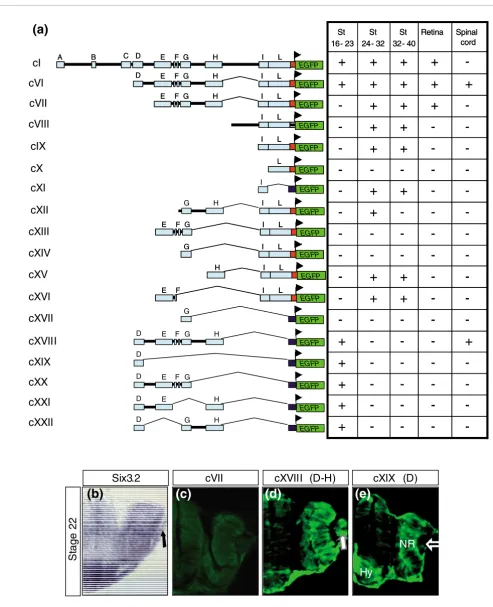
We then sought to determine the functional relevance of the remaining D to L conserved modules. To this end we gener-ated a series of additional constructs (named cVI to cXXII; Figure 4a) based on selective deletion of one or more modules at the time or by including different combinations of a few of them. Transgenesis analysis of these constructs demon-strated that the D module was necessary (cVI to cXVII; Figure 4a,c) and sufficient (cXIX; Figure 4a,e) to drive EGFP expres-sion in all of the anterior neural structures from St16 to St23. In contrast, the D module was necessary but not sufficient
(cXIX; Figure 4e) to control EGFP expression in the lens
pla-code/lens vesicle, as normally observed for the endogenous
olSix3.2 (Figure 4b). Indeed, the activity of modules E to H was further required for EGFP expression in the lens (cVI and cXVIII; compare Figure 4d with Figure 4e), because deletion of either one of them was sufficient to abrogate the reporter expression in the lens ectoderm (cXIX to cXXII; Figure 4a,e),
suggesting that multiple cis-regulatory sequences spread
along these four modules contribute to olSix3.2 expression in
this tissue. This is somewhat in contrast with the apparently
simpler regulation of olSix3.2 distribution in the early neural
tissue, which mostly depends on the D block.
Different constructs used to generate stable transgenic lines and corresponding distribution of EGFP reporter in expected olSix3.2 expression domains
Figure 4 (see following page)
Different constructs used to generate stable transgenic lines and corresponding distribution of EGFP reporter in expected olSix3.2 expression domains. (a) Drawings to the left of the panel are schematic representations of the different constructs (cI and cVI to cXXII) used to generate stable transgenic lines, whereas the tables to the right summarize the presence (+) or absence (-) of enhanced green fluorescent protein (EGFP) reporter expression
comm en t re v ie w s re ports refer e e d re sear ch de p o si te d r e se a rch interacti o ns inf o rmation
Figure 4 (see legend on previous page)
EGFP
A B C D E F G H I L
EGFP
A B C D E F G H II LL
EGFP
I L
D E F G H
EGFP
I L
I L
D E F G H
EGFP
I L
E F G H
EGFP
I L
I L
E F G H
EGFP I L I L G H EGFP I L
E F G
EGFP
I L
I L
E F G
EGFP I L G EGFP I L I L G EGFP I L H EGFP I L I L H EGFP I L E F EGFP I L I L E F EGFP
-+
-+
-+
-+
-+
-+
-+
+
-+
+
-+
+
-+
+
+
Retina-+
-+
+
+
+
+
St 24- 32-+
+
+
+
+
St 32- 40-+
+
-+
Spinal cord St 16- 23-+
-+
-+
-+
-+
-+
-+
+
-+
+
-+
+
-+
+
+
Retina-+
-+
+
+
+
+
St 24- 32-+
+
+
+
+
St 32- 40-+
+
-+
Spinal cord St 16- 23 I L EGFP I L I L EGFP I L EGFP I L I L EGFP L EGFP L EGFPD E F G H
EGFP D EGFP I G EGFP
St
a
g
e
2
2
Six3.2
(a)
(b)
cXIX (D)
(e)
cXVIII (D-H)
(d)
cVII
C
cI
cVI
cVII
cVIII
cXI
cXIII
cXIV
cXV
cXVI
cXVII
cXVIII
cXIX
cXII
cIX
cX
D E F G
EGFP
D E H
EGFP
D G H
[image:7.612.58.551.82.689.2]Notably, modules D to H (cXVIII; Figure 4a) were also suffi-cient to induce caudal expansion of reporter expression, with a pattern identical to that observed in the absence of the A module (Figure 3e-g), indicating that modules I and L do not contribute to this expansion or to early expression of the gene.
During organogenesis, appropriate expression of olSix3.2 requires the combined activity of two silencers, one enhancer, and two putative 'silencer blockers' To determine whether these last two modules were
function-ally relevant to any other aspect of olSix3.2 expression, we
designed a number of constructs in which modules I and L were assayed separately (cX and cXI), in conjunction (cIX),
and combined with the olSix3.2 endogenous promoter (cX) or
with the minimal tyrosine kinase promoter (cXI). Injections
of cX were not associated with EGFP expression in any region
of the embryo at any stage (Figure 4a). This indicates that, as in the case of modules B and C, the L block had no enhancer silencer activity relevant to the regulation of olSix3.2, at least in the tested conditions, although its sequence is strongly conserved among all vertebrates. In contrast, the activity of
block I was clearly linked to control of olSix3.2 distribution in
the forebrain starting from St26 onward, when EGFP was
gradually observed, with progressively increasing intensity, first in the telencephalic, then in the hypothalamic, and finally in the thalamic region (Figures 4a and 5c). This reca-pitulates the endogenous expression of the gene (Figure 1q).
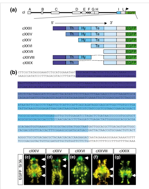
To determine the minimal region of module I involved in the control of this expression, we engineered five 5' to 3' stepwise deletions covering the entire module (cXXIII to cXXVII; Fig-ure 5a). Notably, deletions two, three, and four resulted in
progressive abrogation of EGFP expression in the thalamic,
hypothalamic (Figyre 5b-d), and telencephalic regions (not shown). This strongly suggests that module I contains a 5' to
3' organized succession of cis-regulatory elements that
con-trol the posterior to anterior spatio-temporal organization of
olSix3.2 expression in the developing brain. This interpreta-tion was further supported by the injecinterpreta-tion of two internal deletion constructs (cXXVIII and cXXIX) in which the stretches of nucleotides apparently responsible for hypotha-lamic and telencephalic expression were removed from cXX-III (Figure 5a). Indeed, in 11% (close to transgenic efficiency)
of the embryos analyzed in F0, EGFP fluorescence was not
detected in the telencephalon (cXXVIII; Figure 5f) or in the hypothalamus and telencephalon (cXXIX; Figure 5g), clearly
indicating that deleted elements are the main driver of
olSix3.2 expression in these regions.
The elements contained in the I module appeared to suffice in
terms of regulating late olSix3.2 embryonic expression in the
brain. Nevertheless, we considered whether any additional module could modify their activity. Transgenic embryos car-rying cXIV, in which the G module was combined with the I module, had no reporter expression in the brain (Figure 4a), raising the possibility that the G module contained a 'silencer' that, in turn, could be normally regulated by a 'silencer blocker', as previously proposed [41,42]. Addition of the H block (cXII) proved that this was the case, because its pres-ence restored reporter expression, although only from St26 to St32. Further addition of the E block (cVII, containing E, G, H and I) appeared to overcome the effect of the G silencer
from St32 onward. Thus, proper regulation of late olSix3.2
embryonic expression requires the participation of five differ-ent modules - one enhancer, one silencer, and two silencer blockers - in addition to the silencer activity contained in the distal A module (Figure 6c,d).
When tested alone, block I did not drive EGFP expression in
the differentiating retina, whereas activity of the D block was sufficient to maintain reporter expression only in the
pro-spective neural retina (Figure 4a,d,e). Thus, olSix3.2
expres-sion in the differentiating retina appeared to depend on a combination of modules different from those tested thus far. The search for this code demonstrated that only the combined activity of the E to I modules (cVII; Figure 4a) was effective in supporting EGFP expression in the late developing retina.
Identification and characterization of conserved regions among vertebrate
Altogether these data provide a detailed picture of the
regula-tory code that governs olSix3.2 expression during eye and
brain development in medaka. As summarized in Figure 6, this spatio-temporal code is provided by the combined use of at least seven different modules, all conserved among fishes, with distinct enhancer, silencer, or silencer blocker activities. The next logical question was whether this regulatory
organi-sation was conserved in the Six3 locus of vertebrates other
than fishes.
To address this problem, we used the characterized olSix3.2
regulatory region as a query to search public databases
Module I contains a 5' to 3' organized sequence of cis-regulatory elements that control the posterior to anterior expression of olSix3.2 in brain
Figure 5 (see following page)
comm
en
t
re
v
ie
w
s
re
ports
refer
e
e
d
re
sear
ch
de
p
o
si
te
d r
e
se
a
rch
interacti
o
ns
inf
o
rmation
Figure 5 (see legend on previous page)
Te
Th
Th
(a)
A
B
C
D
E F G H
I L
EGFP
5’
3’
cI
cXXIV
cXXV
cXXVI
EGFP
Hy
Te
EGFP
EGFP
Hy
Te
EGFP
EGFP
EGFP
EGFP
Hy
Te
cXXVII
cXXIII
(b)
cXXVIII
Th
Th
Hy
Hy
EGFP
EGFP
EGFP
Th
EGFP
EGFP
Th
cXXIX
EG
FP
S
t3
6
(c)
(d)
(e)
cXXVI
cXXV
cXXIV
cXXVIII
(f)
cXXIX
(g)
CTTCGCTATAGGGAAATCTGCATGGAAATAATGTGCAGATTGACTTGCTTCCATTCAAAATTCCC GAAGCGATATCCCTTTAGACGTACCTTTATTACACGTCTAACTGAACGAAGGTAAGTTTTAAGGG
GAGTTGTAGTCATTGGTTGTCCATTTGTCCCCCATTTAAAGCTCCCTCTCCCTCACTCCCTCCCC CTCAACATCAGTAACCAACAGGTAAACAGGGGGTAAATTTCGAGGGAGAGGGAGTGAGGGAGGGG
GTCTCTACTAAGCATCTCCAGTCTACATATCTTCTTTAGCTTTAACGAGCCTCGTTAAGATCGCA CAGAGATGATTCGTAGAGGTCAGATGTATAGAAGAAATCGAAATTGCTCGGAGCAATTCTAGCGT
ATAATATTCCACCCTCTAATTGCTCATTCCATTCAGCAGATAGGCGAGCATTGGCTTGTGCCTGA TATTATAAGGTGGGAGATTAACGAGTAAGGTAAGTCGTCTATCCGCTCGTAACCGAACACGGACT
TGCGCGCGGTGCGGTGGGAGGGTTGCTGTGGAGATCCTAGACTCTGATAACCCCCCGTGCGTGCT ACGCGCGCCACGCCACCCTCCCAACGACACCTCTAGGATCTGAGACTATTGGGGGGCACGCACGA
GCACAAGTGGTGAAAGCCTCGCGCTACGTACTGGCTAATGATTGGCACGCTTGACAGTGATTGGC CACGACGTGTTCACCACTTTCGGAGCGCGATGCATGACCGATTACTAACCGTGCGAACTGTCACT
[image:9.612.53.554.78.712.2](Genome Bioinformatics UCSC [University of California, Santa Cruz]) for the ortholog regions in vertebrates other than fishes. This analysis showed that only part of the mod-ules identified in teleosts were conserved among all verte-brate phyla (Figure 7a). Attempts to align each of the A to F modules separately and enlarging the search to the 120 kb
flanking Six3 in the Xenopus laevi, chicken, mouse, and
human genomes were unsuccessful in detecting alignable sequences using the VISTA and multialign software [43,44]. Thus, only the G and L modules were highly conserved and similarly organized in all genomes, whereas the sequences that constitute the H and I modules in fishes were conserved but fragmented in a larger stretch of DNA in the other genomes analysed (Figure 7b), with the exception of the mar-supial opossum, in which the I block was co-linear with that
of fishes (data not shown). In spite of fragmentation, trans-genic embryos, carrying the human sequence that included the G module and the dispersed H and I sequences (Figure
7c), exhibited spatio-temporal EGFP expression in the
devel-oping brain identical to that observed in the equivalent medaka genomic region (Figure 7d-i). In addition, reporter expression was observed in the lens placode/vesicle. This
suggested that although control of at least part of Six3
expres-sion in the brain has been conserved, its regulation during lens development has undergone a reorganization of the
appropriate cis-regulatory elements during evolution (data
not shown).
Although the human construct (h-cI) we injected drove EGFP
expression only in the late olSix3.2 expression domain,
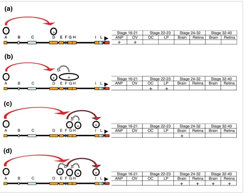
[image:10.612.59.560.85.488.2]Summary of the regulatory code that control the entire expression of olSix3.2
Figure 6
Summary of the regulatory code that control the entire expression of olSix3.2. (a) Early expression of olSix3.2 in the forebrain and eye depends on enhancers in module D and a silencer activity (activities) in module A. (b) olSix3.2 expression in the lens placode requires multiple elements distributed along modules D to H. (c) During organogenesis, correct olSix3.2 expression requires the activity of different enhancer arranged in a 5'to 3' mode within module I. The activity of I is repressed by module G, which, in turn, is neutralized initially by module H and at later stages (d) by the combined activity of the E and H silencers. Module A is necessary at all stages analyzed to prevent reporter expansion to caudal central nervous system.
(b)
F GH
+ +
A B C D E I L
-+
A B C D E F G H I L
- +
A B C D E F G H I L
-(a)
(c)
F GH
+
A B C D E I L
- - +
(d)
F GH
+
A B C D E I L
- + - +
Stage 16-21 Stage 22-23 Stage 24-32 Stage 32-40 ANP OV OC LP Brain Retina Brain Retina
ANP OV OC LP Brain Retina Brain Retina
ANP OV OC LP Brain Retina Brain Retina
ANP OV OC LP Brain Retina Brain Retina
+
+
+
+
+
+
+
+
+
Stage 16-21 Stage 22-23 Stage 24-32 Stage 32-40
Stage 16-21 Stage 22-23 Stage 24-32 Stage 32-40
comm
en
t
re
v
ie
w
s
re
ports
refer
e
e
d
re
sear
ch
de
p
o
si
te
d r
e
se
a
rch
interacti
o
ns
inf
o
rmation
ules G to L, we could not exclude that this human region contained regulatory information not readable in fish. Thus, to rule out possible cross-species interferences, we amplified
from genomic DNA the equivalent Xenopus region, in which
the G to L elements are organized as in humans (Figure 7a).
Transgenesis analysis in Xenopus embryos using a construct
containing this fragment (X-cI; Figure 7c) yielded results equivalent to those observed with the human fragment; EGFP reporter expression was detected only at later stages of brain
development in the expected domain of Xsix3.2 expression
(Figure 7j,k). This supports the idea that the regulatory
infor-mation for early Six3 expression in vertebrates other than
fishes reside in as yet unidentified genomic regions.
Discussion
Six3 is an important regulator of vertebrate forebrain devel-opment. Gene regulatory network models predict that the precise spatio-temporal expression pattern of genes funda-mental for embryo development must be orchestrated by the interaction of various regulatory regions [17]. Supporting the model, we functionally demonstrated that the entire
expres-sion of the newly identified olSix3.2 is orchestrated by the
combined use of seven different cis-regulatory modules
(Fig-ure 6) and that at least part of this regulation is conserved in the Six3 locus of vertebrates other than fishes. Two main
'enhancer' modules (D and I) are responsible for olSix3.2
expression at early and late stages of brain development, respectively. Their activity is spatially refined by the function of two 'silencers' and two 'silencer blockers'. In addition,
olSix3.2 expression in the lens ectoderm and in the differen-tiating retina requires the combined activity of five different
cis-regulatory modules. This apparently simple regulation
may hide additional organization, as we have demonstrated
for the I enhancer, in which an organized sequence of cis
-reg-ulatory elements control the posterior to anterior expression of olSix3.2 in the brain.
The availability of different genome sequences and the devel-opment of analytical bioinformatic tools have facilitated
study of cis regulation of a number of genes with evolutionary
conserved roles in vertebrate embryonic development. Some of these studies have focused, as has ours, on a specific gene or a gene cluster, identifying enhancers that are involved in the control of specific expression domains [21,23,25,45-49]. However, possibly because of the size of the genomic regions that are involved, or to the laborious and time consuming use of mice, or the limitations of chick electroporation in validat-ing regulatory activities, these studies have mostly focused on each enhancer as a separate entity, thus missing the effects of possible cooperative activities. Other recent and extremely informative studies, based on medium or small throughput screens in zebrafish, have instead systematically tested the autonomously enhancing function of large numbers of highly conserved noncoding elements positioned in areas
surround-cation only of a fraction of them [21,39]. Because each element is tested in an unconstrained context, negative regu-lators as well as modulatory functions of surrounding endog-enous elements are also undetected using these approaches [39]. In contrast, possibly benefiting from the high transgen-esis efficiency of the medaka fish [50] and its compact genome, we were able to assign enhancer, silencer, and mod-ulatory functions to the majority of the highly conserved
non-coding elements surrounding the Six3 gene in fishes. Testing
different combinations of these elements, we have also estab-lished their required interactions for proper expression of the gene. Thus, to our knowledge, we provide the first description of the regulatory code necessary for the expression of a verte-brate gene and offer a unique framework to define the entire
interplay of trans-acting factors that control the evolutionary
conserved use of Six3 during forebrain development.
Teleosts are the most diverse class of vertebrates with a huge variety of different species; they are characterized by broad size range and dynamic organization of genomes, which are the result of an initial genome duplication followed by subse-quent independent evolution of the different lineages [51,52]. Comparison of divergent teleost genomes largely separated in the phylogenetic tree, such as the medaka and zebrafish genomes (approximately 115 to 200 million years [28]), is thus a powerful tool with which to study gene regulatory mechanisms. Adopting this strategy, we identified a cluster of
potential regulatory modules in the Six3 locus, which were
barely identifiable in a comparison among mammalian genomes (compare Figure 2a with Figure 7a). VISTA analysis of the available genomic sequences flanking the homologous
vertebrate Six3 genes revealed several blocks of highly
con-served noncoding sequences in the gene surroundings. Although a few of these blocks were located downstream of the coding sequence (data not shown), we demonstrated that
the pattern of olSix3.2 expression could be recapitulated by
4.5 kb of genomic sequence flanking the 5' end of the gene. This conclusion is based on a relatively efficient (roughly 20% of injected embryos) and highly reproducible (basically 100%, albeit with different EGFP intensity, thus excluding chromo-somal position effects) transgenic analysis using three inde-pendent and stable medaka lines generated for all of the constructs we tested. Thus, we are fairly confident that we
identified the main regulatory region for olSix3.2, although
we cannot entirely exclude the possibility that additional or duplicated regulatory elements positioned in untested regions may contribute to a refinement of the main
expres-sion domain. Indeed, redundant cis-regulatory elements have
been reported to control specific expression domains in
dif-ferent genes, including Otx2, Shh, and Sox2 [22,23,25].
According to our analysis, the regulatory region of olSix3.2 is
relatively compact as compared with those reported for other
genes that are involved in neural development, such as Sox2,
Figure 7 (see legend on next page)
H
(a)
St
2
4
St
2
6
St
3
3
Bright field EGFP
-794 -739
human CAACAA-AAT CCCCTC-TCT AATTGCACAT CCCATTCAAC AACTCTGCCC AGGG--CTT
chimp CAACAA-AAT CCCCGC-TCT AATTGCACAT CCCATTCAAC AACTCTGCCC AGGG--CTT
mouse CAA-AACACT CCGTGTAACG AATAGCTCGC TGCATTCAAC TACTCTACTC CTATTCCTT
Rattus CAA-AACACT CCTGCTAACTAATTGCTTATCGCATTCAACTACTCTATGC CTATTCCTT
opossum CAATAATA-T TCCACCCTCT AATTGCTCAT CCCATTCAAC AAATACGTGC ACACTGCTT
Xenopus CAATAATA-T TCCACCCACT AATTGCTCAT CCCATTCAACAATCACATTA TTTGATCCC
fugu CAATAATA-T TCCACCCTCT AATTGCTCAT TCCATTCAGC AGATAGGCGA GCATGCCTT
Medaka CAATAATA-T TCCACCCTCT AATTGCTCAT TCCATTCAGC AGATAGGCGA GCATGGCTT
-538 -483
human GCT-AAGTGG TAAAACCGTC ---CTTACGT TCTCGGTATT GATTGGCAGG GC–TGACAGT
chimp GCT-AAGTGG TAAAACCGTC ---CTTACGT TCTCGGTATT GATTGGCAGG GC–TGACAGT
mouse GCT-AAGTGG TAAAACCGTC ---CTTACGT TCTCAGTATT GATTGGCAGG GC–TGACAGT
Rattus GCT-AAGTGG TAAAACCTTC ---CTTACGT ACTCAGTATT GATTGGCAGG GC–TGACAGA
opossum GCT-AAGTGG GAAAAACCTT C--CTTACGT ACTCATTATT GATTGGCAGT GC–TGACAGT
Xenopus GG--AAGTGG TAACTGCAAG ---TGACG TCGTCGCCGT TGATTGGCAG AGGTGACAGT
fugu GCACAAGTGG TGAAAGCCTC GCGC-TACGT ACTGGCTAAT GATTGGCA-C GCTTGACAGT
Medaka GCACAAGTGG TGAAAGCCTC GCGC-TACGT ACTGGCTAAT GATTGGCA-C GCTTGACAGT
(b)
(c)
(d)
(i)
(g)
(e)
(f)
(h)
A B C
I L
EGFP G
D E F G H I L
EGFP Me daka
Human
cI
h-cl
Me
d
a
k
a
vs
H
u
m
a
n
M
o
u
s
e
Op
o
s
s
u
m
a
Xe
n
o
p
u
s
F
u
g
u
r.
100%
50% 100%
50% 100%
50% 100%
50% 100%
50%
0 Kb 0,4
0,8 1,2
1,6
2
*
*
**
0,40,4-1951 -1902
human TTAGGATAAT TAT-TTCAGC TTTATTGAGGGCAGATTAGT TGAAGTCTGG
chimp TTAGGATAAT TAT-TTCAGC TTTATTGAGGGCAGATTAGT TGAAGTCTGG
mouse TTAGGATAAT TAT-TTCAGC TTTATTGAGGGCAGATTAGT TGAAGTCTGG
Rattus TTAAGATAAT TAT-TTCAGC TTTATTGAGGGCAGATTAGT TGAAGTCTGG
opossum ATCCCCTA-- GATCTTTAGC TTTAACGAGGCTCG--TAAA AAAAAACA--
Xenopus TTAGGATAAT GAT-TTCAGC TTTATTGAGGGCAGATTAGT AGCAGTCTCG
fugu TCTACATA-T CTTCTTTAGC TTTAACGAGACTCG-TTAAG AT---C
Medaka TCTACATA-T CTTCTTTAGC TTTAACGAGCCTCG-TTAAG AT---C
St
3
5
(j)
Xsix3.2 EGFP
*
*
**
EGFP
EGFP Xenopus
X-cI
G H I L [image:12.612.57.560.82.718.2]comm
en
t
re
v
ie
w
s
re
ports
refer
e
e
d
re
sear
ch
de
p
o
si
te
d r
e
se
a
rch
interacti
o
ns
inf
o
rmation
region of 10, 100, and even 1,000 kb away from their promot-ers have been reported [23-27,53,54], even in the compact
Fugu genome [46]. Genes with complex patterns of expres-sion are predicted to have more regulatory elements and occupy significantly more space in the genome than those with simpler expressions that are restricted to populations of cells with similarities or shared identity [55]. It is thus
possi-ble that the compactness of the olSix3.2 regulatory region
might reflect the association that exists among the main ter-ritories in which the gene is expressed. Indeed, the specifica-tion of telencephalic and eye fields appears to be closely
linked [2], and the initial expression of olSix3.2 in both
regions appears to depend on the activity of a single enhancer element (D) and a distal silencer (A), which constrains the expression domain to the anteriormost neural tube. This hypothesis could also explain why the combined activities of five different modules (D to H) are instead needed to control expression in the lens placode, which is the only non-neural
domain of olSix3.2 expression. Nevertheless, compactness
does not appear to be, at least in this case, a reflection of sim-plicity, because each of the conserved modules may include additional regulatory organization. This is the case of module I, which is the main enhancer involved in the late embryonic expression of the gene. Stepwise and internal deletions of this module have revealed a peculiar organization, in a 5' to 3'
direction, of a series of cis-regulatory elements that are
required for the posterior to anterior spatio-temporal
expres-sion of olSix3.2 in the thalamus, hypothalamus, and
telen-cephalon. The activity of the I module is refined by a silencer, G, the activity of which is modulated by two silencer blockers that act in a temporal sequence, thus establishing an elaborate control code. Furthermore, although the L module
per se has no activity, we cannot totally exclude the possibility that this module might contribute, together with modules E to H, to the regulation of I, because it was present in the con-structs used for this analysis.
Alternatively, the short-range regulation of olSix3.2 may be
linked to the chromosomal localization of the Six genes,
which are organized in two evolutionarily conserved clusters
[56]. Although the expression of the other Six family
mem-bers (Six1, Six2, Six4, and Six5) is mostly associated with tis-sues of mesodermal and ectodermal origin [3], it is possible that genes within the same cluster (Six4, Six1, and Six6) will
share a few regulatory elements, which might have imposed constrains against rearrangement during evolution [57].
In silico comparison identified ten conserved modules in the
teleost Six3 locus. Transgenic analysis in medaka
demon-strated clear regulatory activity for seven of them, whereas
modules B, C, and L did not influence EGFP reporter
expres-sion. Although these modules might have subtle regulatory activities below the resolution of our analysis, their conserva-tion could reflect other important roles in gene transcripconserva-tion control, such as regulation of chromatin structure or - in the particular case of module L - they may contribute to minimal promoter functions.
The regulatory region we have studied belongs to a newly
identified medaka Six3 gene, namely olSix3.2. Genomic
organization and phylogenetic analysis suggests that olSix3.2
is more closely related to the mammalian Six3 than the
previ-ously identified olSix3.1 [12]. Like its mammalian homolog
[4], olSix3.2 is strongly expressed in various forebrain regions
where its paralog is not expressed. Our comparative expres-sion study suggests that the combination of expresexpres-sion
domains of olSix3.1, olSix3.2, and the related olSix6
corre-spond to the combined tissue distribution observed for the
mouse and chick Six3 and Six6 [32-34], with a preponderant
expression of olSix3.1 in the eye, of olSix3.2 in the
telen-cephalic and thalamic regions, and of olSix6 in the
hypothala-mus. Genetic abrogation studies in mice demonstrated that
Six3 is necessary for the formation of forebrain, which is
absent in homozygous embryos [5]. Genetic deletion of Six6
instead is associated with pituitary defects, absence or hypo-plasia of the optic nerves, and chiasm and alteration in neural
retina proliferation [58]. How the functions of olSix3.1,
olSix3.2, and olSix6 relate to those described in the mouse for
Six3 and Six6 is still unresolved and knock-down analysis of all three genes in medaka will be necessary to address this
issue. Thus far, morpholino-based knock-down of olSix3.1
results in forebrain and eye defects, including loss of optic stalk markers [9], whereas preliminary analysis indicates that
olSix3.2 morphants are characterized by strong ventral fore-brain defects with minor eye malformations (De la Torre A, Conte I, Bovolenta P, unpublished observations), suggesting
that the two olSix3 paralogs may cover Six3 as well as part of
the mouse Six6 functions, a possibility that is also supported
Modules G, H, and I are functionally conserved in humans
Figure 7 (see previous page)
by the phylogenetic position of olSix3.1, which falls almost in
between the Six3 and Six6 branches of the Six gene family
(Additional data file 2).
Comparative analysis of the regulatory code of the three medaka genes currently ongoing in our laboratory might be useful in complementing these studies by providing insights into the sub-functionalization or neo-functionalization of
olSix3.1, olSix3.2, and olSix6 as compared with their mam-malian counterparts. Furthermore, they will help to elucidate
whether Six3 and Six6 have arisen from the duplication of a
common ancestor, as previously proposed [56], possibly duplicating at least part of their regulatory region. This is an important point because, with the comparison parameters used, we were unable to identify in other vertebrate species the conservation and distribution of the A to F regulatory modules characterized in fishes. This is particularly impor-tant for the A and D modules, which are the main regulators
of early Six3 expression in fishes. Informatics searches of
cor-responding regions in mammalian genomes yielded no clear information, suggesting that these modules might be present outside the regions that we analyzed or they might have evolved differently in other vertebrate genomes, making their search even more difficult than that of the H and I modules. Alternatively, these modules may represent a new acquisition of olSix3.2 caused by teleost genome duplication.
In our study, we demonstrated strong functional conserva-tion between fishes and other vertebrates only for the G, H,
and I modules, which control late expression of olSix3.2. The
sequences that compose the H and I modules in fishes were intermixed and differently arranged in other vertebrate genomes, although their function was strongly conserved
when assayed in medaka and Xenopus transgenesis. This
sug-gests that sequences from different vertebrates are activated by common transcription factors, although the binding sites for these factors might be distributed, oriented, or repre-sented in different numbers among species. An additional explanation for the different arrangement of the H and I modules might be species-specific nucleotide modifications, which have been proposed to contribute to gene transcrip-tional evolution [59-61].
Conservation of regulatory function between human and fish in the absence of clear sequence conservation has previously
been reported also for the RET gene. In this case, lack of
correlation between the two events was even more marked,
and different in silico analysis designed to detect shorter
stretches of sequence similarities or the existence of inversion and rearrangement failed to detect alignable sequences [62]. Thus, our data, together with few additional observations [63-65], strongly support the idea proposed by Fisher and colleagues [62] that some relevant regulatory information might be conserved among species at a level that is not detect-able using genomic sequence alignment.
Conclusion
Our study established the cis-regulatory code required for the
proper expression of olSix3.2 and demonstrates that there is
a need to test different combinations of highly conserved
putative cis-regulatory regions to elucidate how each
con-served element contributes to the spatio-temporal control of gene expression. In fact, one limitation of previous studies that have used transgenic analysis to test the function of highly conserved noncoding sequences is the identification of single enhancers uprooted from possible interactions with the remaining regulatory elements. Our comprehensive
description of the olSix3.2 regulatory code is now a powerful
starting point from which to define the entire interplay of
trans-acting factors that control the evolutionarily conserved
use of Six3 during forebrain development. From a broader
perspective, this type of information will be necessary to elu-cidate the composition and evolution of vertebrate gene regu-latory networks, as compared with those of invertebrates such as Drosophila and sea urchin, in which this type of informa-tion is accumulating at a much faster pace [17].
Materials and methods
Microinjection and establishment of transgenic lines
Adult and embryonic medaka fishes (Oryzia latipes) from the
Cab inbred strain were used throughout the study. Fertilized eggs were collected immediately and incubated at 4 to 10°C in Yamamoto's embryo rearing medium to suppress further development [66]. DNA was prepared using a High Pure Plas-mid Isolation Kit (Roche, Basel, Switzerland). DNA injections
(10 ng/μl DNA in ISceI enzyme reaction) were performed as
previously described [50]. Embryos were staged according to the method proposed by Iwamatsu [66], raised to sexual maturity, and transgenic founder fishes were identified by out-crossing to wild-type fishes. Transcriptional activation of the constructs was monitored by EGFP expression observed in living embryos under UV fluorescent stereo-microscopy
(Leica Microsystems, Wetzlar, Germany). Xenopus laevis
embryos were obtained and raised as described previously
[21]. Xenopus transgenesis was performed following the
same procedures as used for the medaka embryos.
Whole-mount in situ hybridization
Whole-mount in situ hybridizations were performed as previ-ously described using digoxigenin labelled riboprobes [29].
Anti-sense and sense riboprobes for medaka olSix3.1,
olSix3.2, and olSix6 and the Xenopus Xsix3.2 were used. A minimum of 40 embryos were hybridized for each marker
and condition. In toto hybridized embryos were
photo-graphed, embedded in gelatine/albumine block, and further sectioned using a vibratome (Leica Microsystems, Wetzlar, Germany).
Sequence analysis
The vertebrate Six3 genomic sequences were retrieved from
comm
en
t
re
v
ie
w
s
re
ports
refer
e
e
d
re
sear
ch
de
p
o
si
te
d r
e
se
a
rch
interacti
o
ns
inf
o
rmation
genomic DNA using the following primers: olSix3 forward
CCTCATTAAATGTCGCTAAC, and olSix3 reverse
cgcctaatgacac cagcctc. Sequence alignments were performed using the VISTA [43] and Multalign programs [44], which are available at the corresponding websites [69,70]. The criterion used for comparisons was a minimum 75% nucleotide iden-tity with a window size of over 100 bp. Phylogenetic analysis was performed using the PHYLIP package [71]. The results were plotted using the Tree-view software package [72].
olSix3.2 protein sequences were scanned for motifs using online software available at HGMP [73] and NCBI [74].
Isolation of olSix3.2 cDNA
Total RNAs from medaka embryos at different stages were isolated by RNAzol B (Campro Scientific, Berlin, Germany) and treated with Dnase I (Invitrogen, Carlsbad, CA). RT-PCR reactions were performed using SUPERSCRIPT II (Invitro-gen, Carlsbad, CA), as described previously [75]. PCR using
olSix3.2 specific primers was performed using 2 μl of the reverse transcription reaction as a template with the High Fidelity PCR system (Roche, Basel, Switzerland).
Oligonucle-otide primers used to isolate olSix3.2 cDNA are listed in
Addi-tional data file 4.
Plasmid constructions
A 4.5 kb region of olSix3.2 genomic sequence containing nine
nucleotides (corresponding to the first three amino acids) of the coding region was cloned in frame with EGFP reporter gene into the pSKII-ISceI-EGFP vector [50], to create the cI
construct. Xenopus and human sequences were amplified
from corresponding genomic DNA and cloned in the pSKII-ISceI-EGFP vector with the same strategy. The medaka
deleted constructs pSix3.2ΔXhoI (cII), pSix3.2ΔXhoI-NsiI
(cVIII), and pSix3.2ΔXbaI-HindIII (cIX) were obtained by
digesting the pSix3.2-4.5 kb construct using the indicated
enzymes. All the other deleted constructs (pSix3.2Δel1,
pSix3.2Δel2, pSix3.2Δel3, pSix3.2Δel4, pSix3.2Δel5,
pSix3.2Δel6, and pSix3.2Δel7; cXXIII to cXXIX) were
obtained by PCR amplification from pSix3.2-4.5 kb and then cloned into pSKII-ISceI-EGFP vector. The A, B, and C mod-ules were deleted by restriction enzyme digestion (A, NarI/ KpnI; B, BtsI/BglII; and C, BamHI/ClaI) and inserted (in sense and anti-sense orientations) into the polylinker of
pSix3.2ΔXhoI (cIII to cV), pSix3.2Δel1 and
pSKII-ISceI-Tk-EGFP (containing the tyrosine kinase minimal promoter) vectors to test their potential regulatory activity. All of the other modules were amplified and cloned (in sense and
anti-sense orientations) into the polylinker of pSix3.2Δel1 (cVI to
cVII, cX, and cXII to cXVI) and pSKII-ISceI-Tk-EGFP (cXI and cXVII to cXXII). The primer sequences used to generate these constructs are shown in Supplementary Table I. All con-structs were verified by automated sequencing.
The following additional data are available with the online version of this manuscript. Additional data file 1 is a Figure
reporting the amino acid sequence alignment of Six3 genes
from different vertebrate species. Additional data file 2 is a
figure illustrating the phylogenetic tree of the SIX family.
Additional data file 3 provides the precise nucleotide sequences of modules A to L described in the report. Addi-tional data file 4 is a table listing the sequences of the primers used to amplify the DNA fragments, which were used to design the different constructs described in the report.
Additional data file 1
Amino acid sequence alignment of Six3 genes from different verte-brate species
Presented is a figure reporting the amino acid sequence alignment of Six3 genes from different vertebrate species
Click here for file Additional data file 2
Phylogenetic tree of the SIX family
Presented is a figure illustrating the phylogenetic tree of the SIX
family
Click here for file Additional data file 3
Precise nucleotide sequences of modules A to L described in the report
Presented are the precise nucleotide sequences of modules A to L described in the report.
Click here for file Additional data file 4
Sequences of the primers used to amplify the DNA fragments Presented is a table listing the sequences of the primers used to amplify the DNA fragments, which were used to design the differ-ent constructs described in the report.
Click here for file
Acknowledgements
We are grateful to Drs WA Harris, H Kondoh, and M Manzanares for crit-ical reading of the manuscript, and to Dr J Wittbrodt and members of our laboratory for many helpful suggestions. We wish to thank I Dompablo for excellent technical assistance. This study was supported by grants from Spanish Ministerio de Educación y Ciencia (BFU-2004-01585) and in part by the EU (QLG3-CT-2001-01460) and the HFSPO (RGP0040/2001-M) to PB. A Telethon Foundation (GFP03007) and MEC (SB2003-0182) fellowships supported the postdoctoral work of IC.
References
1. Wilson SW, Houart C: Early steps in the development of the forebrain. Dev Cell 2004, 6:167-181.
2. Esteve P, Bovolenta P: Secreted inducers in vertebrate eye development: more functions for old morphogens. Curr Opin Neurobiol 2006, 16:13-19.
3. Rodríguez de Córdoba S, Gallardo ME, Lopez-Rios J, Bovolenta P: The human Six family of homeobox genes. Curr Genomics 2001, 2:231-242.
4. Conte I, Morcillo J, Bovolenta P: Comparative analysis of Six 3 and Six 6 distribution in the developing and adult mouse brain. Dev Dyn 2005, 234:718-725.
5. Lagutin OV, Zhu CC, Kobayashi D, Topczewski J, Shimamura K, Puelles L, Russell HR, McKinnon PJ, Solnica-Krezel L, Oliver G: Six3 repression of Wnt signaling in the anterior neuroectoderm is essential for vertebrate forebrain development. Genes Dev 2003, 17:368-379.
6. Wallis DE, Muenke M: Molecular mechanisms of holoprosencephaly. Mol Genet Metab 1999, 68:126-138. 7. Pasquier L, Dubourg C, Gonzales M, Lazaro L, David V, Odent S,
Encha-Razavi F: First occurrence of aprosencephaly/atelen-cephaly and holoprosenaprosencephaly/atelen-cephaly in a family with a SIX3 gene mutation and phenotype/genotype correlation in our series of SIX3 mutations. J Med Genet 2005, 42:e4.
8. Liu W, Lagutin OV, Mende M, Streit A, Oliver G: Six3 activation of Pax6 expression is essential for mammalian lens induction and specification. EMBO J 2006, 25:5383-5395.
9. Carl M, Loosli F, Wittbrodt J: Six3 inactivation reveals its essen-tial role for the formation and patterning of the vertebrate eye. Development 2002, 129:4057-4063.
10. Bernier G, Panitz F, Zhou X, Hollemann T, Gruss P, Pieler T: Expanded retina territory by midbrain transformation upon overexpression of Six6 (Optx2) in Xenopus embryos. Mech Dev 2000, 93:59-69.
11. Kobayashi M, Nishikawa K, Suzuki T, Yamamoto M: The homeobox protein Six3 interacts with the Groucho corepressor and acts as a transcriptional repressor in eye and forebrain formation. Dev Biol 2001, 232:315-326.
12. Loosli F, Winkler S, Wittbrodt J: Six3 overexpression initiates the formation of ectopic retina. Genes Dev 1999, 13:649-654. 13. Lopez-Rios J, Tessmar K, Loosli F, Wittbrodt J, Bovolenta P: Six3 and
Six6 activity is modulated by members of the groucho family. Development 2003, 130:185-195.