0095-1137/06/$08.00⫹0 doi:10.1128/JCM.44.4.1413–1418.2006
Copyright © 2006, American Society for Microbiology. All Rights Reserved.
Use of PCR and Reverse Line Blot Hybridization Assay for
Rapid Simultaneous Detection and Serovar Identification
of
Chlamydia trachomatis
Likuan Xiong,
1,2Fanrong Kong,
1Hua Zhou,
2and Gwendolyn L. Gilbert
1*
Centre for Infectious Diseases and Microbiology, Institute of Clinical Pathology and Medical Research, Westmead, NSW, Australia,1and Institute of Molecular Biology, Centre for Prevention and Control of Sexually Transmitted Disease,
Shenzhen Chronic Disease Hospital, Shenzhen, Guangdong Province, People’s Republic of China 5180202
Received 26 August 2005/Returned for modification 12 October 2005/Accepted 27 January 2006
The aim of this study was to develop and evaluate multiplex and nested PCR-reverse line blot (RLB) hybridiza-tion assays for detechybridiza-tion and serovar identificahybridiza-tion ofChlamydia trachomatis. Two sets of primers targeting the VD2 region of theomp1gene and one set targeting the cryptic plasmid were designed for use in multiplex (both targets) and nested PCR (omp1 only). For the RLB assay, labeled omp1 amplicons were hybridized to a membrane containing probes specific for 15C. trachomatisserovars. The assays were used to test 429 clinical specimens, which had been previously tested forC. trachomatisusing the COBAS AMPLICOR system. Specimens were tested without knowledge of the COBAS AMPLICOR result. Of 205 specimens that were positive by COBAS AMPLICOR, 201 (98%) were positive by multiplex PCR-RLB and 188 (92%) were also positive byomp1nested PCR-RLB. In addition, three of 224 COBAS AMPLICOR-negative specimens were positive by omp1 nested PCR-RLB. One hundred sixty-six of 191 (87%) specimens in whichC. trachomatisserovars were identified contained only one serovar and 25 (13%) contained two or three serovars. Serovars D, E, and F were found in 31 (16%), 83 (43%), and 51 (27%) specimens, respectively. Serovar E (41%) was the most commonly identified single serovar. Serovars J and K were found alone uncommonly (<2% each), but 18 of 25 (72%) specimens with multipleC. trachomatisserovars contained one or both (10 specimens) of these serovars. The nested (ompI) PCR-RLB is a specific and sensitive method for simultaneous detection and serovar identification ofC. trachomatis, which can reliably identify mixedC. trachomatis
serovars. It is suitable for use in epidemiological studies.
Chlamydia trachomatisis one of the most common sexually transmissible pathogens. An estimated 92 million new cases occur worldwide each year (19). This is probably an underes-timate, becauseC. trachomatisinfection in men and women is often asymptomatic, and unrecognized infections are a reser-voir for sexual transmission.
C. trachomatisinfections can be diagnosed by cell culture, immunofluorescence (IF), enzyme immunoassay (EIA), direct DNA hybridization, and PCR. Laboratory diagnosis of chla-mydial infection by culture is limited by the fact that collection of urethral swabs is unacceptable to many asymptomatic men. PCR, using various gene targets, including the cryptic plasmid,
omp1 (which encodes the major outer membrane protein, MOMP), and rRNA genes, is more sensitive than culture, EIA, or IF (4, 7). Moreover, urine specimens can be used for PCR, which are more convenient to collect and more acceptable to patients.
Serotyping ofC. trachomatisis unnecessary to make a clin-ical diagnosis of chlamydial infection. However, it is useful for epidemiologic research, investigation of person-to-person transmission, and study of differences in clinical manifestations or responses to treatment between serovars. While most C. trachomatisinfections are due to single serovars, up to 15% of
infections have been reported to involve two or more (1–3, 7,13–16). IF and EIA are commonly used for serotyping and detection of multiple serotypes in C. trachomatis cultures. PCR, plus restriction fragment length polymorphism (RFLP) analysis or DNA sequencing ofomp1amplicons, is needed to identify serovars directly from clinical specimens (2), but nei-ther can reliably detect mixed infections (6, 7, 17, 22). Recently the combination of PCR with reverse dot blot or reverse line blot (RLB) assays has been described (12, 17). In this study, we modified these methods to detect 15C. trachomatisserovars in a variety of clinical specimens from men and women.
MATERIALS AND METHODS
Reference strains.The following reference strains were used in this study:C. trachomatisserotype H ATCC UR-898,Chlamydia pneumoniaeTWAR strain TW-183,Chlamydia psittaciserotype ATCC VR-628,Ureaplasma urealyticum
serovar 1 ATCC 27813,Ureaplasma urealyticumserovar 4 ATCC 27816, Myco-plasma hominisATCC 23114,Mycoplasma genitaliumATCC 33530 (G37), Gard-nerella vaginalisATCC 14018, andNeisseria gonorrhoeaeWHO A.
Clinical specimens.Four hundred twenty-nine specimens, which had been referred to the Centre for Infectious Diseases and Microbiology for diagnosis of
C. trachomatisinfection between January 2004 and February 2005, were selected (by staff from the diagnostic laboratory) so as to ensure a fairly even distribution ofC. trachomatis-positive and -negative specimens. They consisted of first voided urine (309 specimens), rectal swabs (2 specimens), and cervical swabs (118 specimens).
COBAS AMPLICOR testing.Specimens were kept at⫺20°C until testing and then thawed, resuspended in 500l wash buffer, vortexed vigorously, and incu-bated at 37°C for 30 min. After centrifugation at 13,000⫻gfor 15 min, the supernatant was discarded, 250lC. trachomatislysis buffer was added, and after another 15-min incubation at 37°C, 250lC. trachomatisspecimen diluent was added to the lysate. The contents of the tubes were mixed by vortexing, centri-* Corresponding author. Mailing address: Centre for Infectious
Dis-eases and Microbiology (CIDM), Institute of Clinical Pathology and Medical Research (ICPMR), Westmead Hospital, Darcy Rd., West-mead, New South Wales 2145, Australia. Phone: (612) 9845-6255. Fax: (612) 9893-8659. E-mail: [email protected].
1413
on May 16, 2020 by guest
http://jcm.asm.org/
fuged at 13,000⫻gfor 10 min, and incubated at 95°C for 10 min. All specimens were tested forC. trachomatisusing the COBAS AMPLICOR (Amplicor; Roche Diagnostics Australia Pty. Limited Systems, Castle Hill, Australia), as described previously (9–11) and according to the manufacturer’s instructions. DNA ex-tracts were frozen at⫺20°C until required for further testing in this study, in which they were tested without knowledge of the COBAS AMPLICOR result.
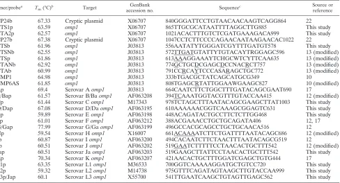
Probe and primer design.Two sets of primers targeting the VD2 region ofC. trachomatisand one set targeting the cryptic plasmid were designed or modified from previous publications. For each of 15 serovars, serovar-specific oligonucle-otide probes, based on the published VD2 region ofomp1sequences, and two probes based on published cryptic plasmid sequences ofC. trachomatis, were designed or modified (Table 1). All probes and primers were checked for spec-ificity against all sequences in GenBank by using QueryBD, WebANGIS GCG, SeqSearch, and the Browse code in the Australian National Genomic Informa-tion Services (ANGIS) programs (http://www1.angis.org.au/WebANGIS/WebFM). Probes were designed to have similar melting temperatures (Tm) of more than 59°C, and their lengths varied from 19 to 27 bp. Oligonucleotide probes were labeled with an amine group at the 5⬘end, and primers were labeled with biotin at the 5⬘end (Sigma-Aldrich, St. Louis, Mo.), respectively.
Multiplex PCR amplification. Amplification was performed using primers CP24b/CP27b for the cryptic plasmid and CTSb/CTAb and CTSNb/CTANb for
omp1. The amplifications were performed using a 25-l reaction mixture con-taining 10 mM Tris-HCl (pH 8.3), 50 mM KCl, 1.5 mM MgCl2, 0.1% Triton
X-100, 200M (each) deoxynucleoside triphosphate (dNTP), 25 pmol of each primer, 1 U of HotStart DNA polymerase (QIAGEN, Pty. Ltd., Doncaster, Australia), and 10l of DNA. The thermal profile involved initial denaturation for 15 min at 96°C and 35 cycles with the following steps: 0.5 min of denaturation at 94°C, 0.5 min of annealing at 60°C, and 1 min of extension at 72°C, with a final extension for 10 min at 72°C.
Nested PCR amplification.Amplification of VD2omp1was performed with outer primers CTSb and CTAb in a 25-l reaction mixture containing 10 mM Tris-HCl (pH 8.3), 50 mM KCl, 1.5 mM MgCl2, 0.1% Triton X-100, 200M
(each) dNTP, 25 pmol of each primer, 1 U of DNA polymerase (Promega, Pty. Ltd., Annandale, Australia), and 10l of DNA. The thermal profile involved initial denaturation for 15 min at 96°C and 35 cycles with the following steps: 0.5 min of denaturation at 94°C, 0.5 min of annealing at 60°C, and 1 min of extension at 72°C, with a final extension for 10 min at 72°C. Ten microliters of the primary
PCR product was used for a secondary PCR, which was prepared and run using the same reagents and conditions as the primary PCR, except that the (inner) primers used were CTSNb and CTANb. Strict procedures to avoid specimen contamination and carryover were followed when performing nested PCRs. All DNA extractions were done in a room dedicated to PCR use.
PCR products were electrophoresed on a 1.5% agarose gel and analyzed using SRBR Safe DNA gel stain (Molecular Probes Europe BV, Leiden, The Neth-erlands).
RLB assay. The RLB assay was performed using a system that has been described previously (21). Briefly, slots (Miniblotter 45; Immunetics) were filled with 150l of two to three different concentrations of each probe solution (0.3125, 0.625, and 1.25 pmol). Each PCR product was denatured and immedi-ately chilled on ice. Hybridization was performed at 60°C for 60 min. The washed membrane was incubated in peroxidase-labeled streptavidin conjugate (Roche, Mannheim, Germany) at 42°C for 45 min. Then the washed membrane was incubated in chemiluminescence blotting substrate (ECL Direct System; Roche) for 1 min, and the membrane was covered with Hyperfilm X-ray film (Amer-sham) for detection of chemiluminescence. The film was exposed for 7 min.
Sequencing and sequence searching.C. trachomatis omp1amplicons, gener-ated by nested PCR, were sequenced as described previously (21), using primers CMP1 and CMP6AS (Table 1) and Applied Biosystems (Foster City, Calif.) BigDye terminator chemistry on an ABI Prism 373 DNA sequencer. Sequences were identified using the FastA program group accessed through WebANGIS.
RESULTS
[image:2.585.54.542.81.345.2]Sensitivities of multiplex and nested PCR-RLB.The results of multiplex and nested PCR—in which amplicons were iden-tified by RLB—are shown in Table 2. Three specimens that had been negative in the COBAS AMPLICOR assay were positive in the nested PCR-RLB and identified by hybridiza-tion with the corresponding probes as serovar E. Assuming that all 208 positive PCR results of all three assays are true positives, the relative sensitivities of COBAS AMPLICOR,
TABLE 1. Primers and probes used forC. trachomatisPCR-RLB and DNA sequencing
Primer/probea T
m(°C)b Target
GenBank
accession no. Sequence
c Source or
reference
CP24b 67.33 Cryptic plasmid X06707 840GGGATTCCTGTAACAACAAGTCAGG864 22
CTS1p 63.59 omp1 X06707 865TTGCGCATAATTTTAGGCTTG885 This study
CTA2p 62.57 omp1 X06707 1021ACACTTTGTCTCGATGAAAGACA999 This study
CP27b 67.38 Cryptic plasmid X06707 1047CCTCTTCCCCAGAACAATAAGAACAC1022 22
CTSb 61.96 omp1 J03813 556AATATYTGGGATCGYTTTGATGT578 This study
CTSNb 62.55 omp1 J03813 572TTGATGTATTYTGTACAYTRGGAGC596 13 (modified)
CTSp 61.86 omp1 J03813 613AAAGGAAAYTCHGCWTCYTTCAA635 13 (modified)
CTANb 62.92 omp1 J03813 774GCTGCDCGAGCDCCNACRCT757 13 (modified)
CTAb 60.99 omp1 J03813 791CCRCAYTCCCASARAGCTGC772 13 (modified)
CMP1 64.98 omp1 J03813 333bTGACGCTATCAGCATGCG349 10
CMP6AS 61.85 omp1 J03813 808TGAGCRTATTGGAAWGAAGC827 10 (modified)
Ap 69.4 Serovar Aomp1 J03813 664CAATCTTCTGGCTTTGATACAGCGAAT690 17
B/Bap 61.57 Serovar B/Baomp1 AF063208 394TCAAATGGTACGTTTGTACCAA415 12 (modified)
Cp 61.44 Serovar Comp1 M17343 978TCTAGCTTTAATACAGCGAAGCTTAT1003 This study
D/Dap 67.08 Serovar D/Daomp1 AF063195 610AAAAAACGGTCAAAGCGGAGTC631 This study
Ep 59.89 Serovar Eomp1 AF063198 448ACAGATACTGCCTTCTCTTGG468 This study
Fp 61.01 Serovar Fomp1 AF063212 388ACGAAACCTGCTGCAGATA406 12, 17
G/Gap 77.99 Serovar G/Gaomp1 AF063199 496GCCACGCAGCCTGCTGCAACA516 12
Hp 59.54 Serovar Homp1 X16007 661ACAAAATCTTCTGATTTTAATACAGC686 12 (modified)
Ip 60.87 Serovar Iomp1 AF063200 494CACAATCTTCTAACTTTAATACAGCG519 12
Jp 60.51 Serovar Jomp1 AF063202 519GAATCTTTTTCCTAACACTGCTTT542 12 (modified)
Jap 60.51 Serovar Jaomp1 AF063203 519GAAGCTTATTCCTAACACTGCTTT542 This study
Kp 70.34 Serovar Komp1 AF063207 421AACACTGCTTTGGATCGAGCTGTG444 17
L1p 63.35 Serovar L1omp1 M36533 700GGTCAAAAAGGATGCTGTCC720 This study
L2p 59.32 Serovar L2omp1 M14738 975GTTTCAGATAGTAAGCTTGTACCAA999 This study
L3p/Jap 60.1 Serovar L3omp1 X55700 541TTGAATCAAGCTGTAGTTGAGC562 This study
ap indicates biotin-labeled probe, and b indicates biotin-labeled primer. bMelting temperatures provided by manufacturer.
cNumbers relate to positions in GenBank sequences. Underlined sequences indicate modifications compared with published sequences.
on May 16, 2020 by guest
http://jcm.asm.org/
multiplex PCR-RLB, and nested PCR-RLB for detection ofC. trachomatiswere 98.6%, 96.6%, and 91.8%, respectively.
In the multiplex PCR-RLB, the cryptic plasmid was more likely to be amplified thanomp1(Table 2; Fig. 1). The serovar was identified by multiplex PCR-RLB in only 149 of 201 (74%)
C. trachomatis-positive specimens. However, theomp1nested PCR-RLB, using probes for each serovar, amplified 188 of 201 (94%) specimens positive by multiplex PCR as well as 3 of 224 that were negative by COBAS AMPLICOR and multiplex PCR (Table 2; Fig. 2). It was more sensitive than multiplex PCR-RLB for serovar identification and more convenient than sequencing.
Specificity of C. trachomatis typing assay. There were no cross-reactions between C. trachomatisprobes and DNA ex-tracts from the panel of other potential urogenital pathogens (see Materials and Methods). The specificities of both the mutiplex and nested PCR-RLB assays were further evaluated against individualC. trachomatisserovars (C, D, F, E, G, H, I, J, and K)—confirmed by sequencing (see below)—all of which hybridized with the corresponding probe with no cross-reac-tion with other probes.
DNA sequencing was performed on omp1amplicons from 23 selected specimens, of which 10 contained single serovars and 11 contained multiple serovars (Table 3). DNA sequenc-ing confirmed the results of PCR-RLB for all specimens in which a single serovar was identified but was uninterpretable for specimens with multiple serovars. Eleven of 12 amplicons from single serovars hadomp1sequences identical to the cor-responding sequences in GenBank; the other was a variant serovar C sequence, with 5 nucleotide differences compared with the GenBank sequence.
Typing of C. trachomatis. Of 208 C. trachomatis-positive specimens, 191 (92%) were successfully typed byomp1nested PCR-RLB (Table 4). SingleC. trachomatisserovars were de-tected in 166 (87%) and two or three serovars in 25 (13%) positive specimens. Overall, serovars D, E, and F were found in 15%, 41%, and 26% of specimens (including mixed), respec-tively. Serovar E was the serovar most commonly identified alone (39%). Serovar H, J, or K was identified in only 6 of 166 (3.6%) specimens with single serovars (or 3% of all positive specimens), but these serovars were found together or with other serovars in 19 of 25 (76%) specimens with mixed sero-vars (or 10% of all positive specimens). This discrepancy was most marked for serovar K, which was found alone in only 1 of 166 (0.6%) specimens with single serovars compared with 15 of
25 (60%) of those with mixed serovars (P⬍0.001) (or 1 versus 15 of 191 positive specimens;P⬍0.001). The equivalent fig-ures for serovar J were 3 of 166 (1.8%) versus 11 of 25 (44%) (P⬍0.001) (or 3 versus 11 of 191;P⫽0.03).
[image:3.585.304.541.138.617.2]FIG. 1. Hybridization, with 18 probes, of multiplex PCR products from a representative sample of 23C.trachomatis-positive specimens. From top to bottom, probes are as shown in numbered rows. The probe concentrations in each set of three rows (top to bottom) are 0.3125, 0.625, and 1.25 pmol (or 0.625 and 1.25 pmol when there are only two rows per probe). Lanes 1, 7, 8, 11, 15, 17, 19, and 20 are negative; lanes 2 to 6, 9 to 10, 12 to 13, 16, 18, and 21 to 23 are cryptic plasmid positive; lanes 21 and 22 areomp1(CTS1p) negative and so cannot be subtyped; lanes 2 and 10 areomp1positive but cannot be subtyped; results for the remaining lanes are as follows: 3, D; 4, F; 5, D; 6, D and E; 9, F; 12, J; 13, I;16, D; 18, E; and 23, D.
TABLE 2. Multiplex and nested PCR results in 429 clinical specimens previously tested forChlamydia trachomatisby
COBAS Amplicor assay
No. with COBAS Amplicor cp
result
Result bya:
Multiplex PCR-RLB (cp andomp1)
Nested PCR-RLB (omp1)
Positiveb
Negative Positive Negative
Positive, 205 201 4 188 17
Negative, 224 0 224 3 237
aPCR targets are shown in parentheses: cp, cryptic plasmid;omp1, outer membrane protein gene.
bBoth the cryptic plasmid andomp1were amplified from 149 specimens; the cryptic plasmid only was amplified from 52 specimens.
on May 16, 2020 by guest
http://jcm.asm.org/
DISCUSSION
We have developed a PCR-RLB hybridization assay for simul-taneous detection and genotyping ofC. trachomatiswhich is faster and more convenient than other serotyping methods such as PCR sequencing, PCR-RFLP, and culture-based methods using
anti-sera. First we used multiplex PCR to amplifyomp1and the cryptic plasmid and then hybridized biotin-labeled amplicons to sero-type-specificomp1and plasmid probes, respectively. When mul-tiplex PCR was negative orC. trachomatiscould not be typed, we used nested PCR to amplifyomp1and hybridized biotin-labeled amplicons to the serotype-specificomp1probes.
Nucleic acid amplification is now considered to be the most sensitive and specific method for screening and diagnosis ofC.
trachomatisinfections. The sensitivity of PCR varies, depend-ing on the target and, in particular, whether there are one or more copies. Mahony et al. compared the sensitivities of five PCR assays—including two targeting the cryptic plasmid (of which there are 10 copies, on average), two targetingomp1
[image:4.585.109.471.65.384.2](single copy), and one targeting rRNA genes (multiple cop-ies)—by testing serial dilutions of C. trachomatisDNA and genitourinary tract specimens (9–11). The plasmid PCRs can detect as little as 0.1 fg ofC. trachomatisplasmid DNA and 10 fg of total cellular DNA (which is equivalent to⬃0.01 inclu-sion-forming unit [IFU], whereas the twoomp1PCRs and one rRNA PCR could only detect the equivalent of 1, 100, and 10 IFU or 0.1 pg, 10 pg, and 1 pg, respectively, of cellular DNA.
FIG. 2. Hybridization, with 18 probes, of nested PCR products from a representative sample of 23C.trachomatis-positive specimens. From top to bottom are probes CTS1p (concentrations 1, 2, and 3),CP24p (concentrations 1 and 2), CP27p (concentrations 1 and 2), Ap (concentrations 1 and 2), Bp (concentrations 1 and 2),Cp (concentrations 1 and 2), Dp (concentrations 1, 2, and 3), Ep (concentrations 1, 2, and 3), Fp (concentrations 1, 2, and 3), Gp (concentrations 1, 2, and 3), Hp (concentrations 1, 2, and 3), Ip (concentrations 1, 2, and 3), Jp (concentrations 1, 2, and 3), Kp (concentrations 1, 2, and 3), Ja (concentrations 1 and 2), L1 (concentrations 1 and 2), L2 (concentrations 1 and 2), and L3 (concentrations 1 and 2). Concentrations 1, 2, and 3 represent probe concentrations 0.325, 0.625, and 1.25pmol, respectively. Results for lanes are as follows: 3, E; 4, E and F; 5, H and K; 6, G; 7, G; 8, E; 10, F; 11, F; 13, G; 14, F; 15, E;16, F; 17, E; 19, G; 23, D; 25, F; 28, G; 29, K; 31, C and I; 34, D and E; 35, E; 37, E; 38, E; 39, F; 40, C, J, and K; 42, E; and 43, D.
TABLE 3. Results ofC. trachomatisPCR-RLB assay and DNA sequencing in 23 selectedomp1-positive specimens
RLB results (n) No. of
specimens DNA sequencing result(s)
Single infections 12 1 each of C, D, E (3),
F, G, H, I, J (2), K
Results corresponded with those of RLB; 1 serovar C specimen was sequence variant
Multiple infections 11 2 serovars: C/I, D/E,
J/K (4), H/K
7 Uninterpretable
3 serovars: A⫹C⫹J, F/J/K, C/I/K, D/E/H
4 Uninterpretable
on May 16, 2020 by guest
http://jcm.asm.org/
[image:4.585.43.283.595.725.2]Our multiplex PCR, in which cryptic plasmid and omp1
DNA were amplified from 98% and 78%, respectively, of 205 COBAS AMPLICOR-positive samples, is relatively insensitive for serovar identification, using RLB to detect serovar-specific
omp1sequences. The nested format increased the sensitivity of the omp1 PCR to a level close to that of plasmid PCR; it amplified DNA from 92% of COBAS AMPLICOR-positive samples, as well as 3 of 224 negative samples. This latter find-ing is consistent with previous reports ofC. trachomatisstrains which do not have cryptic plasmids (9–12, 18). Molano et al. (12) reported that nested omp1 PCR-RLB identified C. tra-chomatisin 94% of specimens in which it had been detected by a cryptic plasmid PCR.
Serovars B, L1, L2, and L3 were not identified in our study—as would be expected in the population from which the specimens were derived (mainly women and heterosexual men). However, theoretically, our PCR-RLB typing method should be able to detect and identify these serovars and, if so, could be used for the diagnosis of lymphogranuloma venereum as well as genital chlamydial and other infections due to the trachoma serovars. However, further evaluation is required to confirm this.
The most commonly identified serovars in our study, overall, were D (15%), E (41%), and F (26%) (Table 3). These results are consistent with those in studies of urogenital chlamydial infection elsewhere, although there are some differences in relative proportions in different geographic areas. For exam-ple, in Sweden (7) serovar E (47%) and in Thailand (2) sero-vars F (25%) and D (23%) were the most commonly identified; in Colombia (12) serovars D, F, G, and E together accounted for 74% of isolates, compared with 85% in our study.
The distribution of serovars in our study was similar between specimens from women and (predominantly) heterosexual
men. A recent study in Melbourne, Australia (8), showed sig-nificant differences in serovar distribution between women and men who have sex with men, especially in the relative distri-bution of the two most common serovars, E (17% and 3%, respectively) and D (7% and 54%, respectively). Serovar G was present in higher proportions of specimens (17% and 26% in women and men who have sex with men, respectively) than in our study (4%). The number of specimens tested in the Mel-bourne study was relatively small, and mixed serovars were not detected (serovars were identified by sequencing). Although difficult to interpret, these differences suggest that further in-vestigation of the relative distribution of serovars in different patient groups would be useful.
In our study, 13% ofC. trachomatis-positive specimens con-tained two or three serovars. Using identical probes, Molano et al. reported that 9% of specimens from women in Colombia contained more than one C. trachomatisserovar (12). Many other studies from different parts of the world (12–15, 20–22) have shown small proportions of specimens with mixed sero-vars (2 to 15%) (1, 5, 6, 13, 14). Using a reverse dot blot assay, Stothard (17) was able to identify bothC. trachomatisserovars in eight artificially mixed specimens and seven of eight clini-cally mixed specimens (each containing two serovars), as iden-tified by immunofluorescence. However, as in our study, indi-vidual serovars were poorly recognized by DNA sequencing, which may depend on the relative proportions of each serovar present. In one study, when DNA sequencing identified mixed infection, reanalysis of the RFLP pattern showed minor bands that had been overlooked but that could be interpreted as representing multiple serovars (17, 18).
The infrequent occurrence of serovars J and K in specimens with single serovars is consistent with other studies. However, as far as we know, the relatively high frequency of these sero-vars together or with others has not been previously reported. The explanation for this phenomenon is not clear. Perhaps serovars K and J are relatively low-grade pathogens, which can only persist in the presence of, or act synergistically with, other serovars. Further investigation is required to confirm these findings, which were only possible because of the ability of the PCR-RLB method to identify mixed serovars.
Our RLB assay facilitates the detection of multiple serovars that are difficult to identify by the more commonly used PCR-RFLP and sequencing methods. This is important to advance our knowledge of the natural history ofC. trachomatis infec-tions and to determine whether there are significant differ-ences in infectivity, immune response, treatment response, or reinfection rates between C. trachomatis serovars (2). Our
omp1nested PCR-RLB method will be useful in large epide-miological studies. Compared with sequencing or RFLP, it can analyze large numbers of clinical samples relatively rapidly and inexpensively without specialized instruments.
ACKNOWLEDGMENTS
We thank Susan Alderson, Terry Flood, and Beverley Horne for COBAS AMPLICOR testing forC. trachomatisand selection of spec-imens. We thank Ilya Henner for help in DNA sequencing.
REFERENCES
[image:5.585.42.283.89.322.2]1.Bandea, C. I., K. Kubota, T. M. Brown, P. H. Kilmarx, V. Bhullar, S. Yanpaisarn, P. Chaisilwattana, W. Siriwasin, and C. M. Black.2001. Typing ofChlamydia trachomatisstrains from urine samples by amplification and TABLE 4. Serovar distribution in 191C. trachomatis omp1-positive
urogenital specimens
Serovar
No. of specimens containing serovar(s)
%a Men
(n⫽94)
Women (n⫽97)
Total (n⫽191)
Single 82 84 166 87
C 1 1 2 1
D 15 13 28 15
E 34 40 74 39
F 25 21 46 24
G 4 4 8 4
H 0 1 1 0.5
I 0 2 2 1
J 2 1 3 2
K 1 1 2 1
Multiple 12 13 25 13
C⫹I 0 1 1 0.5
D⫹E 1 1 2 1
E⫹F 1 1 2 1
H⫹K 3 2 5 3
J⫹K 5 4 9 5
A⫹C⫹J 1 1 2 1
C⫹J⫹K 1 0 1 0.5
D⫹E⫹H 0 1 1 0.5
F⫹G⫹K 0 1 1 0.5
a
Values are percentages of total numbers ofC. trachomatis-positive specimens (n⫽191).
on May 16, 2020 by guest
http://jcm.asm.org/
sequencing the major outer membrane protein gene (omp1). Sex. Transm. Infect.77:419–422.
2.Brunham, R., C. Yang, I. Maclean, J. Kimani, G. Maitha, and F. Plummer.
1994.Chlamydia trachomatisfrom individuals in a sexually transmitted dis-ease core group exhibit frequent sequence variation in the major outer membrane protein (omp1) gene. J. Clin. Investig.94:458–463.
3.Brunham, R. C., M. Laga, J. N. Simonsen, D. W. Cameron, R. Peeling, J. McDowell, H. Pamba, J. O. Ndinya-Achola, G. Maitha, and F. A. Plummer.
1990. The prevalence ofChlamydia trachomatisinfection among mothers of children with trachoma. Am. J. Epidemiol.132:946–952.
4.Claas, H. C., W. J. Melchers, I. H. de Bruijn, M. de Graaf, W. C. van Dijk, J. Lindeman, and W. G. Quint.1990. Detection ofChlamydia trachomatisin clinical specimens by the polymerase chain reaction. Eur. J. Clin. Microbiol. Infect. Dis.9:864–868.
5.Dean, D., and K. Millman.1997. Molecular and mutation trends analyses of omp1 alleles for serovar E ofChlamydia trachomatis. Implications for the immunopathogenesis of disease. J. Clin. Investig.99:475–483.
6.Dean, D., E. Oudens, G. Bolan, N. Padian, and J. Schachter.1995. Major outer membrane protein variants ofChlamydia trachomatisare associated with severe upper genital tract infections and histopathology in San Fran-cisco. J. Infect. Dis.172:1013–1022.
7.Jurstrand, M., L. Falk, H. Fredlund, M. Lindberg, P. Olce´n, S. Andersson, K. Persson, J. Albert, and A. Ba¨ckman.2001. Characterization ofChlamydia trachomatis omp1genotypes among sexually transmitted disease patients in Sweden. J. Clin. Microbiol.39:3915–3919.
8.Lister, N. A., S. N. Tabrizi, C. K. Fairley, A. Smith, P. H. Janssen, and S. Garland.2004. Variability of theChlamydia trachomatis omp1gene detected in samples from men tested in male-only saunas in Melbourne, Australia. J. Clin. Microbiol.42:2596–2601.
9.Mahony, J. B., K. E. Luinstra, M. Tyndall, J. W. Sellors, J. Krepel, and M. Chernesky.1995. Multiplex PCR for detection ofChlamydia trachomatisand
Neisseria gonorrhoeaein genitourinary specimens. J. Clin. Microbiol. 33:
3049–3053.
10.Mahony, J. B., K. E. Luinstra, J. Waner, G. McNab, H. Hobranzska, D. Gregson, J. W. Sellors, and M. A. Chernesky.1994. Interlaboratory agree-ment study of a double set of PCR plasmid primers for detection of Chla-mydia trachomatisin a variety of genitourinary specimens. J. Clin. Microbiol.
32:87–91.
11.Mahony, J. B., X. Song, S. Chong, M. Faught, T. Salonga, and J. Kapala.
2001. Evaluation of the NucliSens Basic Kit for detection ofChlamydia trachomatisandNeisseria gonorrhoeaein genital tract specimens using nu-cleic acid sequence-based amplification of 16S rRNA. J. Clin. Microbiol.
39:1429–1435.
12.Molano, M., C. J. L. M. Meijer, S. A. Morre´, R. Pol, and A. J. C. van den Brule.2004. Combination of PCR targeting the VD2 ofomp1and reverse line blot analysis for typing of urogenitalChlamydia trachomatisserovars in cervical scrape specimens. J. Clin. Microbiol.42:2935–2939.
13.Morre´, S. A., R. Moes, I. Van Valkengoed, J. P. Boeke, J. T. M. van Eijk, C. J. L. M. Meijer, and A. J. C. van den Brule.1998. Genotyping of Chla-mydia trachomatisin urine specimens will facilitate large epidemiological studies. J. Clin. Microbiol.36:3077–3078.
14.Morre´, S. A., J. M. Ossewaarde, J. Lan, G. J. J. van Doornum, J. M. M. Walboomers, D. M. MacLaren, C. J. L. M. Meijer, and A. J. C. van den Brule.1998. Serotyping and genotyping of genitalChlamydia trachomatis
isolates reveal variants of serovars Ba, G, and J as confirmed byomp1
nucleotide sequence analysis. J. Clin. Microbiol.36:345–351.
15.Morre´, S. A., J. M. Ossewaarde, P. H. M. Savelkoul, J. Stoof, C. J. L. M. Meijer, and A. J. C. van den Brule.2000. Analysis of genetic heterogeneity inChlamydia trachomatisclinical isolates of serovars D, E, and F by ampli-fied fragment length polymorphism. J. Clin. Microbiol.38:3463–3466. 16.Morre´, S. A., L. Rozendaal, I. G. M. van Valkengoed, A. J. P. Boeke, P. C.
Voorst Vader, J. Schirm, S. de Blok, J. A. R. van Den Hoek, G. J. J. van Doornum, C. J. L. M. Meijer, and A. J. C. van den Brule.2000. Urogenital
Chlamydia trachomatisserovars in men and women with a symptomatic or asymptomatic infection: an association with clinical manifestations? J. Clin. Microbiol.38:2292–2296.
17.Stothard, D. R.2001. Use of a reverse dot blot procedure to identify the presence of multiple serovars inChlamydia trachomatisurogenital infection. J. Clin. Microbiol.39:2655–2659.
18.Stothard, D. R., J. A. Williams, B. Van Der Pol, and R. B. Jones.1998. Identification of aChlamydia trachomatisserovar E urogenital isolate which lacks the cryptic plasmid. Infect. Immun.66:6010–6013.
19.Taylor-Robinson, D.2000. Genitourinary chlamydial infections. Int. J. STD AIDS11:272.
20.van de Laar, M. J., Y. T. van Duynhoven, J. S. Fennema, J. M. Ossewaarde, A. J. van den Brule, G. J. van Doornum, R. A. Coutinho, and J. A. van Den Hoek.1996. Differences in clinical manifestations of genital chlamydial in-fections related to serovars. Genitourin. Med.72:261–265.
21.van den Brule, A. J. C., R. Pol, N. Fransen-Daalmeijer, L. M. Schouls, C. J. L. M. Meijer, and P. J. F. Snijders.2002. GP5⫹/6⫹PCR followed by reverse line blot analysis enables rapid and high-throughput identification of human papillomavirus genotypes. J. Clin. Microbiol.40:779–787. 22.Yang, C. L., I. Maclean, and R. C. Brunham.1993. DNA sequence
poly-morphism of theChlamydia trachomatis omp1gene. J. Infect. Dis.168:1225– 1230.