0095-1137/05/$08.00
⫹
0
doi:10.1128/JCM.43.10.5102–5110.2005
Copyright © 2005, American Society for Microbiology. All Rights Reserved.
Development of Multiplex PCRs for Detection of Common Viral
Pathogens and Agents of Congenital Infections
C. J. McIver,
1,2,3C. F. H. Jacques,
2S. S. W. Chow,
5S. C. Munro,
2,5G. M. Scott,
2,3,5J. A. Roberts,
1M. E. Craig,
4,6and W. D. Rawlinson
1,2,3,5*
Department of Microbiology, South Eastern Area Laboratory Service, Prince of Wales Hospital, New South Wales 2031, Australia
1;
School of Biotechnology and Biomolecular Sciences,
2School of Medical Sciences,
3and School of Women’s and Children’s Health,
4University of New South Wales, Kensington, New South Wales 2031, Australia; Virology Division,
Department of Microbiology, Prince of Wales Hospital, New South Wales 2031, Australia
5; and
Department of Paediatrics, St George Hospital, New South Wales 2217, Australia
6Received 25 March 2005/Returned for modification 5 May 2005/Accepted 7 July 2005
Potential causes of congenital infection include
Toxoplasma gondii
and viruses such as cytomegalovirus
(CMV), enterovirus, hepatitis C virus, herpes simplex virus types 1 and 2 (HSV-1 and -2), human herpesvirus
types 6, 7, and 8, lymphocytic choriomeningitis virus, parvovirus, rubella virus, and varicella-zoster virus.
Testing for each of these agents using nucleic acid tests is time consuming and the availability of clinical
samples such as amniotic fluid or neonatal blood is often limited. The aim of this study was to develop
multiplex PCRs (mPCRs) for detection of DNA and RNA agents in the investigation of congenital infection and
an mPCR for the viruses most commonly requested in a diagnostic virology laboratory (CMV, Epstein-Barr
virus, enterovirus, HSV-1, HSV-2, and varicella-zoster virus). The assays were assessed using known
pathogen-positive tissues (cultures, placentae, plasma, and amniotic fluid) and limits of detection were determined for
all the agents studied using serial dilutions of plasmid targets. Nested PCR was performed as the most
sensitive assay currently available, and detection of the amplicons using hybridization to labeled probes and
enzyme-linked immunosorbent assay detection was incorporated into three of the four assays. This allowed
detection of 10 to 10
2copies of each agent in the samples processed. In several patients, an unexpected infection
was diagnosed, including a case of encephalitis where HSV was the initial clinical suspicion but CMV was
detected. In the majority of these cases the alternative agent could be confirmed using reference culture,
serology, or fluorescence methods and was of relevance to clinical care of the patient. The methods described
here provide useful techniques for diagnosing congenital infections and a paradigm for assessment of new
multiplex PCRs for use in the diagnostic laboratory.
Nucleic acid testing has allowed more sensitive and specific
detection of infectious agents and is being increasingly adopted
by diagnostic laboratories. The technology is particularly useful
in virology as it can replace conventional culture methods that
are often expensive and labor intensive, detect fastidious
or-ganisms such as hepatitis C virus (HCV), detect
low-copy-number agents such as herpes simplex virus (HSV) in
cerebro-spinal fluid, and improve turn-around times for detection of
treatable agents such as herpesviruses (30, 42, 48, 50). In the
clinical and diagnostic setting, accurate and rapid diagnosis of
the causative agent of disease is paramount. Testing for various
agents using multiple primer sets in multiplex PCR (mPCR)
reactions is an innovation that offers significant benefits in
costs, time and accurate diagnosis (20, 35). Furthermore, for
any given clinical syndrome there a number of candidate
agents that may be implicated, particularly with regard to
con-genital infection.
In the diagnostic setting, standardization of assays, use of
quality controlled (usually commercially available) reagents,
extensive validation of the assays used, and sensitive detection
using standard techniques are all required. Standardization of
PCRs with improved ease of use has resulted from the
avail-ability of commercial master mixes that include hot-start
Taq
polymerase and novel formulations to enhance amplification
(20). These properties were utilized in the development of
applied mPCRs that are simple to prepare and can be
vali-dated and individualized for the clinical situation to maximize
efficacy for diagnostic use (12, 29, 42, 43, 48).
There is wide range of putative agents implicated in
congen-ital infections (4, 8, 24, 25, 33, 34) and clinical samples such as
amniotic fluid and neonatal blood may be limited. The aim of
this study was to develop and validate mPCRs that would
facilitate this testing. Three nested mPCRs were designed to
detect the majority of agents commonly associated with
con-genital infections (VDL01, VDL03, and VDL04). In addition,
a generic nested mPCR was developed for the detection of six
viruses commonly tested in a routine diagnostic laboratory
(VDL05). As far as possible, identical preparation and
condi-tions were employed to minimize complexity, facilitate use in a
high volume, routine diagnostic laboratory, and allow rapid
design and implementation of additional mPCR tests.
MATERIALS AND METHODS
Samples.Amniotic fluid collected during the first trimester of pregnancy, placenta, culture isolates, and clinical specimens were provided by the Virology Diagnostic and Research Laboratories of the Microbiology Department (South Eastern Area Laboratory Services), Prince of Wales Hospital. Ethics committee
* Corresponding author. Mailing address: Department of
Microbi-ology, South Eastern Area Laboratory Service, Prince of Wales
Hos-pital, High Street, Randwick, New South Wales 2031, Australia. Phone:
61-2-93829113. Fax: 61-2-93984275. E-mail: [email protected].
5102
on May 15, 2020 by guest
http://jcm.asm.org/
approval and patient consent was obtained for the examination of amniotic fluid and placentae. The presence of infectious agents was previously determined by one or more other methods including culture in MRC5 (human embryonic lung tissue) and LLCMK2 (monkey kidney tissue cells); serology: Biotrin parvovirus B19 immunoglobulin G (IgG) enzyme immunoassay (Biotrin International, Ire-land), Biotrin parvovirus B19 IgM enzyme immunoassay (Biotrin International, Ireland); or monoplex nucleic acid testing: Cobas HCV Amplicor Monitor 2.0 (Roche) and CMV Oligodetect (Light Diagnostics) and developmental mPCRs (described below).
Extraction. DNA and RNA were extracted from amniotic fluid using the MiniElute viral spin kit (QIAGEN) following manufacturer’s instructions. Phe-nol-chloroform extraction methods based on those of Chomczynski and Sacchi (15) and Sambrook et al. (45) were used to extract RNA and DNA from placenta, respectively. Extractions from cultures and clinical samples were per-formed using High Pure viral nucleic acid kit (Roche, Germany) and COBAS HCV extraction Amplicor Monitor (Roche, Germany), and using semiauto-mated extraction on robots (MagnaPure, Roche, Germany, or BioRobot M8, QIAGEN, Germany) as indicated in Table 2. Extracts were stored at⫺20°C for less than 1 month before testing.
Developmental multiplexes for congenital diseases.Three mPCRs designated VRL01, VRL03, and VRL04 were developed by the Virology Research Labo-ratory of this department for systematic screening of amniotic fluid and placentae for congenital diseases. VRL01 was designed as a screen for DNA agents in-cludingToxoplasma gondii, herpes simplex virus types 1 and 2 (HSV-1 and -2), cytomegalovirus (CMV), parvovirus, and varicella-zoster virus; VRL03 for CMV, human herpesvirus (HHV)-6, HHV-7, and HHV-8; and VRL04 for RNA vi-ruses: lymphocytic choriomeningitis virus, rubella virus, hepatitis C virus (HCV), and enterovirus.
Unless indicated the primers used were derived from previous publications (Table 1). The potential cross-reactivity of the oligonucleotides and target spec-ificity was elucidated using the basic local alignment search tool (BLAST) pro-gram on the National Centre for Biotechnology Information (NCBI) website (http://www.ncbi.nlm.nih.gov) (38).
For the VRL01 array (DNA agents) the final PCR mixture for a 50-l reaction (first and second rounds) contained 1⫻Taqbuffer (Promega), 2 mM MgCl2
(Promega), 0.2 mM deoxynucleoside triphosphates, 0.20M of each primer (T. gondii, HSV, CMV, parvovirus, varicella-zoster virus) (outer and inner sense), 1.5 UTaqpolymerase (Promega), and 5l of extracted template or first-round product. First-round amplification conditions included denaturation at 94°C for 2 min followed by 30 cycles of 94°C for 45 seconds, 60°C for 45 seconds, and 72°C for 1 min; 7 min final extension at 72°C; and a 4°C hold. The second-round conditions included denaturation at 94°C for 2 min followed by 30 cycles of 94°C for 30 seconds, 60°C for 45 seconds, and 72°C for 45 seconds; 7 min final extension at 72°C; and a 4°C hold.
The final PCR mixture for VRL03 array (herpes viruses) in a 50-l reaction included 1⫻ Taqbuffer (Promega); 3.5 mM MgCl2 (Promega); 2 mM
de-oxynucleoside triphosphate mixture; 0.16M of CMV and HHV-8 primers, 0.30 M HHV-6 and 0.20M HHV-7 primers (outer and inner sense); 2.5 UTaq polymerase (Promega); and 4l of template (both rounds). Amplification con-ditions for both rounds were the same and included denaturation at 94°C for 2 min followed by 30 cycles of 94°C for 30 seconds, 58°C for 40 seconds, and 72°C for 50 seconds; 3 min final extension at 72°C; and a 4°C hold.
The final reverse transcription (RT)-PCR mixture (first round) for the VRL04 array (RNA viruses) in a 50-l reaction contained 1⫻Taqbuffer (Promega); 2 mM MgCl2(Promega), 0.4 mM deoxynucleoside triphosphates; 0.01 mM
dithio-threitol; 0.40M of rubella virus, hepatitis C virus, and enterovirus primers, and 0.80M of lymphocytic choriomeningitis virus primers (outer and inner sense); 3 U avian myeloblastosis virus reverse transcriptase (Promega); 1.5 UTaq poly-merase (Promega); and 10l of extracted template. Amplification conditions included a reverse transcription step at 42°C for 40 min; denaturation at 94°C for 2 min followed by 30 cycles of 94°C for 45 seconds, 55°C for 30 seconds, and 72°C for 45 seconds; 7 min final extension at 72°C; and a 4°C hold. The composition of the second-round PCR for VRL04 array was the same as for the VRL01 array using the same primer concentrations (above) and 5l of first-round product was used (Promega). Amplification conditions included denaturation at 94°C for 2 min; followed by 30 cycles of 94°C for 30 seconds, 55°C for 30 seconds, and 72°C for 30 seconds; 7 min final extension at 72°C; and a 4°C hold.
Glyceraldehyde-3-phosphate dehydrogenase PCR detection was used to vali-date extraction and was performed in parallel to mPCR. The glyceraldehyde-3-phosphate dehydrogenase primers were not included in the mPCR because of observed interference with reaction kinetics and cross-reaction.
Amplicons for the above methods were detected by electrophoresis.
Applied multiplexes.The above multiplexes (VRL01, VRL03, and VRL04) were modified for application as a screening tool in a diagnostic laboratory and are designated VDL01, VDL03, and VDL04, respectively. Modifications in-cluded using commercial master mixes, different primer concentrations and cycling conditions. The modified Herpes virus mPCR (VDL03) did not include detection of CMV. In addition, a multiplex designated VDL05 was developed for the detection of viral agents most commonly requested in our diagnostic labo-ratory, based upon review of 6 years testing (data not shown): HSV-1, HSV-2, CMV, varicella-zoster virus, Epstein-Barr virus, and enterovirus.
The same primers for each agent above were used in these modified mPCRs using the following concentrations of each outer and inner sense primer in a final PCR volume of 50l: VDL01: 0.10M of eachT. gondii, HSV, CMV, parvo-virus, and varicella-zoster virus; VDL03: 0.38M HHV-6, 0.25M HHV-7, and 0.2M HHV-8; VDLO4: 0.13 M of rubella virus, hepatitis C virus, and enterovirus, and 0.25M of lymphocytic choriomeningitis virus; and VDL05: 0.10M of each (HSV, CMV, varicella-zoster virus, Epstein-Barr virus, and enterovirus). The composition of the master mixes and the cycling conditions for the first and second-round reactions for each of the applied multiplexes are identical.
The first-round amplification reaction for all mPCRs utilized the QIAGENOneStep RT-PCR kit (QIAGEN) as the master mix for reverse tran-scription and PCR amplification. The use of a common master mix for RNA and DNA agents simplifies the procedures for a diagnostic laboratory. The reaction components were prepared in accordance with the manufacturer’s instructions for a 50l reaction and consisted of 10.5l of RNase-free water, 10.0l of buffer, 2.0l of deoxynucleoside triphosphate mix, 5l of primer mix, 2.0l QIAGEN OneStep RT-PCR enzyme mix, 0.5l AmpErase (uracil N-glycosy-lase) (Applied Biosystems), and 20l of template. Cycling conditions included a reverse transcription step at 50°C for 30 min; denaturation at 95°C for 15 min; then 35 cycles of 94°C for 45 seconds and 57°C for 45 seconds; and 72°C for one min; 7-min final extension at 72°C; and a 4°C hold. A culture of enterovirus virus was included in each VDL04 run to control the reverse transcription step.
The second-round master mix (50l) comprised of 17.8l RNase-free water, 25l of AmpliTaqGold PCR Master Mix (Applied Biosystems), 5l of primer mix, 0.2l Digoxigenin-11-dUTP (digoxigenin) (Roche, Germany), and 2l of first-round product. PCR was performed with denaturation at 95°C for 5 min followed by 35 cycles of 94°C for 20 seconds, 57°C for 20 seconds and 72°C for 20 seconds; 7 min final extension at 72°C; and a 4°C hold.
Plasmid controls for applied multiplexes.Plasmid constructs of the target genes were used as reaction controls and to measure the limit of detection for each agent. First-round amplification products (Table 1) were separately cloned using the pGEM-T Easy Vector System II (Promega) and constructs extracted using the Wizard PCR Preps DNA purification system (Promega). Genomic concentration was calculated using absorbance measurements at 260 nM (␥) (Beckman Du, National Technologies Laboratories). Sequences were verified on an ABI 3730 DNA capillary sequencer using the ABI PRISM Big Dye kit (Perkin-Elmer) and elucidated using NCBI BLAST (as above). The limit of detection for each target was defined as the lowest dilution detected of a series of serially diluted (1:10) plasmid constructs of the target sequence.
Plasma samples spiked with plasmid constructs were used as qualitative pos-itive controls (high range) for the agents found rarely in clinical samples (HHV-6, HHV-7, HHV-8, lymphocytic choriomeningitis virus, and rubella virus) and two common agents (hepatitis C virus and enterovirus). Cultures of JM109 bacterial cells containing plasmid constructs (above) were grown overnight on horse blood agar plates (Oxoid, United Kingdom) (37°C) and suspended in 0.85% saline to give an absorbance of 0.75 at 640 nM (␥) in a Spectronic 20 spectrophotometer (Milton Roy). This suspension is equivalent to 108
to 109
CFU/ml (36) of which 125l of two different clone suspensions (including either enterovirus or hepatitis C virus) were added to 400l of plasma (Red Cross blood donation) before extraction. Doubling dilutions of plasmid constructs in different plasma were also prepared to test a low range of 103to 105copies/
reaction of either HHV-6, HHV-7, HHV-8, lymphocytic choriomeningitis virus, rubella virus, hepatitis C virus, or enterovirus.
Amplicon detection for applied multiplexes.Products were visualized by gel electrophoresis. Additionally, the remainder of the reaction (45l) from the VDL01, VDL04 and VDL05 mPCRs was used to confirm the identity of ampli-cons by hybridization with biotin-labeled oligonucleotide probes (Proligo, Aus-tralia) (Table 1), followed by enzyme-linked immunosorbent assay (ELISA) detection of the digoxigenin-labeled products using PCR ELISA (digoxigenin detection) (Roche, Germany). The manufacturer’s instructions for the detection reaction were modified by using Amplicor wash buffer (Roche, Germany) and 3,3⬘,5,5⬘-tetramethylbenzidine (Sigma) to substitute the wash buffer and horse-radish peroxidase substrate provided, respectively. These changes were found to
on May 15, 2020 by guest
http://jcm.asm.org/
TABLE 1. Oligonucleotides used in mPCRs
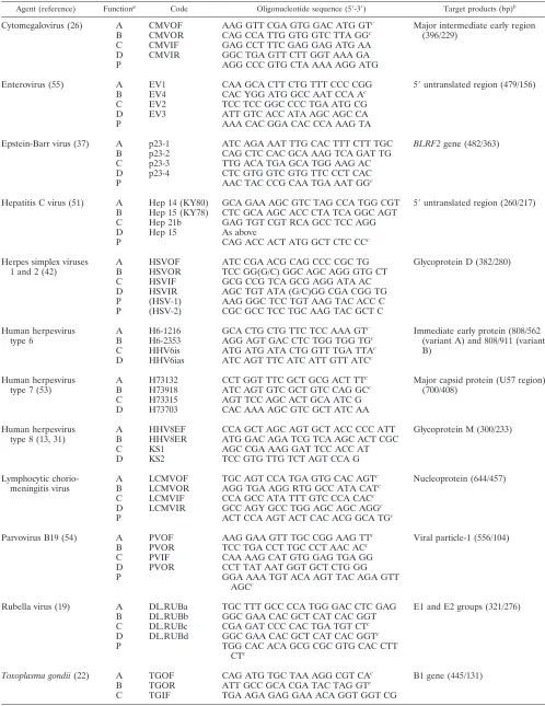
Agent (reference) Functiona Code Oligonucleotide sequence (5⬘-3⬘) Target products (bp)b
Cytomegalovirus (26)
A
CMVOF
AAG GTT CGA GTG GAC ATG GT
cMajor intermediate early region
(396/229)
B
CMVOR
CAG CCA TTG GTG GTC TTA GG
cC
CMVIF
GAG CCT TTC GAG GAG ATG AA
D
CMVIR
GGC TGA GTT CTT GGT AAA GA
P
AGG CCC GTG CTA AAA AGG ATG
Enterovirus (55)
A
EV1
CAA GCA CTT CTG TTT CCC CGG
5
⬘
untranslated region (479/156)
B
EV4
CAC YGG ATG GCC AAT CCA A
cC
EV2
TCC TCC GGC CCC TGA ATG CG
D
EV3
ATT GTC ACC ATA AGC AGC CA
P
AAA CAC GGA CAC CCA AAG TA
Epstein-Barr virus (37)
A
p23-1
ATC AGA AAT TTG CAC TTT CTT TGC
BLRF2
gene (482/363)
B
p23-2
CAG CTC CAC GCA AAG TCA GAT TG
C
p23-3
TTG ACA TGA GCA TGG AAG AC
D
p23-4
CTC GTG GTC GTG TTC CCT CAC
P
AAC TAC CCG CAA TGA AAT GG
cHepatitis C virus (51)
A
Hep 14 (KY80)
GCA GAA AGC GTC TAG CCA TGG CGT
5
⬘
untranslated region (260/217)
B
Hep 15 (KY78)
CTC GCA AGC ACC CTA TCA GGC AGT
C
Hep 21b
GAG TGT CGT RCA GCC TCC AGG
D
Hep 15
As above
P
CAG ACC ACT ATG GCT CTC CC
cHerpes simplex viruses
1 and 2 (42)
A
HSVOF
ATC CGA ACG CAG CCC CGC TG
Glycoprotein D (382/280)
B
HSVOR
TCC GG(G/C) GGC AGC AGG GTG CT
C
HSVIF
GCG CCG TCA GCG AGG ATA AC
D
HSVIR
AGC TGT ATA (G/C)GG CGA CGG TG
P
(HSV-1)
AAG GGC TCC TGT AAG TAC ACC C
P
(HSV-2)
CGC GCC TCC TGC AAG TAC GCT C
Human herpesvirus
type 6
A
H6-1216
GCA CTG CTG TTC TCC AAA GT
cImmediate early protein (808/562
(variant A) and 808/911 (variant
B)
B
H6-2353
AGG AGT GAC CTC TGG TGG TG
cC
HHV6is
ATG ATG ATA CTG GTT TGA TTA
cD
HHV6ias
ATC AGT TTC ATC ATT GTT ATC
cHuman herpesvirus
type 7 (53)
A
H73132
CCT GGT TTC GCT GCG ACT TT
cMajor capsid protein (U57 region)
(700/408)
B
H73918
ATC AGT GTC GCT GTC CAG GC
cC
H73315
AGT TCC AGC ACT GCA ATC G
D
H73703
CAC AAA AGC GTC GCT ATC AA
Human herpesvirus
type 8 (13, 31)
A
HHV8EF
CCA GCT AGC AGT GCT ACC CCC ATT
Glycoprotein M (300/233)
B
HHV8ER
ATG GAC AGA TCG TCA AGC ACT CGC
C
KS1
AGC CGA AAG GAT TCC ACC AT
D
KS2
TCC GTG TTG TCT AGT CCA G
Lymphocytic
chorio-meningitis virus
A
LCMVOF
TGC AGT CCA TGA GTG CAC AGT
cNucleoprotein (644/457)
B
LCMVOR
AGG TGA AGG RTG GCC ATA CAT
cC
LCMVIF
CCA GCC ATA TTT GTC CCA CAC
cD
LCMVIR
GCC AGY GCC TGG AGC AGC AGG
cP
ACT CCA AGT ACT CAC ACG GCA TG
cParvovirus B19 (54)
A
PVOF
AAG GAA GTT TGC CGG AAG TT
cViral particle-1 (556/104)
B
PVOR
TCC TGA CCT TGC CCT AAC AC
cC
PVIF
CAA AAG CAT GTG GAG TGA GG
D
PVOR
CCT TAT AAT GGT GCT CTG GG
P
GGA AAA TGT ACA AGT TAC AGA GTT
AGC
cRubella virus (19)
A
DL.RUBa
TGC TTT GCC CCA TGG GAC CTC GAG
E1 and E2 groups (321/276)
B
DL.RUBb
GGC GAA CAC GCT CAT CAC GGT
C
DL.RUBc
CGA GAT CCC CAC TGA TGT CT
cD
DL.RUBd
GGC GAA CAC GCT CAT CAC GGT
cP
TGG CAC ACA GCG CGC GTG CAC CTT
CT
cToxoplasma gondii
(22)
A
TGOF
CAG ATG TGC TAA AGG CGT CA
cB1 gene (445/131)
B
TGOR
ATT GCC GCA CGA TAC TAG GT
cC
TGIF
TGA AGA GAG GAA ACA GGT GGT CG
Continued on facing page
on May 15, 2020 by guest
http://jcm.asm.org/
consistently offer clear distinction between blank and positive control absorbance values (data not shown).
Confirmatory tests for applied multiplexes.Results of other tests that may have been performed on mPCR-positive clinical samples were collated. Such tests include: culture (as above), enterovirus direct fluorescent antibody panels (Light Diagnostics), HSV 1/HSV 2 direct specimen direct fluorescent antibody (Trinity Biotech, Ireland), Merifluor VZV direct fluorescent antibody (Merid-ian), Cobas CMV Amplicor Monitor (Roche, Germany), CMV IgG AXSYM System (Abbott, Germany), CMV IgM ELISA (DiaSorin, Italy), HerpeSelect 1 ELISA IgG (Focus Technologies), HerpeSelect 2 ELISA IgG (Focus Technol-ogies), herpes simplex 1 and 2 IgM ELISA (Diagnostic Systems Laboratories), parvovirus (as above), Enzygnost anti-varicella-zoster virus/IgG (Dade Behring), and Enzygnost anti-varicella-zoster virus/IgM (Dade Behring).
Statistics.Sensitivity is defined as the ability of the mPCR to give a positive finding for samples previously established as positive by an alternate method. Similarly, specificity is the reproducibility of an established negative finding. Both measures are expressed as percentages and for values less than 100%, estimation of the population parameter (95% confidence interval [CI]) were calculated using a method for proportions (GraphPad InStat).
RESULTS
Screening of congenital agents using developmental
multi-plexes.
Infectious agents were not detected in 191 amniotic
fluid samples screened by the developmental mPCRs with
VRL01 (DNA) and VRL04 (RNA) arrays. Furthermore,
her-pes viruses (CMV and HHV-6, -7, and -8) were not detected in
15 of placentae screened using the VRL03 mPCR. Samples
with sufficient volume remaining were used for the assessment
of the applied mPCRs.
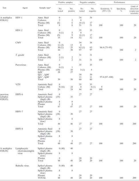
Assessment of applied multiplexes.
A summary of the
as-sessment of the four mPCRs: VDL01, VDL03, VDL04, and
VDL05 is shown in Table 2. For all agents tested, the
multi-plexes showed a sensitivity of
ⱖ
95% or a 95% CI that includes
values in this range, and a specificity of 100%. Plasmid
con-structs for the four mPCRs diluted from 0.2 to 200 000 copies
per reaction (10
0to 10
6copies/ml) showed a limit of detection
range from 2 to 200 copies per reaction (10
1to 10
3copies/ml).
All cultures and spiked samples were positively identified using
multiplexes. A notable discrepancy was observed for CMV
detection in VDL01 where only 78% of PCR-proven
CMV-positive samples were detected. In contrast, a higher sensitivity
was observed for CMV detection using VDL05 when different
extracts were used than those for VDL01 evaluation.
During the post-PCR probe detection stage, several isolates
of enterovirus cross-reacted with the HSV-1 probe (data not
shown). Furthermore, weak reactions sometimes occurred
be-tween HSV-2 probes and HSV-1 amplicons. Given these
ob-servations, probe hybridization was used only to confirm the
identity of electrophoretic bands and this was particularly
use-ful when nonspecific amplification products such as primer
dimers were present.
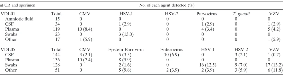
Since this assessment, VDL01 and VDL05 have been used in
our routine diagnostic laboratory for six months during 2004 to
2005 (Table 3). Using VDL01 an agent was detected in 13.5%
of samples tested and 15 of 17 (88.2%) mPCR-positive
speci-mens were confirmed by additional tests (including repeat tests
on consecutive samples, culture, antigen detection by direct
fluorescent antibody, CMV quantification and IgM serology).
Similarly, the overall detection rate of the VDL05 was 22.2%
of all samples and 35/37 (94.6%) were confirmed by additional
testing (as above). During this period of diagnostic use
alter-native agents to that clinically suspected were detected for
some cases. These episodes included six requests for HSV that
were positive for CMV (a corneal ulcer swab and two
cerebro-spinal fluid specimens), varicella-zoster virus (a swab of facial
cellulitis), and Epstein-Barr virus (two conjunctival swabs);
Epstein-Barr virus detected in two specimens of bronchial
washings where CMV testing was requested and one stool
sample where enterovirus was requested; and four specimens
where varicella-zoster virus was requested that were found to
be positive for HSV-1 (swab of skin lesion) and HSV-2 (swab
of skin vesicle and two blisters).
DISCUSSION
[image:4.585.46.540.80.219.2]Although monoplex PCR and real-time assays have
consid-erable benefits in targeting detection of specific organisms,
they do not necessarily allow detection of the causative agent,
due to the specificity of the primer sets used. Increasingly,
detection of the causative agent using multiplexes in
respira-tory specimens (16, 50), gastrointestinal specimens (7), the eye
(14), conditions causing lymphadenopathy (39), cerebrospinal
fluid (11, 49) are replacing pathogen specific (versus clinical
conditions specific) detection (32). Both forms of multiplex
testing are useful–the VDL03 (herpesviruses) and VDL04
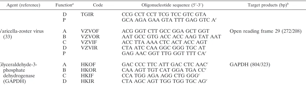
TABLE 1—
Continued
Agent (reference) Functiona Code Oligonucleotide sequence (5⬘-3⬘) Target products (bp)b
D
TGIR
CCG CCT CCT TCG TCC GTC GTA
P
GCA AGA GAA GTA TTT GAG GTC A
cVaricella-zoster virus
(33)
A
VZVOF
ACG GGT CTT GCC GGA GCT GGT
Open reading frame 29 (272/208)
B
VZVOR
AAT GCC GTG ACC ACC AAG TAT AAT
C
VZVIF
ACC TTA AAA CTC ACT ACC AGT
D
VZVIR
CTA ATC CAA GGC GGG TGC AT
P
GAG AAC GGT TTG GGT TTT CA
cGlyceraldehyde-3-phosphate
dehydrogenase
(GAPDH)
A
B
C
D
HKOF
HKOR
HKIF
HKIR
GAC CCC TTC ATT GAC CTC AAC
cCAA AGT TGT CAT GGA TGA CC
cCCA TGG AGA AGG CTG GGG
cCTA AGC AGT TGG TGG TGC AG
cGAPDH (804/323)
a
A, outer sense primer; B, outer antisense primer; C, inner sense primer; D, inner antisense primer; P, probe.
b
First-round product/second-round product.
c
Sequence was designed in-house.
on May 15, 2020 by guest
http://jcm.asm.org/
TABLE 2. Assessment of applied multiplex PCRs
Test Agent Sample typea
Positive samples Negative samples Performance
No. tested
No. positive
No. tested
No. negative
Sensitivity, % (95% CI)
Specificity, %
Limit of detection (copies per
reaction)
DNA multiplex
(VDL01)
HSV-1
Amn. fluid
0
34
34
Cultures
2
2
0
Plasma (M)
(6)
6
16 (1)
17
Total
8
8
51
51
100
100
20
HSV-2
Amn. fluid
0
35
35
Cultures (M)
4 (1)
5
0
Plasma (M)
(9)
9
16 (1)
17
Total
14
14
52
52
100
100
20
CMV
Amn. fluid
0
34
34
Cultures (M)
11 (2)
13
0
Plasma (M)
30 (1)
25
62 (1)
63
86.4 (73–95)
Total
44
38
97
97
100
2
T. gondii
Amn. fluid
0
31
31
Cultures (M)
1 (1)
2
Total
2
2
31
31
100
100
2
Parvovirus
Amn. fluid
0
35
35
Cultures (M)
0 (1)
1
Plasma (M)
(10)
10
3 (1)
4
IgM
⫹28
27
IgG
⫺, IgM
⫺30
30
IgG
⫹, IgM
⫺11
11
97.4 (87–100)
Total
39
38
80
80
100
20
VZV
Amniotic fluid
0
35
35
Culture (M)
8 (16)
24
0
8 (1)
9
Total
24
24
44
44
100
100
20
Herpesvirus
multiplex
(VDL03)
HHV-6
Amniotic fluid
0
27
27
Spiked plasma
(high) (B)
(35)
35
Spiked plasma
8
8
Plasma
4
4
Total
47
47
27
27
100
100
20
HHV-7
Amniotic fluid
0
27
27
Spiked plasma
(high) (B)
(38)
38
Spiked plasma
(low)
8
8
Total
46
46
27
27
100
100
200
HHV-8
Amniotic fluid
0
27
27
Spiked plasma
(high) (B)
(38)
38
Spiked plasma
(low)
8
8
Plasma
2
2
Total
48
48
27
27
100
100
200
RNA multiplex
(VDL04)
Lymphocytic
choriomeningitis
virus
Spiked plasma
(high) (B)
0 (40)
40
Spiked plasma
(low)
8
8
Plasma
0
20
20
Total
48
48
20
20
100
100
20
Rubella virus
Spiked plasma
(high) (B)
0 (40)
40
Spiked plasma
(low)
8
8
Plasma
0
20
20
TOTAL
48
48
20
20
100
100
200
Continued on facing page
on May 15, 2020 by guest
http://jcm.asm.org/
(RNA viruses) multiplexes used here were designed to allow
more efficient testing of infrequently requested agents, thereby
allowing more rapid turn-around times, and more efficient
testing of commonly requested agents that were determined on
the basis of requesting, rather than on the basis of any common
pathologies (VDL05 mPCR).
The four applied mPCRs described here were developed for
use in a large routine diagnostic laboratory. Appropriate to this
setting, we aimed to develop methods of simplicity, robustness
and minimal complexity without compromise to sensitivity and
specificity. To achieve this, we used common protocols for
master mixes (for both the RNA and DNA agents),
thermo-cycling conditions for first and second-round reactions and
amplicon detection. The RT-PCR was also used for the DNA
agents for conformity of methodology to reduce complexity
between methods and may have enhanced sensitivity by
trans-forming RNA transcripts, though this is speculative and was
not investigated. In VDL01, VDL04 and VDL05 mPCRS,
ura-cil
N
-glcosylase was incorporated in the first-round reaction to
lower the risk of cross contamination linked to carryover of
digoxigenin labeled amplicons.
[image:6.585.47.536.84.483.2]We used hot-start PCR to minimize nonspecific reactions
such as primer dimers when annealing occurs during precycling
temperatures (20). Furthermore, nested PCRs were used to
enhance sensitivity and specificity. To improve sensitivity in
specimens of low genomic content, we used 20
l of template
in a 50-
l reaction. These applied mPCRs generally showed
high sensitivity when testing a variety of specimens of differing
genomic content that were extracted by different methods and
were either recently extracted or had been stored at
⫺
20°C for
up to one month. The only exception was the sensitivity
(86.4%) for CMV by VDL01 for which the limit of detection
was 2 copies per reaction. It is likely that measurements of
sensitivity for this virus may have been compromised by testing
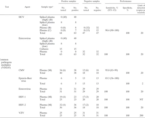
TABLE 2—
Continued
Test Agent Sample typea
Positive samples Negative samples Performance
No. tested
No. positive
No. tested
No. negative
Sensitivity, % (95% CI)
Specificity, %
Limit of detection (copies per
reaction)
HCV
Spiked plasma
(high) (B)
0 (40)
40
Spiked plasma
(low)
8
8
Plasma (B)
0 (8)
8
0 (32)
32
Plasma (C)
0 (8)
7
0 (15)
15
98.4 (90–100)
Total
64
63
47
47
100
20
Enterovirus
Spiked plasma
(high) (B)
0 (40)
40
Spiked plasma
(low)
8
8
Cultures
15
15
Plasma
0
0
32
32
Total
63
63
32
32
100
100
20
Common
pathogens
multiplex
(VDL05)
CMV
Plasma (M)
34 (6)
38
12 (6)
18
95.0 (83–99)
Total
40
38
18
18
100
20
Epstein-Barr
virus
Plasma
6
5
13
13
83.3 (36–100)
Total
6
5
13
13
100
2
Enterovirus
Plasma
31
31
29
29
Total
31
31
29
29
100
100
20
HSV-1
Plasma (M)
19 (4)
23
17 (3)
20
Total
23
23
20
20
100
100
NT
HSV-2
Plasma (M)
32 (4)
36
17 (2)
18
Total
36
36
18
18
100
100
20
VZV
Plasma
25
25
31
31
Total
25
25
31
31
100
100
200
a
Samples designated plasma are clinical samples that have been previously tested by another method. Unless indicated, samples were extracted by HPA (Roche). The number extracted by an alternative method is given in parentheses. The number of samples extracted using a MagNaPure LC (total nucleic acid isolation kit) (Roche, Germany) (M), BioRobot M8 (viral isolation kit) (QIAGEN, Germany) (B), or COBAS Amplicor (Roche) (C) is shown in parentheses. Amn., amniotic.
on May 15, 2020 by guest
http://jcm.asm.org/
predominantly stored extracts. The small number of samples
tested for Epstein-Barr virus would account for the low
sensi-tivity measured for detection of this virus. However, the
sen-sitivity of the mPCRs for other agents was
ⱖ
95.0% and no
false-positives were recorded.
The specificity of the mPCRs is enhanced by using both
electrophoresis and probe-amplicon hybridization methods.
The multiplexes were optimized to reduce spurious
amplifica-tion products such as due to the formaamplifica-tion of primer dimers
and nonspecific interaction with nucleic acid rich extracts.
Post-PCR probe-hybridization assisted in identifying
ampli-cons when nonspecific electrophoresis products were present.
However, probes were not developed for the VDL03 multiplex
as it tests for only three agents (HHV6, -7, and -8) and the
products were easily discernible by electrophoresis. Given our
observations of cross-hybridization between enterovirus and
the HSV-1 probe and between the HSV-2 probe and HSV-1
amplicons, post-PCR detection by this method was used to
confirm the electrophoresis band and not used independently,
i.e., as a means of increasing sensitivity of detection.
The use of VDL01 and VDL05 mPCRs in our diagnostic
laboratory over a six month period not only enhanced our
validation, but showed the resourcefulness of the array of
de-tectable agents by each method-demonstrated by detection
rates of 13.5% and 22.2%, respectively (Table 3). The VDL01
mPCR was developed for detection of common intrauterine
infections caused by DNA containing agents, and
demon-strated its utility as a diagnostic screen for other infections such
as viral meningitis and skin lesions. The array for VDL05 was
selected on the basis of commonly requested pathogens and
evident by the high detection rate, implementation has
re-sulted in cost efficiencies and timely reporting of results.
Fur-ther diagnostic benefits were demonstrated when alternate
agents to that clinically suspected were detected. This had
occurred in 11 requests, including requests for varicella-zoster
virus in swabs of skin lesions when HSV-1 and HSV-2 were
detected, and requests for HSV in cerebrospinal fluid when
CMV was detected.
Both HSV-1 and HSV-2 infection cause dermatomal
vesic-ular lesions not unlike those caused by varicella-zoster virus
(52). However, CMV infection of the central nervous system is
rare and can occur in infants following intrauterine infection
(6). Epstein-Barr virus was detected in a variety of specimens,
including: conjuctival swabs (
n
⫽
2), bronchial washings (
n
⫽
2), plasma (
n
⫽
6), and feces (
n
⫽
1). This virus can be isolated
from oropharyngeal washings or from circulating lymphocytes
of 80 to 90% of patients with infectious mononucleosis (46).
However, serendipitous detection should receive careful
con-sideration given the ubiquity of virus shedding in both healthy
persons and in those with unrelated illnesses (46).
The motivation for the development of these novel mPCRs
was for the screening of amniotic fluid for pathogens known to
cause fetal loss (miscarriages and stillbirths). In Australia,
common fetal pathogens include CMV, HSV, rubella virus,
T.
gondii
and VZV (1, 9, 10, 28, 40). To our knowledge, there is
no universal screening for any of these infections, and
diagno-sis is often difficult, particularly as detailed viral testing in this
country is rarely performed in intrauterine deaths (40), in
neonates and even in postnatal death from sudden infant death
syndrome (23, 41).
The availability of these mPCRs in the routine diagnostic
laboratory will enable more frequent testing of a broad
spec-trum of agents implicated in congenital disease including
vi-ruses whose association is suspected but has not been
estab-lished in Australia. lymphocytic choriomeningitis virus is
well-established as a cause of congenital disease in the United
States and Europe (5) but not yet evident in this country (47).
HHV-6, HHV-7 and HHV-8 are infectious agents with
possi-ble associations with congenital anomalies and stillbirth, on the
basis of case reports, plausible animal models, or detection in
placental or uterine tissue of affected and unaffected babies (2,
25, 34). Antenatal infections with enterovirus have been
asso-ciated with neurodevelopmental delay (21), and infant diabetes
(17, 27). Transplacental transmission of hepatitis C virus is
uncommon although the risk of transmission may increase
when the mother is coinfected with human immunodeficiency
virus (6).
Failure to detect infectious agents in the amniotic fluids
tested by the developmental mPCRs reflects the rarity of
con-genital infections in a population of healthy pregnant women.
Furthermore, amniotic fluid collected during the first trimester
may be too early for detection of agents such as CMV (18) and
T. gondii
(44). The applied mPCRs that have been adapted for
routine use will enable testing to be undertaken more
fre-quently and on a larger scale, and where recent infection is
suspected by illness or seroconversion.
[image:7.585.46.541.79.211.2]CMV is the most common cause of intrauterine infection
and most studies of the clinical significance of viral detection in
TABLE 3. Detection of agents using VDL01 and VDL05 (applied multiplexes) in the routine diagnostic laboratory
mPCR and specimen No. of each agent detected (%)
VDL01
Total
CMV
HSV-1
HSV-2
Parvovirus
T. gondii
VZV
Amniotic fluid
15
0
0
0
0
0
0
CSF
34
0
1 (2.9)
0
1 (2.9)
0
1 (2.9)
Plasma
119
10 (8.4)
0
0
4 (3.4)
0
5 (4.2)
Swabs
23
0
3 (13.0)
0
0
0
0
Other
17
1 (5.9)
0
0
0
0
1 (5.9)
VDL05
Total
CMV
Epstein-Barr virus
Enterovirus
HSV-1
HSV-2
VZV
CSF
144
3 (2.1)
5 (3.5)
10 (6.9)
0
3 (2.1)
1 (0.7)
Plasma
136
10 (7.4)
8 (5.9)
0
0
0
0
Swabs
128
0
2 (1.6)
0
16 (12.5)
9 (7.0)
17 (13.2)
Other
51
0
5 (9.8)
2 (3.9)
2 (3.9)
3 (5.9)
6 (11.8)
on May 15, 2020 by guest
http://jcm.asm.org/
amniotic fluid has been done with this virus. A recent study
(24) showed with 100% probability that the presence of
ⱖ
10
3genome equivalents/ml predicted mother-child infection, and
ⱖ
10
5genome equivalents predicted the development of a
symptomatic infection. The limit of detection of this agent in
VDL01 and VDL05 appears to be appropriately sensitive for
the prediction of these clinical outcomes and should be
aug-mented by quantitative PCR assessment.
The techniques used here have allowed mPCR detection of
congenital agents from genomic material extracted from
am-niotic fluid (3) from plasma and has been compared with
de-tection of known virus in clinical samples, cultures, or plasma
spiked with plasmids (Table 2). This work also represents a
standard approach to the assays, using commercial agents, and
use of consistent and thorough assessment of these assays to
give accurate figures for sensitivity, specificity and limit of
detection (Table 2). The use of nested PCR and RT-PCR has
meant these assays typically detect down to 10
1to 10
2copies of
the etiological agent, an important element of detection in
congenital infections, and infections of the central nervous
system and cerebrospinal fluid particularly (14, 49). Although
not examined in this study, a potential use of the mPCRs would
be the detection of congenital agents in dried blood spots
retrospectively collected from children postnatally diagnosed
with conditions such as deafness after birth which is detectable
months to years after birth (4).
The increase in diagnostic capacity of these mPCRs offers
the cost benefits of less reagents and consumables, and
im-proved turn-around time. Furthermore, the development
en-ables testing for a wide range of agents using a small volume of
clinical sample. The automation of the extraction process as
used in this study (Table 2) further enhances efficiency. These
mPCRs have suitable performance characteristics for the
de-tection of a broad range of agents associated with congenital
and other infections. The use of a common methodology is
conducive to routine screening of small sample volumes,
in-clusive of the rarer agents such as lymphocytic
choriomenin-gitis virus, and HHV-6, HHV-7, and HHV-8. The increase in
testing will enhance our understanding of the role played by
these agents in congenital disease within our epidemiologic
setting. Furthermore, the benefits observed from the use of
mPCRs in a routine diagnostic laboratory such as VDL05 for
the detection of commonly tested viruses is the motivation for
continuing development of other organ-specific mPCRs.
ACKNOWLEDGMENTS
We thank Gwen Lewis, Michael Fennell, the Virology Diagnostic
Laboratory, and the South Eastern Laboratory Services for assistance.
REFERENCES
1.Armstrong, L., D. Isaacs, and N. Evans.2004. Severe neonatal toxoplasmosis after third trimester maternal infection. Paediatr. Infect. Dis. J.23:968–969. 2.Ashshi, A. M., P. E. Klapper, and R. J. Cooper.2003. Detection of human cytomegalovirus, human herpesvirus type 6 and human herpesvirus type 7 in urine specimens by multiple PCR. J. Infect.47:59–64.
3.Avidor, B., G. Efrat, M. Weinberg, Z. Kra-oz, J. Satinger, S. Mitrani-Rosen-baum, Y. Yaron, L. Shulman, M. Tepperberg-Oikawa, D. Wolf, S. A. Berger, S. Liptz, E. Mendelson, and M. Giladi. 2004. Insight into the intrinsic sensitivity of the PCR assay used to detect CMV infection in amniotic fluid specimens. J. Clin. Virol.29:260–270.
4.Barbi, M., S. Binda, V. Primache, S. Caroppo, P. Dido, P. Guidotti, C. Corbetta, and D. Melotti.2000. Cytomegalovirus DNA detection in Guthrie cards: a powerful tool for diagnosing congenital infection. J. Clin. Virol.
17:159–165.
5.Barton, L. L., and M. B. Mets.2001. Congenital lymphocytic choriomenin-gitis virus infection: decade of rediscovery. J. Infect. Dis.33:370–374. 6.Benenson, A. S. (ed.).1995. Control of communicable diseases manual.
American Public Health Association, Washington, D.C.
7.Beuret, C. 2004. Simultaneous detection of enteric viruses by multiplex real-time RT-PCR. J. Virol. Methods115:1–8.
8.Burg, J. L., C. M. Grover, P. Pouletty, and J. C. Boothroyd.1989. Direct and sensitive detection of a pathogenic protozoan, Toxoplasma gondii, by poly-merase chain reaction. J. Clin. Microbiol.27:1787–1792.
9.Burgess, M., and J. Forrest.November 2001. Congenital and neonatal vari-cella. APSU-RACP (Paed. Div.) [Online.] http://apsu.inopsu.com. 10.Burgess, M., J. Forrest, C. A. Jones, P. McIntyre.11thFebruary, 2005.
Congenital rubella. Reporting of communicable disease conditions under surveillance by the APSU, 1 January to 30 June 2003. Commun. Dis. Intell. 28(4). [Online.] http://www.health.gov.gov.au.
11.Calvario, A., A. Bozzi, M. Scarasciulli, C. Ventola, R. Seccia, D. Stomati, and B. Brancasi.2002. Herpes Consensus PCR test: a useful diagnostic approach to the screening of viral diseases of the central nervous system. J. Clin. Virol.
25(Suppl. 1):S71–78.
12.Candotti, D., J. Temple, S. Owusu-Ofori, and J. P. Allain.2004. Multiplex real-time quantitative RT-PCR assay for hepatitis B virus, hepatitis C virus, and human immunodeficiency virus type 1. J. Virol. Methods118:39–47. 13.Chang, Y., E. Cesarman, M. S. Pessin, F. Lee, J. Culpepper, D. M. Knowles,
and P. S. Moore.1994. Identification of Herpesvirus-like DNA sequences in AIDS-associated Kaposi’s sarcoma. Science266:1865–1869.
14.Chichili, G. R., S. Athmanathan, S. Farhatullah, N. Gangopadhyay, S. Ja-lali, G. Pasricha, and S. Sharma.2003. Multiplex polymerase chain reaction for the detection of herpes simplex virus, varicella-zoster virus and cytomeg-alovirus in ocular specimens. Curr. Eye Res.27:85–90.
15.Chomczynski, P., and N. Sacchi.1987. Single-step method of RNA isolation by acid guanidinium thiocyanate-phenol-chloroform extraction. Anal. Bio-chem.162:156–159.
16.Coiras, M. T., J. C. Aguilar, M. L. Garcia, I. Casas, and P. Perez-Brena.
2004. Simultaneous detection of fourteen respiratory viruses in clinical spec-imens by two multiplex reverse transcription neSted-PCR assays. J. Med. Virol.72:484–495.
17.Craig, M. E., N. J. Howard, M. Silink, and W. D. Rawlinson.2003. Reduced frequency of HLA DRB1*03-DQB1*02 in children with type 1 diabetes associated with enterovirus RNA. J. Infect. Dis.187:1562–1570.
18.Donner, C., C. Liesnard, F. Brancart, and F. Rodesch.1994. Accuracy of amniotic fluid testing before 21 weeks’ gestation in prenatal diagnosis of congenital cytomegalovirus infection. Prenat. Diagn.14:1055–1059. 19.Eggerding, F. A., J. Peters, R. K. Lee, and C. B. Inderlied.1991. Detection
of rubella virus gene sequences by enzymatic amplification and direct se-quencing of amplified DNA. J. Clin. Microbiol.29:945–952.
20.Elnifro, E. M., A. M. Ashshi, R. J. Cooper, and P. E. Klapper.2000. Mul-tiplex PCR: optimization and application in diagnostic virology. Clin. Micro-biol. Rev.13:559–570.
21.Euscher, E., J. Davis, I. Holzman, and G. J. Nuovo.2001. Coxsackie virus infection of the placenta associated with neurodevelopmental delays in the newborn. Obstet. Gynecol.98:1019–1026.
22.Franzen, C., M. Altfeld, P. Hegener, P. Hartmann, G. Arendt, H. Jab-lonowski, J. Rockstroh, V. Diehl, B. Salzberger, and G. Fa¨tkenheuer.1997. Limited value of PCR for detection ofToxoplasma gondiiin blood from human immunodeficiency virus-infected patients. J. Clin. Microbiol.
35:2639–2641.
23.Gleeson, M., R. L. Clancy, A. J. Cox, S. A. Gulliver, S. T. Hall, and D. M. Cooper.2004. Mucosal immune responses to infections in infants with acute life threatening events classified as ‘near-miss’ sudden infant death syn-drome. FEMS Immunol. Med. Microbiol.42:105–118.
24.Guerra, B., T. Lazzarotto, S. Quarta, M. Lanari, L. Bovicelli, A. Nicolosi, and M. P. Landini.2000. Prenatal diagnosis of symptomatic congenital cytomegalovirus infection. Am. J. Obstet Gynecol.183:476–482.
25.Hall, C. B., M. T. Caserta, K. C. Schnabel, C. Boettrich, M. P. McDermott, G. K. Lofthus, J. A. Carnahan, and S. Dewhurst.2004. Congenital infections with human herpesvirus 6 (HHV6) and human herpesvirus 7 (HHV7). J. Pe-diatr.145:472–477.
26.Halwachs-Baumann, G., M. Wilders-Truschnig, G. Enzinger, M. Eibl, W. Linkesch, H. J. Dornbusch, B. J. Santner, E. Marth, and H. H. Kessler.2001. Cytomegalovirus diagnosis in renal and bone marrow transplant recipients: the impact of molecular assays. J. Clin. Virol.20:49–57.
27.Horwitz, M. S., L. M. Bradley, J. Harbertson, T. Krahl, J. Lee, and N. Sarvetnick. 1998. Diabetes induced by coxsackie virus: initiation by by-stander damage and not molecular mimicry. Nat. Med.4:781–785. 28.Jones, C. A., D. Isaacs, P. McIntyre, T. Cunningham, S. Garland.11th
February, 2005. Neonatal herpes simplex virus infection. Reporting of com-municable disease conditions under surveillance by the APSU, 1 January to 30 June 2003. Commun. Dis. Intell. 28(4). [Online.] http://www.health.gov .gov.au.
29.Kirschberg, O., Schuttler, C., Repp, R., and S. Schaefer.2004. A multiplex-PCR to identify hepatitis B virus–enotypes A-F. J. Clin. Virol.29:39–43. 30.Liolios, L., A. Jenney, D. Spelman, T. Kotsimbos, M. Catton, and S.
on May 15, 2020 by guest
http://jcm.asm.org/
selingh.2001. Comparison of a multiplex reverse transcription-PCR-enzyme hybridization assay with conventional viral culture and immunofluorescence techniques for the detection of seven viral respiratory pathogens. J. Clin. Microbiol.39:2779–2783.
31.Lock, M. J., P. D. Griffiths, and V. C. Emery. 1997. Development of a quantitative competitive polymerase chain reaction for human herpesvirus 8. J. Virol. Methods64:19–26.
32.Mackay, I. M., T. Gardam, K. E. Arden, S. McHardy, D. M. Whiley, E. Crisante, and T. P. Sloots.2003. Co-detection and discrimination of six human herpesviruses by multiplex PCR-ELAHA. J. Clin. Virol.28:291–302. 33.Mahalingam, R., M. Wellish, W. Wolf, A. N. Dueland, R Cohrs, A. Vajai, and D. Gilden.1990. “Latent varicella-zoster viral DNA in the human trigeminal and thoracic ganglia.” N. Engl. J. Med.323:627–631.
34.Mantina, H., C. Kankasa, W. Klaskala, B. Brayfield, J. Campbell, Q. Du, G. Bhat, F. Kasolo, C. Mitchell, and C. Wood.2001. Vertical transmission of Kaposi’s sarcoma-associated herpesvirus. Int. J. Cancer94:749–752. 35.Markoulatos, P., N. Siafakas, and M. Moncany.2002. Multiplex polymerase
chain reaction: a practical approach. J. Clin. Lab. Anal.16:47–51. 36.McIver, C. J., and J. W. Tapsall.1991. In vitro susceptibilities of clinical
isolates of cysteine-requiringEscherichia colito twelve antimicrobial agents. Antimicrob. Agents Chemother.35:995–338.
37.Meerbach, A., B. Gruhn, R. Egerer, U. Reischl, F. Zintl, and P. Wutzler.
2001. Semiquantitative PCR analysis of Epstein-Barr virus DNA in clinical samples of patients with EBV-associated diseases. J. Med. Virol.65:348–357. 38.National Center for Biotechnology Information.2005. Basic local alignment search tool (BLAST). National Library of Medicine and National Institutes of Health. [Online.] http://www.ncbi.nlm.nih.gov.
39.Nopponpunth, V., S. Changrad, A. Rakyuu, J. Nertsawange, W. Chansupit, and Y. Poovorawan.2003. Design of degenerate primers for multiplex nest-ed-PCR detection of human lymphotropic herpesvirus. Southeast Asian J. Trop. Med. Public Health34:120–125.
40.Rawlinson, W., D. Trincado, G. Scott, S. Munro, P. Palasanthiran, M. Ferson, D. Smith, G. Higgins, M. Catton, A. McGregor, D. Dwyer, and A. Kesson.11thFebruary, 2005. Congenital cytomegalovirus infection.
Report-ing of communicable disease conditions under surveillance by the APSU, 1 January to 30 June 2003. Commun. Dis. Intell. 28 (4). [Online.] http://www .health.gov.gov.au.
41.Raza, M. W., and C. C. Blackwell.1999. Sudden infant death syndrome, virus infections and cytokines. FEMS Immunol. Med. Microbiol.25:85–96. 42.Read, S. J., and J. B. Kurtz.1999. Laboratory diagnosis of common viral
infections of the central nervous system by using a single multiplex PCR screening assay. J. Clin. Microbiol.37:1352–1355.
43.Read, S. J., J. L. Mitchell, and C. G. Fink.2001. LightCycler multiplex PCR for the laboratory diagnosis of common viral infections of the central ner-vous system. J. Clin. Microbiol.39:3056–3059.
44.Rommand, S., M. Wallon, J. Franck, P. Thulliez, F. Peyron, and H. Dumon.
2001. Prenatal diagnosis using polymerase chain reaction on amniotic fluid for congenital toxoplasmosis. Obstet. Gynecol.97:296–300.
45.Sambrook, J., E. F. Fritsch, and T. Maniatis.1989. Molecular cloning: a laboratory manual, 2nd ed. Cold Spring Harbor Laboratory Press, Cold Spring Harbor, N.Y.
46.Schooley, R. T.2000. Epstein-Barr virus (infectious mononucleosis), p. 1599– 1613.InG. L. Mandell, J. E. Bennett, and R. Dolin (ed.), Principles and practice of infectious diseases. Churchill Livingstone, Philadelphia, PA. 47.Singleton, G. R., A. L. Smith, G. R. Shellam, N. Fitzgerald, and W. J. Muller.
1993. Prevalence of viral antibodies and helminthes in field populations of house mice (Mus domesticus) in southeastern Australia. Epidemiol. Infect.
110:399–417.
48.Syrmis, M. W., D. M. Whiley, M. Thomas, I. M. Mackay, J. Williamson, D. J. Siebert, M. D. Nissen, and T. P. Sloots.2004. A sensitive, specific, and cost-effective multiplex reverse transcriptase-PCR assay for the detection of seven common respiratory viruses in respiratory samples. J. Mol. Diagn.
6:125–131.
49.Tarrago, D., C. Quereda, and A. Tenorio.2003. Different cytomegalovirus glycoprotein B genotype distribution in serum and cerebrospinal fluid spec-imens determined by a novel multiplex nested PCR. J. Clin. Microbiol.
41:2872–2877.
50.Templeton, K. E., S. A. Scheltinga, M. F. Beersma, A. C. Kroes, and E. C. Claas.2004. Rapid and sensitive method using multiplex real-time PCR for diagnosis of infections by influenza A and influenza B viruses, respiratory syncytial virus, and parainfluenza viruses 1, 2, 3, and 4. J. Clin. Microbiol.
42:1564–1569.
51.White, P. A., X. Zhai, I. Carter, Y. Zhao, and W. Rawlinson.2000. Simplified hepatitis C virus genotyping by heteroduplex mobility analysis. J. Clin. Mi-crobiol.38:477–482.
52.Whitely, R. J.2000. Varicella-zoster virus, p. 1580–1586.InG. L. Mandell, J. E. Bennett, and R. Dolin (ed.), Principles and practice of infectious diseases. Churchill Livingstone, Philadelphia, Pa.
53.Yalcin, S., T. Karpuzoglu, G. Suleymanlar, G. Mutlu, T. Mukai, T. Yamamoto, Y. Isegawa, and K. Yamanishi.1994. Human herpesvirus 6 and human herpesvirus 7 infections in renal transplant recipients and healthy adults in Turkey. Arch. Virol.136:183–190.
54.Zerbini, M., M. Musiani, G. Gentilomi, S. Venturoli, G. Gallinella, and R. Morandi.1996. Comparative evaluation of virological and serological meth-ods in prenatal diagnosis of Parvovirus B19 fetal hydrops. J. Clin. Microbiol.
34:603–608.
55.Zoll, G. J., W. J. G. Melchers, H. Kopecka, G. Jambroes, H. J. A. van der Poel, and J. M. D. Galama.1992. General primer-mediated polymerase chain reaction for detection of enteroviruses: application for diagnostic rou-tine and persistent infections. J. Clin. Microbiol.30:160–165.