Copyright © 2001, American Society for Microbiology. All Rights Reserved.
Evaluation of Typing of
Vibrio parahaemolyticus
by Three PCR
Methods Using Specific Primers
HIN-CHUNG WONG*ANDCHIH-HSUEH LIN
Department of Microbiology, Soochow University, Taipei, Taiwan 111, Republic of China
Received 20 June 2001/Returned for modification 26 August 2001/Accepted 8 September 2001
Vibrio parahaemolyticus is a halophilic bacterium frequently involved in human outbreaks of
seafood-associated gastroenteritis. For epidemiological purposes, different molecular typing methods, such as pulsed-field gel electrophoresis (PFGE) or ribotyping, have been developed for this pathogen; however, these methods are mostly labor-intensive and time-consuming. In this work, we designed and evaluated three rapid PCR typing methods for this pathogen using primers designed on the basis of the following specific sequences: conserved ribosomal gene spacer sequence (RS), repetitive extragenic palindromic sequence (REP), and enterobacterial repetitive intergenic consensus sequence (ERIC). Typing patterns and clustering analysis indicated that these methods apparently differentiatedV.parahaemolyticusstrains from reference strains of interspecificEscherichia coli,V.cholerae, andV.vulnificusand were also valuable in subspecies typing of this pathogen. Forty domestic strains ofV.parahaemolyticus, representing a wide range of PFGE patterns, were grouped into 15, 27, and 27 patterns, with discrimination indexes of 0.91, 0.97, and 0.98, by RS-, REP-, and ERIC-PCR, respectively. The discriminative abilities of these PCR methods closely approached or even exceeded those of PFGE and ribotyping. REP-PCR is preferable to ERIC-PCR because of the greater repro-ducibility of its fingerprints, while RS-PCR may be a practical method because it generates fewer amplification bands and patterns than the alternatives.
Vibrio parahaemolyticusis a halophilic gram-negative bacte-rium that causes acute gastroenteritis in humans. Food poison-ing caused by this pathogen is generally associated with the consumption of contaminated seafood; this organism is a cru-cial food-borne pathogen in Taiwan, Japan, and other coastal countries with high rates of seafood consumption (17). Clinical manifestations include diarrhea, abdominal cramps, nausea, vomiting, headache, fever, and chills, with incubation periods ranging from 4 to 96 h (4, 9).
Isolates ofV.parahaemolyticuscan be differentiated by se-rotyping, and 13 O groups and 71 K types have been identified (7). Serotyping is generally unable to differentiate all isolates originating from different regions or sources. Reliable molec-ular methods for strain typing would significantly aid epidemi-ological investigations. Recently, several molecular methods were developed for the subspecies typing ofV. parahaemolyti-cus, namely, pulsed-field gel electrophoresis (PFGE) (33), ri-botyping (29), and random amplified polymorphic DNA (RAPD) analysis (30). The PFGE method usingSfiI digestion is reliable, achieves high discrimination efficiency, and has been applied to typing ofV.parahaemolyticusstrains in many situations, such as the first pandemic O3:K6 strains (32), food poisoning outbreaks (28), environmental strains from seafood (31), and nosocomial outbreaks (12). However, the whole pro-cess takes several days to complete. Compared with PFGE, RAPD analysis has the merits of being less labor-intensive and faster to complete (30). Nevertheless, RAPD analysis, or the arbitrarily primed PCR method, which is based on short oli-gonucleotide primers, is impaired by lower discrimination
ef-ficiency (16, 30) and is complicated by variations in band in-tensity and the lack of reproducibility of certain minor bands (21).
By using a 22-mer primer specific for the enterobacterial repetitive intergenic consensus sequence (ERIC), Marshall et al. found that ERIC-PCR is useful for evaluating genetic and epidemiological relationships among V. parahaemolyticus
strains (14). Besides ERIC-PCR, PCR methods based on the highly conserved ribosomal gene spacer sequence (RS) and the 38-bp repetitive extragenic palindromic sequence (REP) in
Enterobacteriaceaeand other bacteria have been used for the typing of pathogenic bacteria (25, 26). To develop a reliable rapid subspecies typing method forV. parahaemolyticus, the application of these three PCR methods (RS-, REP-, and ERIC-PCR) for typing 41 strains representing different PFGE patterns was evaluated.
MATERIALS AND METHODS
Bacterial strains.Forty strains ofV.parahaemolyticusisolated from outbreaks
in Taiwan during 1993 and 1994 and representing different PFGE patterns were analyzed here (28). Clinical strain ST550, O4:K13 and Kanagawa phenomenon positive and originating from Japan, was used as a reference strain (34). Esch-erichia coliJM109,V.cholerae569B, andV.vulnificusCCRC12905 were used as interspecies reference strains. These bacterial cultures were stored at⫺80°C in tryptic soy broth (Difco Laboratories, Detroit, Mich.) containing 20% glycerol with no supplementary NaCl forE.coliand 3% NaCl for theVibriocultures. The
Vibriostock cultures were incubated in tryptic soy broth–3% NaCl at 37°C, agitated at 160 rpm for about 16 h, and streaked on tryptic soy agar–3% NaCl. TheE.colistock was cultured in Luria-Bertani broth medium (Difco) at 37°C, shaken at 160 rpm for about 16 h, and streaked on Luria-Bertani medium with 1.5% agar.
Preparation of genomic DNA.Colonies on agar plates were picked, and their
genomic DNA was isolated by the small-scale preparation method of Sambrook et al. (20), suspended in 10 mM Tris hydrochloride buffer–1 mM EDTA (pH 7.5), and stored at⫺20°C until required.
PCR primers.Three sets of amplification oligonucleotide primers were
syn-thesized. For RS-PCR, a pair of 15-mer primers (L1, 5⬘-CAA GGC ATC CAC * Corresponding author. Mailing address: Department of
Microbi-ology, Soochow University, Taipei, Taiwan 111, Republic of China. Phone: (886)2-28819471, ext. 6852. Fax: (886)2-28831193. E-mail: [email protected].
4233
on May 15, 2020 by guest
http://jcm.asm.org/
CGT-3⬘, and G1, 5⬘-GAA GTC GTA ACA AGG-3⬘) was designed on the basis of the spacer sequences of 16S and 23S ribosomal DNAs (8). For REP-PCR, the primers contained multiple nucleotides at ambiguous positions in the consensus REP. The following pair of 18-mer primers was used for REP-PCR: REP-1D, 5⬘-NNN RCG YCG NCA TCM GGC-3⬘, and REP-2D, 5⬘-RCG YCT TAT CMG GCC TAC-3⬘, where M is A or C, R is A or G, Y is C or T, and N is any nucleotide (25). For ERIC-PCR, a pair of 22-mer primers (ERIC1R, 5⬘-ATG TAA GCT CCT GGG GAT TCA C-3⬘, and ERIC2, 5⬘-AAG TAA GTG ACT GGG GTG AGC G-3⬘) was designed on the basis of the core repeated sequence of ERIC (27).
Amplification conditions.Optimized PCR conditions were developed to
pro-duce reproducible fingerprints forV.parahaemolyticusstrains.V. parahaemolyti-cusstrain ST550 was used as a reference strain in every PCR experiment and was resolved in every electrophoresis gel, while the PCR assays were repeated three
[image:2.587.48.539.83.564.2]times with otherV.parahaemolyticusstrains to ensure reproducibility. PCR amplifications were conducted with a buffer (50 mM KCl, 1.5 mM MgCl2, 10 mM Tris HCl [pH 8.8], 1% Triton X-100) containing 200M each dATP, dCTP, dGTP, and dTTP, 50 pmol of primers, and 100 ng of template DNA in a final volume of 50l. Amplification was performed with a thermal cycler, Personal Cycler 20 (Biometra Biomedizinische Analytik Gmbh, Gottingen, Germany). All manipulations were conducted using dedicated DNA-free pipettes in a sterile field to minimize contamination risk. The reaction mixture was overlaid with a drop of sterile mineral oil and incubated in the thermal cycler at 95°C for 7 min. Then, 1.0 U of DyNAZyme II thermostable DNA polymerase (Finnzymes Oy, Espoo, Finland) was added, and the mixture was amplified at different temper-ature settings. RS-PCR was performed via denaturation at 90oC for 30 s, an-nealing at 55oC for 1 min, and extension at 70oC for 5 min; REP-PCR was performed via denaturation at 90oC for 30 s, annealing at 45oC for 1 min, and TABLE 1. Subspecies typing patterns determined for different strains ofV. parahaemolyticusby different molecular methodsa
Pattern determined by:
Strain Location of isolation Date of isolation(yr/mo/day) Serotype RS-PCR ERIC-PCR REP-PCR PFGEb Ribotypingb RAPD analysisb
A1 K P D3 A7 C5 677 Taipei 1994/6/30 K8
A1 K P D3 A7 C5 679 Peng-Hu 1994/6/30 K8
A1 K P D3 A7 C5 680 Peng-Hu 1994/6/30 K8
A2 F1 F2 B2 E5 B1 323 Miao-Li Unknown ND
A2 G1 E A5 E2 D1 166 Taichung 1992/10/5 K29
A2 G2 D2 A5 G2 C3 168 Taipei 1992/9/28 ND
A2 H1 D1 A4 F1 D2 302 Miao-Li 1993/6/14 K29
A2 I G A6 F2 C3 197 Kaohsiung 1992/10/22 ND
A2 J C1 E1 C3 C5 283 Kaohsiung 1993/7/4 K3
A3 B3 I2 ND ND ND ST550 Japan Unknown K13
A3 G3 I1 G2 E1 B2 134 Kaohsiung 1992/9/20 ND
A3 L P D3 A7 C5 690 Peng-Hu 1994/6/30 ND
A3 N Q C4 B2 E1 757 Tai-nan 1994/12/13 K68
A3 N Q C4 B2 E1 758 Tai-nan 1994/12/13 K68
A3 N Q C4 B2 E1 759 Tai-nan 1994/12/13 K68
A3 P P B3 A2 C5 742 Ping-Tung 1994/10/15 K8
A4 Q N E2 F1 D2 554 Taichung 1993/10/2 K29
B A1 L1 O1 C2 B1 402 Tai-nan 1993/7/28 K12
B A1 L3 E3 A5 B1 355 Taipei 1993/6/27 ND
B A2 L1 O1 C2 B1 403 Tai-nan 1993/7/28 K12
B M N A6 F1 D2 415 Chia-Yi 1993/7/29 K29
B O1 M C3 A1 E1 418 Chia-Yi 1993/7/29 ND
B O2 M C5 G1 E1 436 Yun-lin 1993/8/3 K60
C H1 D1 A4 F1 D2 304 Miao-Li 1993/6/14 K29
C H1 N A6 F1 D2 314 Miao-Li 1993/6/14 K29
D1 F2 A2 H1 E3 C5 325 Miao-Li Unknown ND
D1 P R H3 E3 E1 718 Tao-Yuan 1994/9/16 K41
D1 P R H1 E3 E1 720 Tao-Yuan 1994/9/16 K41
D2 C1 A1 H1 E3 E1 199 Taipei 1992/10/2 K41
E A1 H H1 I B2 182 Tai-Tung 1992/10/16 ND
F E J B2 H1 C5 272 Kaohsiung 1993/1/4 K6
G D F1 C2 A2 C3 145 Kaohsiung 1992/9/26 ND
H1 B1 L2 B1 A3 C3 364 Kaohsiung 1993/7/11 ND
H1 B2 K2 B1 A3 C3 626 Kaohsiung 1994/4/15 ND
H1 C2 K1 G3 D2 C3 473 Kaohsiung 1993/8/10 K15
H1 C2 K1 B1 A3 C3 474 Kaohsiung 1993/8/10 K15
H1 C2 K1 B1 A3 C3 487 Kaohsiung 1993/8/10 K15
H1 C3 O J G2 E1 434 Yun-lin 1993/8/3 K60
H2 G1 S F3 E1 C3 135 Kaohsiung 1992/9/26 ND
I ND B B1 A3 C4 702 Kaohsiung 1994/7/30 ND
J H2 C2 K C1 C3 308 Miao-Li 1993/6/14 ND
aND, not determined.
bData are from references 28 to 30..
on May 15, 2020 by guest
http://jcm.asm.org/
extension at 65oC for 5 min; and ERIC-PCR was performed via denaturation at 90oC for 30 s, annealing at 52oC for 1 min, and extension at 70oC for 5 min. Following 30 reaction cycles, all the reaction mixtures were further incubated at 70oC for an additional 10 min.
Gel electrophoresis.Following PCR, 10l of the reaction mixture was mixed
with 2l of loading buffer (20). The mixture was electrophoresed in a horizontal 2% agarose gel (10 by 15 cm) in Tris-borate buffer at 100 V for 30 min. The process was continued at 75 V until the bromophenol blue tracking dye ap-proached the front of the running gel. The amplified DNA bands were visualized following ethidium bromide staining and photographed under UV light. A mix-ture of lambda DNA digested withHindIII and X174 DNA digested with
HaeIII (Finnzymes) was used to mark molecular masses.
Similarities among patterns. The size of each band was determined via
Stratascan 7000 densitometry with one-dimensional analysis software (Strat-agene, La Jolla, Calif.). Data were coded as 0 (negative) or 1 (positive). Follow-ing the method described by Martin-Kearley et al. (15), hierarchical cluster analysis was performed using the average linkage method with the squared Euclidean distance measure. The dendrogram was produced using the program SPSS for Windows, Release 6.0 (SPSS Inc., Chicago, Ill.) (15, 30). Finally, the discriminative abilities of different typing methods were calculated using the method of Hunter and Gaston (6).
RESULTS
The 40 domestic strains of V. parahaemolyticusused here have been previously examined and grouped into 22, 20, or 8 patterns through PFGE, ribotyping, or the RAPD method, respectively (Table 1). Strains with differences of one or more amplification bands were differentiated into different patterns here.
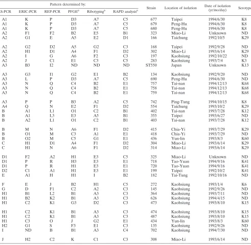
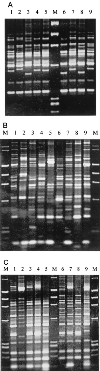
In RS-PCR, 8 to 15 amplified bands with sizes of between 330 and 1,000 bp were found in theV.parahaemolyticusstrains. Bands ranging from 350 to 720 bp could be easily observed on the electrophoresis gel. Specifically, six amplified bands with molecular sizes of 350, 420, 610, 720, 750, and 870 bp were common in all strains (Fig. 1A), and two amplified bands (350 and 720 bp) occurred in allV.parahaemolyticusstrains but not inE.coli,V.cholerae, andV.vulnificus. All 41V. parahaemo-lyticusstrains were grouped into 15 patterns, with A3 (17.1% of the total number of strains) being the predominant pattern (Fig. 2 and Table 2).
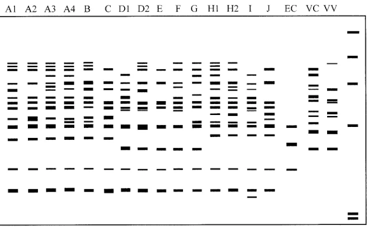
In REP-PCR, between 13 and 26 amplified bands ranging in size from 160 to 3,000 bp were discernible in theV. parahae-molyticusstrains. Several amplified bands with molecular sizes of 200, 470, 500, 600, 640, 805, and 1,355 bp were common in most strains, while only the 805-bp band was present in allV.
parahaemolyticus strains (Fig. 1B). The V. parahaemolyticus
[image:3.587.78.248.76.706.2]strains were grouped into 27 patterns, with P (12.2% of the
FIG. 1. Amplification fingerprints of some V. parahaemolyticus
strains with RS-, REP-, and ERIC-PCR methods. (A) RS-PCR. Lanes:
1, strain 134 (pattern A3); 2, strain 135 (pattern H2); 3, strain 145 (pattern G); 4, strain 166 (pattern A2); 5, strain 168 (pattern A2); 6, strain 182 (pattern E); 7, strain 197 (pattern A2); 8, strain 199 (pattern D2); 9, strain ST550 (pattern A3); M, molecular size markers (from top to bottom, 1,353, 1,078, 872, 603, 310, and 281 bp). (B) REP-PCR. Lanes: 1, strain 134 (pattern I1); 2, strain 135 (pattern S); 3, strain 145 (pattern F1); 4, strain 166 (pattern E); 5, strain 168 (pattern D2); 6, strain 182 (pattern H); 7, strain 197 (pattern G); 8, strain 199 (pattern A1); 9, strain ST550 (pattern I2); M, molecular size markers (from top to bottom, 2,322, 2,027, 1,353, 1,078, 872, 603, 310, 281, and 271 bp). (C) ERIC-PCR. Lanes: 1, strain 355 (pattern A1); 2, strain 364 (pat-tern B1); 3, strain 402 (pat(pat-tern A1); 4, strain 403 (pat(pat-tern A2); 5, strain 415 (pattern M); 6, strain 418 (pattern O1); 7, strain 434 (pattern C3); 8, strain 436 (pattern O2); 9, strain ST550 (pattern B3); M, molecular size markers (from top to bottom, 2,027, 1,353, 1,078, 872, 603, 310, 281, and 271 bp).
on May 15, 2020 by guest
http://jcm.asm.org/
total number of strains) being the predominant one (Fig. 3 and Table 2).
In ERIC-PCR, 12 to 25 amplified bands with sizes ranging between 160 and 1,690 bp were easily discernible in the V.
parahaemolyticusstrains. Several bands with molecular sizes of 270, 320, 520, 560, 660, 780, 900, 950, and 1,355 bp were common in most strains, while 270-, 520-, 660-, and 950-bp bands were present in allV.parahaemolyticusstrains (Fig. 1C). The 39 domestic strains plus the reference strain from Japan were grouped into 27 patterns. Patterns A1, C2, H1, and P, each comprising three strains, were the most predominant patterns (7.5% of the total number of strains) (Fig. 4 and Table 2).
The clonal relationships among these V. parahaemolyticus
strains were examined through cluster analysis of the PCR-generated patterns and are presented in dendrograms (Fig. 5, Fig. 6, and Fig. 7). Following cluster analysis, different patterns were arbitrarily classified into different types with strain dis-similarity values of 5 or more (33). Each type consisted of one to seven different patterns (Table 1). Compared with the in-terspecies reference strains, all theV.parahaemolyticusstrains
were closely related, according to analysis by the PCR meth-ods; they differed significantly from the reference strains ofE.
coli,V.cholerae, andV.vulnificus, having dissimilarity values of 17 or more (Fig. 5 to 7). Strains ofV.parahaemolyticus belong-ing to one or closely related PFGE patterns were generally grouped into closely related patterns by these PCR methods. Also, strains determined by one of these PCR methods to belong to strongly dissimilar patterns were generally noted as being dissimilar by the other two PCR methods (Fig. 5 to 7).
DISCUSSION
Several molecular methods have been developed and as-sessed for the typing of V. parahaemolyticus. PFGE is the method favored in our laboratory, owing to its highly repro-ducible fingerprints and strong discriminative ability (12, 31, 32). However, the ability of this method may be impaired by a high proportion of nontypeable isolates (23%), owing to DNA degradation during endonuclease digestion or other steps (14). A DNA degradation problem has been encountered with some strains in our laboratory, but these strains were successfully typed by repeating the experiment (28). The cause of DNA degradation is unknown; however, careful processing to avoid shearing interference and the use of a suboptimal enzyme reaction temperature of 37°C (optimum temperature of 50°C forSfiI) may have reduced the level of DNA degradation in our study. Furthermore, difficulties were recently encountered with the typing of several strains collected from Japan and America in 2000, with the nontypeable rate reaching about 7% (unpublished data). Therefore, a combination of methods may be required to achieve the complete typing of different V.
parahaemolyticusstrains.
[image:4.587.108.478.77.305.2] [image:4.587.43.285.631.719.2]PCR typing methods using specific primers designed on the basis of the repeated and conserved sequences in bacteria and FIG. 2. Diagram of amplification patterns ofV. parahaemolyticuswith RS-PCR. EC,E.coli; VC,V.cholerae; VV,V.vulnificus. The rightmost lane contains the molecular size markers described in the legend to Fig. 1A.
TABLE 2. Comparison of different molecular methods used in the subspecies typing ofV. parahaemolyticus
Method No. of: % of strains with thepredominant pattern Discriminationindexa Strains Patterns
PFGE 40 22 12.5 0.96
Ribotyping 40 20 12.5 0.95
RAPD 40 8 25.0 0.84
RS-PCR 41 15 17.1 0.91
REP-PCR 41 27 12.2 0.97
ERIC-PCR 40 27 7.5 0.98
aSimpson’s index of diversity (14).
on May 15, 2020 by guest
http://jcm.asm.org/
more stringent annealing conditions display more promising fingerprints than RAPD analysis (11). Spacer regions within the 16S and 23S genes in prokaryotic rRNA genetic loci exhibit significant length and sequence polymorphisms in different species and are flanked by highly conserved sequences (8). Multiple copies of these loci occur in bacteria (24). Therefore,
[image:5.587.97.484.87.329.2]amplification using primers designed on the basis of these flanking sequences will generate polymorphic fingerprints which can be used to distinguish bacterial strains at the species and subspecies levels (1, 2, 8). RS-PCR has been applied to typing of species from many genera, includingListeria, Staph-ylococcus, andSalmonella(8, 10), but had not yet been applied FIG. 3. Diagram of amplification patterns ofV. parahaemolyticuswith REP-PCR. EC,E.coli; VC,V.cholerae; VV,V.vulnificus. The rightmost lane contains the molecular size markers described in the legend to Fig. 1B.
FIG. 4. Diagram of amplification patterns ofV. parahaemolyticuswith ERIC-PCR. EC,E.coli; VC,V.cholerae; VV,V.vulnificus. The rightmost lane contains the molecular size markers described in the legend to Fig. 1C.
on May 15, 2020 by guest
http://jcm.asm.org/
[image:5.587.68.517.470.699.2]toV. parahaemolyticus. The 16S-23S rRNA intergenic spacer regions ofV.parahaemolyticuscontain different tRNA compo-sitions, and similarities in the nucleotide sequences of the noncoding regions flanked by the tRNA genes have been noted (13).
REP-PCR and ERIC-PCR are both based on the presence of repetitive conserved sequences in bacteria. The REP-PCR method is based on the presence of 38-bp REPs in Enterobac-teriaceae and other bacteria and has been applied to many species (14, 19, 25, 26). With REP-PCR, the fingerprinting profiles differentiate toxigenicV.choleraeO1 strains from non-toxigenic O1 and non-O1 strains, while ERIC-PCR further differentiates toxigenic O1 strains into El Tor and classical biotypes (22). This work is the first to apply the RS-PCR and REP-PCR methods to the typing ofV.parahaemolyticus.
ERIC-PCR is the most widely adopted of the above three PCR typing methods and has been applied to the typing of many species, includingV. cholerae(18, 23) and V. parahae-molyticus. Marshall et al. (14) examined 38 clinical strains ofV.
parahaemolyticusfrom outbreaks on Canada’s Pacific coast and several environmental strains using ERIC-PCR, ribotyping, PFGE, and restriction fragment length polymorphism analysis of the genetic locus encoding the polar flagellum. Six ERIC-PCR patterns were identified by using a single primer for the amplification, and it was concluded that ERIC-PCR and ri-botyping were useful for evaluating genetic and epidemiolog-ical relationships amongV.parahaemolyticusstrains (14).
All three PCR typing methods described here could differ-entiateV.parahaemolyticusfrom other species and effectively differentiate intraspecific strains. The V. parahaemolyticus
strains examined here were deliberately selected to represent a variety of different patterns and have been typed using PFGE, ribotyping, and RAPD analysis. The discriminative ability of
these PCR methods can thus be evaluated and compared with that of other published methods. PFGE, ribotyping, REP-PCR, and ERIC-PCR exhibited an excellent discrimination index of 0.95 or higher (Table 2). Based solely on the discrim-ination index (Simpson’s index of diversity [14]), REP-PCR and ERIC-PCR will be selected as the two best rapid PCR typing methods forV.parahaemolyticus. However, REP-PCR could be the better of the two owing to its higher rate of reproducible fingerprints. In the current study, the PCR assays were repeated three times for eachV.parahaemolyticusstrain, and the reproducibility of the banding patterns was observed. In ERIC-PCR, some of the minor light amplification bands were inconsistent, thus complicating pattern differentiation. Among the three PCR methods, RS-PCR generated fewer amplification bands than REP-PCR and ERIC-PCR and thus fewer subspecies patterns and a slightly lower discrimination index (0.91) (Table 2). However, since the RS-PCR patterns were more easily discernible visually than the REP-PCR or ERIC-PCR patterns, they may be a practical method for rou-tine use.
Although the discriminative ability of these PCR typing methods differed from 0.91 to 0.98, these methods are effective for typing strains from outbreaks. When the typing of strains in each outbreak is examined, the results obtained by these PCR methods mirrored those of the PFGE method for some out-breaks, although they differed slightly for other outbreaks. For FIG. 5. Dendrogram illustrating the clustering of amplification
pat-terns ofV. parahaemolyticuswith RS-PCR. The dendrogram was pro-duced using the squared Euclidean distance measure and average linkage clustering method with the program SPSS for Windows, Re-lease 6.0. The dissimilarity units are arbitrary, being based on the sum of the squared Euclidian distance measure. Strains were arbitrarily grouped into different types. Letters at left designate the patterns.
FIG. 6. Dendrogram illustrating the clustering of amplification pat-terns ofV. parahaemolyticuswith REP-PCR. See the legend to Fig. 5 for details.
on May 15, 2020 by guest
http://jcm.asm.org/
example, the outbreak occurring in Miao-Li on 14 June 1993 was typed as A4, A4, K, and A6 by PFGE, A2, C, J, and C by RS-PCR, H1, H1, H2, and H1 by ERIC-PCR, and D1, D1, C2, and N by REP-PCR for strains 302, 304, 308, and 314, respec-tively (Table 1). In another example, the outbreak occurring in Peng-Hu on 30 June 1994 was typed as D3 by PFGE and P by REP-PCR but as A1 and A3 by RS-PCR and K and L by ERIC-PCR (Table 1). The use of a combination of these PCR methods could achieve even higher discriminative ability when fine and rapid typing is required.
The presence of the repeatable fingerprints in REP-PCR and ERIC-PCR suggested the presence of these repetitive consensus sequences (REP and ERIC) inV.parahaemolyticus. In anotherVibriospecies,V.cholerae, the presence of ERIC has been confirmed to be located near the hemolysin gene. Meanwhile, ERIC of V. cholerae is highly homologous with those found inEnterobacteriaceae. A previous study has spec-ulated that a transpecific genetic exchange has affected a group of E. coli hemolysin genes and that ERIC has thus “hitch-hiked” with the hemolysin gene (5). Besides the presence of ERIC, the possibility of fingerprints being formed by random amplification cannot be excluded, and Gillings and Holley (3) confirmed that ERIC-PCR fingerprints may be thus produced. Gillings and Holley performed PCR with ERIC primers using salmon and lambda DNA templates without ERIC, and the
fingerprints were formed and changed according to different annealing conditions used in the PCR procedure (3).
In conclusion, RS-PCR, REP-PCR, and ERIC-PCR are suitable rapid typing methods for V. parahaemolyticus. All three methods have high discriminative ability, but REP-PCR is superior to ERIC-PCR owing to the better reproducibility of fingerprints produced with this method. Nevertheless, RS-PCR, with a slightly lower discriminative ability, may be a more practical method because fewer amplification bands and pat-terns are generated, simplifying review and interpretation of data.
ACKNOWLEDGMENT
We thank the Department of Health of the Republic of China for financially supporting this research under contract no. DOH90-TD-1075.
REFERENCES
1.Al-Saif, N. M., G. L. O’-Neill, J. T. Magee, J. S. Brazier, and B. I. Duerden.
1998. PCR-ribotyping and pyrolysis mass spectrometry fingerprinting of en-vironmental and hospital isolates ofClostridium difficile. J. Med. Microbiol.
47:117–121.
2.Bidet, P., V. Lalande, B. Salauze, B. Burghoffer, V. Avesani, M. Delmee, A.
Rossier, F. Barbut, and J. C. Petit.2000. Comparison of PCR-ribotyping,
arbitrarily primed PCR, and pulsed-field gel electrophoresis for typing Clos-tridium difficile. J. Clin. Microbiol.38:2484–2487.
3.Gillings, M., and M. Holley.1997. Repetitive elemment PCR fingerprinting
(rep-PCR) using enterobacterial repetitive intergenic consensus (ERIC) primers is not necessarily directed at ERIC elements. Lett. Appl. Microbiol.
25:17–21.
4.Hill, W. E., J. M. Madden, B. A. McCardell, D. B. Shah, J. A. Jagow, W. L.
Payne, and B. K. Boutin.1983. Food-borne enterotoxigenicEscherichia coli:
detection and enumeration by DNA colony hybridization. Appl. Environ. Microbiol.45:1324–1330.
5.Hulton, C. S. J., C. F. Higgins, and P. M. Sharp.1991. ERIC sequences: a
novel family of repetitive elements in the genomes ofEscherichia coli, Sal-monella typhimuriumand other enterobacteria. Mol. Microbiol.5:825–834.
6.Hunter, P. R., and M. A. Gaston.1988. Numerical index of the
discrimina-tory ability of typing systems: an application of Simpson’s index of diversity. J. Clin. Microbiol.26:2465–2466.
7.Iguchi, T., S. Kondo, and K. Hisatsune.1995.Vibrio parahaemolyticusO
serotypes from O1 to O13 all produce R-type lipopolysaccharide: SDS-PAGE and compositional sugar analysis. FEMS Microbiol. Lett.130:287– 292.
8.Jensen, M. A., J. A. Webster, and N. Straus.1993. Rapid identification of
bacteria on the basis of polymerase chain reaction-amplified ribosomal DNA spacer polymorphisms. Appl. Environ. Microbiol.59:945–952.
9.Joseph, S. W., R. R. Colwell, and J. B. Kaper.1983.Vibrio parahaemolyticus
and related halophilic vibrios. Crit. Rev. Microbiol.10:77–123.
10.Lagatolla, C., L. Dolzani, E. Tonin, A. Lavenia, M. Di-Michele, T.
Tom-masini, and C. Monti-Bragadin.1996. PCR ribotyping for characterizing
Salmonellaisolates of different serotypes. J. Clin. Microbiol.34:2440–2443.
11.Liu, P. Y. F., Z. Y. Shi, Y. J. Lau, B. S. Hu, J. M. Shyr, W. S. Tsai, Y. H. Lin,
and C. Y. Tseng.1995. Comparison of different PCR approaches for
char-acterization ofBurkholderia(Pseudomonas)cepaciaisolates. J. Clin. Micro-biol.33:3304–3307.
12.Lu, P.-L., S.-C. Chang, H.-J. C. M. L. Pan, and K.-T. Luh.2001. Application
of pulsed-field gel electrophoresis to the investigation of a nosocomial out-break ofVibrio parahaemolyticus. J. Microbiol. Immunol. Infect. (Taipei)
33:29–33.
13.Maeda, T., N. Takada, M. Furushita, and T. Shiba.2000. Structural variation
in the 16S–23S rRNA intergenic spacers ofVibrio parahaemolyticus. FEMS Microbiol. Lett.192:73–77.
14.Marshall, S., C. G. Clark, G. Wang, M. Mulvey, M. T. Kelly, and W. M.
Johnson.1999. Comparison of molecular methods for typingVibrio
para-haemolyticus. J. Clin. Microbiol.37:2473–2478.
15.Martin-Kearley, J., J. A. Gow, M. Peloquin, and C. W. Greer.1994.
Numer-ical analysis and the application of random amplified polymorphic DNA polymerase chain reaction to the differentiation ofVibriostrains from a seasonally cold ocean. Can. J. Microbiol.40:446–455.
16.Okuda, J., M. Ishibashi, E. Hayakawa, T. Nishino, Y. Takeda, A. K.
Muk-hopadhyay, S. Garg, S. K. Bhattacharya, G. B. Nair, and M. Nishibuchi.
1997. Emergence of a unique O3:K6 clone ofVibrio parahaemolyticusin Calcutta, India, and isolation of strains from the same clonal group from Southeast Asian travelers arriving in Japan. J. Clin. Microbiol.35:3150–3155.
17.Pan, T. M., T. K. Wang, C. L. Lee, S. W. Chien, and C. B. Horng.1997.
FIG. 7. Dendrogram illustrating the clustering of amplification pat-terns ofV. parahaemolyticuswith ERIC-PCR. See the legend to Fig. 5
for details.
on May 15, 2020 by guest
http://jcm.asm.org/
Food-borne disease outbreaks due to bacteria in Taiwan, 1986 to 1995. J. Clin. Microbiol.35:1260–1262.
18.Rivera, I. G., M. A. R. Chowdhury, A. Huq, D. Jacobs, M. T. Martins, and
R. R. Colwell. 1995. Enterobacterial repetitive intergenic consensus
se-quences and the PCR to generate fingerprints of genomic DNAs fromVibrio choleraeO1, O139, and non-O1 strains. Appl. Environ. Microbiol.61:2898– 2904.
19.Rodriguez, B. M., R. J. Hamill, E. D. Houston, P. R. Georghiou, J. E.
Clarridge, R. L. Regnery, and J. E. Koehler.1995. Genomic fingerprinting of
Bartonellaspecies by repetitive-element PCR for distinguishing species and isolates. J. Clin. Microbiol.33:1089–1093.
20.Sambrook, J., E. F. Fritsch, and T. Maniatis.1989. Molecular cloning: a
laboratory manual, 2nd ed. Cold Spring Harbor Laboratory Press, Cold Spring Harbor, N.Y.
21.Samore, M. H., M. Kristjansson, L. Venkataraman, P. C. DeGirolami, and
R. D. Arbeit.1996. Comparison of arbitrarily-primed polymerase chain
re-action, restriction enzyme analysis and pulsed-field gel electrophoresis for typingClostridium difficile. J. Microbiol. Methods25:215–224.
22.Shangkuan, Y. H., H. C. Lin, and T. M. Wang.1997. Diversity of DNA
sequences amongVibrio choleraeO1 and non-O1 isolates detected by whole-cell repetitive element sequence-based polymerase chain reaction. J. Appl. Microbiol.82:335–344.
23.Shangkuan, Y. H., C. M. Tsao, and H. C. Lin.1997. Comparison ofVibrio
choleraeO1 isolates by polymerase chain reaction fingerprinting and ribotyp-ing. J. Med. Microbiol.46:941–948.
24.Srivastava, A. K., and D. Schlessinger.1990. Mechanism and regulation of
ribosomal RNA processing. Annu. Rev. Microbiol.44:105–129.
25.Stern, M. J., G. F. L. Ames, N. H. Smith, E. C. Robinson, and C. F. Higgins.
1984. Repetitive extragenic palindromic sequences: a major component of the bacterial genome. Cell37:1015–1026.
26.Stubbs, S. L., J. S. Brazier, G. L. O’Neill, and B. I. Duerden.1999. PCR
targeted to the 16S–23S rRNA gene intergenic spacer region ofClostridium difficileand construction of a library consisting of 116 different PCR ri-botypes. J. Clin. Microbiol.37:461–463.
27.Versalovic, J., T. Koeuth, and J. R. Lupski.1991. Distribution of repetitive
DNA sequence in eubacteria and application to fingerprinting of bacterial genomes. Nucleic Acids Res.19:6823–6831.
28.Wong, H. C., S.-H. Liu, L.-W. Ku, T. K. Wang, Y.-S. Lee, C.-L. Lee, L.-P.
Kuo, and D. Y. C. Shih.2000. Characterization ofVibrio parahaemolyticus
isolates obtained from Food Poisoning Outbreaks during 1992–1995 in Tai-wan. J. Food Prot.63:900–906.
29.Wong, H. C., C. Y. Ho, L. P. Kuo, T. M. Pan, T. K. Wang, and D. Y. C. Shih.
1999. Ribotyping ofVibrio parahaemolyticusisolates obtained from food poisoning outbreaks in Taiwan. Microbiol. Immunol.43:631–636.
30.Wong, H. C., C. C. Liu, T.-M. Pan, T.-K. Wang, and D. Y. C. Shih.1999.
Molecular typing ofVibrio parahaemolyticusisolates obtained from food poisoning outbreaks in Taiwan by random amplified polymorphic DNA analysis. J. Clin. Microbiol.37:1809–1812.
31.Wong, H. C., S.-H. Liu, and D. P. Liu.1999. Incidence of Highly Genetically
DiversifiedVibrio parahaemolyticusin Seafood Imported from Asian coun-tries. Int. J. Food Microbiol.52:181–188.
32.Wong, H. C., S.-H. Liu, T.-K. Wang, C. L. Lee, C. S. Chiou, D. P. Liu, M.
Nishibuchi, and B.-K. Lee.2000. Characterization ofVibrio parahaemolyticus
O3:K6 from Asia. Appl. Environ. Microbiol.66:3981–3986.
33.Wong, H. C., K.-T. Lu, T.-M. Pan, C.-L. Lee, and D. Y. C. Shih.1996.
Subspecies typing of Vibrio parahaemolyticusby pulsed-field gel electro-phoresis. J. Clin. Microbiol.34:1535–1539.
34.Wong, H. C., P. Y. Peng, and S. L. Lan.1998. Effect of heat shock on the
thermotolerance and protein composition inVibrio parahaemolyticus. Infect. Immun.66:3066–3071.
![N (2 Hydroxyethyl) 2 [3 (p tolyl)triazen 1 yl]benzamide](data:image/gif;base64,R0lGODlhAQABAIAAAP///wAAACH5BAEAAAAALAAAAAABAAEAAAICRAEAOw==)