0095-1137/06/$08.00
⫹
0
doi:10.1128/JCM.01411-06
Copyright © 2006, American Society for Microbiology. All Rights Reserved.
Phenotypic Detection of Methicillin Resistance in
Staphylococcus aureus
by Disk Diffusion Testing and Etest on Mueller-Hinton Agar
䌤
R. Skov,
1* R. Smyth,
2A. R. Larsen,
1A. Bolmstro
ˆm,
3A. Karlsson,
3K. Mills,
3N. Frimodt-Moller,
1and G. Kahlmeter
2National Center for Antimicrobials and Infection Control, Statens Serum Institut, Copenhagen, Denmark
1; Department of
Clinical Microbiology, Central Hospital, Va
¨xjo
¨, Sweden
2; and AB BIODISK, Solna, Sweden
3Received 8 July 2006/Returned for modification 29 August 2006/Accepted 5 October 2006
Cefoxitin is increasingly recommended for detection of methicillin resistance in
Staphylococcus aureus
(MRSA) when using disk diffusion testing. In this study, 95
mecA-
negative
S. aureus
isolates and a highly
genetically diverse collection of
mecA
-positive
S. aureus
types (
n
ⴝ
50) were used to investigate the influence of
technical factors such as disk potency, incubation time, and temperature on Mueller-Hinton agar. The use of
cefoxitin MIC testing by Etest for the same purpose was investigated under similar conditions. For disk
diffusion, the accuracy was high at both 35°C and 36°C using overnight incubation, while incubation at 30°C
or 37°C was associated with slightly lower accuracy. Increasing incubation times from 18 to 24 h did not
improve accuracy at either temperature. Cefoxitin Etest MICs for
mecA
-positive strains were 6 mg/liter or
higher, while cefoxitin Etest MICs for
mecA
-negative strains were
<
4 mg/liter. Our findings suggest that the
current CLSI zone diameter breakpoints should be adjusted from resistance (R)
<
19 mm to R
<
21 mm. In
conclusion, cefoxitin disk diffusion testing and Etest MIC testing can accurately predict the presence of the
mecA
gene in
S. aureus
. Testing can be reliably performed using incubation temperatures of 35 to 36°C and
incubation times of 18 to 22 h. We suggest MRSA interpretive criteria of susceptible (S)
<
4 mg/liter and R >
4 mg/liter, corresponding to S
>
22 mm and R
<
21 mm for the 30-
g disk and S
>
17 mm and R
<
16 mm
for the 10-
g cefoxitin disk. These criteria resulted in only one
mecA-
positive isolate being misclassified as
susceptible.
Infections due to methicillin-resistant
Staphylococcus aureus
(MRSA) are an increasing problem worldwide inside and
out-side of hospitals (3).
Phenotypic detection of MRSA has been problematic ever
since its discovery in the early 1960s. The emergence of
low-level-resistant MRSA clones acquired in the community has
only added to these difficulties.
Detection of the
mecA
gene or its product, penicillin binding
protein (PBP2a), is considered the gold standard (5) for
MRSA confirmation. Recent investigations suggest that disk
diffusion using cefoxitin is superior to most previously
recom-mended phenotypic methods, including oxacillin disk diffusion
and oxacillin screen agar testing (8, 14, 19, 21). In 2005, the
Clinical and Laboratory Standards Institute (CLSI) published
zone diameter (6) breakpoint guidelines for cefoxitin.
How-ever, a number of technical issues remain regarding the use of
cefoxitin as a predictor for methicillin resistance. The CLSI
M100-S15 document stipulates an incubation time of 24 h
unless the isolate has a zone diameter of
ⱕ
19 mm (i.e.,
resis-tant), in which case it can be reported after 18 h of incubation.
However, in a recent publication, performance was equally
good at 18 h of incubation (21).
For methicillin and oxacillin, incubation temperature is
known to affect the test results (12, 17). For oxacillin, a
max-imum of 35°C for testing of staphylococci is specified by CLSI.
For cefoxitin, high accuracy has been found using standard
incubation temperatures, i.e., 35 to 37°C (8, 10, 19–21), but one
study showed relatively low sensitivity at 37°C (2). In a recent
investigation, zone diameters obtained on IsoSensitest agar
with semiconfluent inoculum were dependent on whether the
plates were incubated at 35°C or 36°C (R. Skov, unpublished
results). Two studies have shown marginally improved
accu-racy when tests were performed at 30°C compared to 35°C and
37°C (4, 8). The 10-
g cefoxitin disk has been shown to be
superior to the 30-
g disk with IsoSensitest agar and
semicon-fluent growth (20).
The CLSI M100-S15 document recommends oxacillin MIC
testing for MRSA detection by the MIC method, and no
cri-teria are as yet available for the use of cefoxitin as an
alterna-tive (6).
In this study, the influence of incubation time (18 h and 24 h)
and temperatures (30°C, 35°C, 36°C, and 37°C) on the
perfor-mance of 10- and 30-
g cefoxitin disks and cefoxitin Etest on
Mueller-Hinton agar were evaluated for
mecA-
positive and
mecA
-negative
S. aureus.
MATERIALS AND METHODS
Strains.A total of 146S. aureusstrains were included in the study. All isolates
were tested for the presence of themecAgene by the EVIGENE MRSA
De-tection kit (18) (SSI Diagnostika, Statens Serum Institut, Copenhagen, Den-mark) using the manufacturer’s instructions. The strains selected from a
previ-ously tested collection (20) consisted of 95mecA-negative consecutive blood
culture isolates and 51mecA-positive isolates from different patients. Only
mecA-positive isolates which previously had produced inhibition zones with cefoxitin and had distinctly different pulsed-field gel electrophoresis (PFGE) patterns
* Corresponding author. Mailing address: National Center for
An-timicrobials and Infection Control, Statens Serum Institut, 5
Artil-lerivej, DK-2300 Copenhagen S, Denmark. Phone: 45 3268 8348. Fax:
45 3268 3873. E-mail: [email protected].
䌤
Published ahead of print on 18 October 2006.
4395
on May 16, 2020 by guest
http://jcm.asm.org/
(one or more visible band differences) were included. Two recent clinical
mecA-positive isolates with large cefoxitin inhibition zones and three isolates particu-larly sensitive to small variations in temperature, kindly provided by Derek Brown, Addenbrooke Hospital, Cambridge, United Kingdom, were also in-cluded.
OnemecA-positive isolate was later excluded from the investigation as
popu-lation analysis showed it to be a susceptible phenotype (see the popupopu-lation
profile analysis), leaving a total of 95mecA-negative and 50mecA-positive
iso-lates.
Three reference strains were included for quality control: onemecA-positive
strain (S. aureusATCC 43300) and twomecA-negative strains (S. aureusATCC
25923 and ATCC 29213).
All strains stored at⫺80°C were subcultured on two consecutive days using
5% Danish blood agar (SSI Diagnostika) prior to use.
spatyping and MLST.spatyping and multilocus sequence typing (MLST)
were performed as previously described (7, 11). Thespatypes (t) and MLST
sequence types (ST) were assigned through the Ridom (http://www.ridom.de)
and MLST databases (http://www.mlst.net), respectively. Based onspaand/or
sequence types, the isolates were given a predicted clonal complex annotation.
Susceptibility testing.In the first phase of the study (phase I), all 145 strains were tested by disk diffusion using cefoxitin and Etest (AB Biodisk, Solna, Sweden) using cefoxitin and oxacillin. All methodological variants were assessed using the same inoculum. The inoculum was standardized to 0.5 McFarland
turbidity. Disk diffusion was done with 10-g and 30-g disks (Oxoid,
Basing-stoke, United Kingdom) using Mueller-Hinton BBL II agar (Becton Dickinson, Heidelberg, Germany). Agar plates were incubated overnight (18 to 19 h) in ambient air at 30°C, 35°C, and 36°C in stacks no higher than five plates.
Inhibition zone diameters were read from the back of the agar plate using reflected light and calipers to read to the nearest millimeter at the inner zone
edge. For isolates with a zone size of⬎19 mm after 18 to 19 h for the 30-g
cefoxitin disk at 35°C, all plates were further incubated and read after 24 h as specified in M100-S15 (6).
Etest oxacillin MIC testing was performed according to the manufacturer’s instructions using Mueller-Hinton BBL II agar supplemented with 2% NaCl (wt/vol) and incubation at 35°C for a full 24 h. Etest cefoxitin MIC testing was done using Mueller-Hinton BBL II agar without NaCl supplementation. Plates were incubated in ambient air at 30°C, 35°C, and 36°C. Cefoxitin MICs were read
after 18 to 19 h of incubation. Isolates for which cefotixin MICs wereⱕ4 mg/liter
were incubated further and read after a total of 24 h.
The influence of incubation at 37°C was investigated in phase II using a subset
of 54 strains (35mecA-negative and 19mecA-positive strains) incubated
simul-taneously at 35°C and 37°C. For phase II we selected the most challenging strains, those with the largest cefoxitin zone diameters and those that during phase I had shown results influenced by variations in incubation temperature. For phase II, MICs and zone diameters were read after 18 h and 24 h of incubation.
In both phase I and phase II,S. aureusstrains ATCC 29213, ATCC 25923, and
ATCC 43300 were included for quality control on all test runs. The temperature in the incubators was monitored at 10-min intervals for the entire incubation period using TinytagPlus digital thermometers with accompanying software (Gemini Data Loggers Ltd., Chichester, West Sussex, United Kingdom). The temperature curves showed that with empty incubators the maximum variation
during a 12-h test period was⫾0.25°C (data not shown). With plates on the
shelves, the time to reach the preset temperature on each shelf was dependent on the number of plates placed on the shelf. In phase I, when shelves were full, the temperature only reached the preset temperature at the end of the incubation time. This was true for both temperatures. Each strain was subjected to the same conditions for each of the two temperatures. The temperature at the end of incubation was 34.6 to 34.8°C and 35.6 to 36.4°C when incubators were set at 35°C and 36°C, respectively. In phase II the preset temperatures were reached on all shelves within 4 h, and the temperatures were stationary between 34.7 to 35.6°C and 36.8 to 37.1°C when incubators were set at 36°C and 37°C, respec-tively (detailed data not shown).
Intraassay variation was studied usingS. aureusATCC 25923, ATCC 29213,
and ATCC 43300 tested for 10 days using two different batches of
Mueller-Hinton II agar (BBL) and two different batches of 10-g and 30-g disks
(Oxoid). Each agar plate was read independently by five technologists, giving a total of 50 measurements per Mueller-Hinton agar/disk combination.
Population analysis. Two mecA-positive isolates obtained from Norway (strains 9-8 and 10-22), previously undetectable by any phenotypic cefoxitin method (19, 20), were tested by population analysis profile using various con-centrations of cefoxitin and oxacillin in agar. Colonies from a fresh overnight culture were inoculated into 5 ml tryptic soy broth (SSI Diagnostika) and
incu-bated overnight at 35°C. Tenfold dilutions of the culture were prepared in 0.9%
NaCl, and 20 l from each dilution was spot inoculated in duplicate onto
Mueller-Hinton agar plates containing twofold dilutions of oxacillin (0.25 to 128 mg/liter) and cefoxitin (0.25 to 128 mg/liter). The agar plates were incubated a full 24 h at 35°C, and colonies were counted to plot the population analysis profile. Strain 10-22 showed a highly heterogeneous resistance pattern against oxacillin and displayed colonies of up to 64 mg/liter (data not shown). Since strain 9-8 failed to show any colonies at 4 mg/liter in repeat experiments, even in
the undiluted samples, it was concluded that thismecA-positive isolate was an
oxacillin-susceptible phenotype, and it was omitted from further analysis. This is a well-described phenomenon that can be caused by a defect in one or more enzymes needed for an isolate to express oxacillin resistance to, e.g., one of the
femgenes (1).
Statistics.Statistical differences in MICs obtained at different temperatures and/or incubation times were analyzed by Wilcoxon’s matched pairs test, with
P⬍0.05 considered significant. Distributions were reported by medians and
ranges. For statistical analyses, the Statistica software program (version 7.0; Stat Soft Inc., Tulsa, TX) was used.
RESULTS
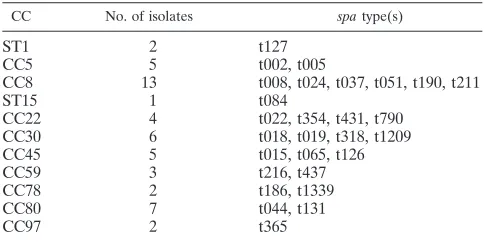
Genotypes.
The 50
mecA
-positive
S. aureus
isolates
repre-sented 28 different
spa
types and 12 different clonal complexes/ST
groups, as shown in Table 1.
MIC.
Etest oxacillin MIC distributions at 35°C after 24 h of
incubation and cefoxitin MIC distributions at 30°C, 35°C, and
36°C after 18 h and 24 h of incubation are shown in Table 2.
Oxacillin MICs for the
mecA-
negative strains were between
0.125 mg/liter and 4 mg/liter (the oxacillin MIC for one isolate
was 4 mg/liter) and for
mecA-
positive strains was between 1
and
⬎
256 mg/liter, with a median of 64 mg/liter. For cefoxitin
the range was 1 to 4 mg/liter (with only one isolate at 1 mg/
liter) and for
mecA-
negative isolates was 4 to
⬎
256 mg/liter,
with a median MIC of 32 mg/liter for
mecA-
positive isolates
regardless of the incubation temperature or time. However,
MICs were significantly lower at 36°C than for incubation at
35°C (
P
⫽
0.006). Similar results were found in phase II (35 to
37°C). There was no difference in MICs obtained at 35°C for
incubation for 18 h or 24 h (not significant;
P
⫽
0.1). There was
no difference between MICs obtained at 30°C and 35°C
incu-bation (not significant;
P
⫽
0.64). Using an MRSA interpretive
breakpoint of susceptible (S)
ⱕ
4 mg/liter and resistant (R)
⬎
4 mg/liter, only one
mecA-
positive isolate (isolate 10-22 from
Norway) was incorrectly categorized as non-MRSA after both
18 h and 24 h of incubation at all temperatures (see also data
from the population profile analysis for this isolate in Material
and Methods). The oxacillin MIC for this isolate was 4 mg/
liter. Another
mecA
-positive isolate (isolate 1748) was
pheno-TABLE 1. Predicted clonal complexes (CC) based on
spa
type of
the 50
mecA-
positive isolates
CC No. of isolates spatype(s)
ST1
2
t127
CC5
5
t002, t005
CC8
13
t008, t024, t037, t051, t190, t211
ST15
1
t084
CC22
4
t022, t354, t431, t790
CC30
6
t018, t019, t318, t1209
CC45
5
t015, t065, t126
CC59
3
t216, t437
CC78
2
t186, t1339
CC80
7
t044, t131
CC97
2
t365
on May 16, 2020 by guest
http://jcm.asm.org/
[image:2.585.300.542.89.210.2]typically sensitive to oxacillin (MIC, 1 mg/liter) but was
resis-tant to cefoxitin (MIC, 16 mg/liter).
Disk diffusion.
Results for cefoxitin 30-
g and the 10-
g
disks incubated 18 to 20 h at 35°C are shown in Fig. 1. Except
for one isolate (isolate 10-22), all
mecA
-positive isolates gave
inhibition zone diameters of
ⱕ
21 mm for the 30-
g disk and
ⱕ
17 mm for the 10-
g disk in both phase I and phase II.
For
mecA
-negative isolates, zone diameters for the 30-
g
disk were
ⱖ
25 mm and
ⱖ
23 mm for phase I and II,
respec-tively, and
ⱖ
17 mm for the 10-
g disk in both phases I and II.
Increasing the incubation time from 18 h to 24 h or the
temperature from 35°C to 36°C did not affect inhibition zone
diameter distributions for either disk for
mecA
-negative or
mecA
-positive isolates (data not shown).
Increasing the temperature from 35°C to 37°C was slightly
more problematic; for the 30-
g disk, a
mecA
-negative strain
was classified as positive, and with the 10-
g disk a
mecA
-positive strain was classified as susceptible. Decreasing the
temperature from 35°C to 30°C made one strain falsely
sus-ceptible by both disks (data not shown).
Repeated testing of quality control strains.
The results for
the 10-day repeated testing of
S. aureus
ATCC 29213 (i.e.,
CLSI quality control strain for routine MIC testing) and
S.
aureus
ATCC 25923 (i.e., CLSI quality control strain for
routine disk diffusion testing) are shown in Table 3 (all
meth-odological variants were tested with the same inoculum
sus-pension).
S. aureus
ATCC 25923, the quality control strain
normally used by the CLSI, on one day (day 5) gave
signifi-cantly different results from the rest of the nine days both for
the 10-
g and the 30-
g disks. Omitting the outlier results
from day 5, the following medians (ranges) were obtained for
S. aureus
ATCC 25923: 25 mm (22 to 28 mm) for the 30-
g
[image:3.585.43.545.79.300.2]disk and 20 mm (18 to 22 mm) for the 10-
g disk. For
S. aureus
TABLE 2. Oxacillin Etest MIC at 35°C after 24 h of incubation and cefoxitin Etest MIC at 30, 35, and 36°C after 18 h and 24 h of incubation
Drug concn (mg/liter)
No. of observations by test type,mecAstatus, time, and tempa
Oxacillin Etest (35°C,
24 h) Cefoxitin Etest
mecA
negative
(n⫽95)
mecA
positiveb
(n⫽49)
30°C 35°C 36°C
mecA
negativeb
(n⫽94) (mecAn⫽50), 18 hpositive
mecAnegative
(n⫽95) mecApositive
(n⫽50), 18 h
mecAnegative
(n⫽95) mecApositive
(n⫽50), 18 h
18 h 24 h 18 h 24 h 18 h 24 h
0.125
2
0.25
13
0.5
50
1
26
1
1
1
2
3
10
10
5
3
6
5
4
1
5
84
84
1
c89
91
1
c89
90
1
c6
d2
5
3
3
8
5
3
2
1
16
4
11
11
15
32
3
8
16
22
64
11
13
9
5
128
6
5
5
2
ⱖ
256
12
4
3
1
aValues in columns are number of observations for each unique variable.
bOne strain did not grow.
cStrain 10-22. After a full 24 h of incubation, the cefoxitin MIC was 4 mg/liter at 30°C, 35°C, and 36°C.
dAll intermediate results are reported to the nearest higher concentration except for 6 mg/liter.
FIG. 1. Zone diameter distribution for 30-
g cefoxitin disk (A) and
10-
g cefoxitin disk (B) against 145
S. aureus
isolates (phase I) and
repeat testing with 54
S. aureus
isolates (phase II) at 35°C and 18 h of
incubation. The current CLSI breakpoint for the 30-
g disk is shown
by a dashed line, and the suggested breakpoints are shown by black
lines in both figures.
䊐
,
mecA
negative, phase I;
u
,
mecA
negative,
phase II;
■
,
mecA
positive, phase I;
o
,
mecA
positive, phase II.
on May 16, 2020 by guest
http://jcm.asm.org/
[image:3.585.300.540.380.662.2]ATCC 29213, tighter zone diameter ranges were obtained for
both disks, giving medians (ranges) of 25 mm (23 to 27 mm) for
the 30-
g disk and 18 mm (17 to 20 mm) for the 10-
g disk.
There was no difference between batches of agar or disks (data
not shown).
DISCUSSION
Cefoxitin disk diffusion testing is now an accepted method
for the detection of methicillin resistance in
S. aureus
by an
increasing number of reference resistance groups, including
CLSI. There are, however, still some unsolved issues, such as
the optimal cefoxitin disk content and incubation temperature
and time. In this study, a highly diverse collection of
mecA
-positive
S. aureus
isolates (12 different clonal complexes) was
used to investigate these technical variants. To increase the
challenge,
mecA
-positive strains without inhibition zones
around the cefoxitin 10-
g disk were not included.
Further-more, only strains with unique PFGE patterns (one or more
visible band differences) were included.
Results obtained suggest that both cefoxitin inhibition zone
diameters and MICs for
mecA-
positive strains are influenced
by incubation temperature, although they are influenced to a
lesser degree than for oxacillin. Most importantly, detection of
MRSA by the cefoxitin-based method was not affected by
tem-perature variations between 35°C and 36°C, while at 37°C disk
diffusion gave one false phenotypic categorization. In the
In-ternational Organization for Standardization (ISO)
methodol-ogy standard for MIC determinations currently under
devel-opment (22), the incubation temperature is specified as 34 to
37°C. For oxacillin testing with staphylococci, there is a note
stating that the temperature should not exceed 35°C. Although
cefoxitin is influenced to a lesser degree, it may be appropriate
to include the same note for cefoxitin testing. No advantage
was seen with the 30°C incubation temperature as proposed by
others (4, 8). Importantly, there was no benefit in extending the
incubation time from 18 h to a full 24 h. This is in agreement
with the findings of Swenson and Tenover (21). It is a major
advantage to clinical laboratories that standard methodology
(medium, inoculum, incubation time, and temperature) can be
used for the detection of MRSA.
For the 30-
g disk, our findings suggest MRSA interpretive
breakpoints of S
ⱖ
22 mm and R
ⱕ
21 mm, i.e., 2 mm larger
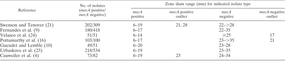
than the criteria published by the CLSI (6). This proposal is
supported by the results of several previous investigations
(Ta-ble 4) in which
mecA
-negative isolates all have had zone
di-ameters of 22 mm or more, and occasionally
mecA
-positive
isolates exhibiting zone diameters of 20 and 21 mm have been
reported (4, 9, 10, 16, 21, 23, 24).
For the 10-
g disk, the results support corresponding
inter-pretive breakpoints of S
ⱖ
17 mm and R
ⱕ
16 mm. The
contents of both disks had comparable sensitivity and
specific-ity for detection of MRSA. The lower disk content produces
smaller zones and thereby reduces the interference with results
for other antibiotic disks tested on the same agar plate. The
higher disk content may have a slightly higher specificity, as
reflected by the larger gap between
mecA
-positive and
mecA
-negative zone diameter results.
[image:4.585.41.542.79.217.2]In our study, cefoxitin MIC testing with Etest was accurate
TABLE 3. Intraassay variation for 10-
g and 30-
g cefoxitin disks for
S. aureus
ATCC 29213 and ATCC 25923
S. aureusstrain Disk size
(g)
Temp (°C)
No. of observations with indicated zone diam (mm)a
16 17 18 19 20 21 22 23 24 25 26 27 28
ATCC 29213
10
35
16 (1)
90 (4)
70 (6)
4 (9)
10
37
11 (1)
85 (7)
77 (11)
7 (1)
ATCC 25923
10
35
(1)
(4)
(9)
19 (6)
78
37
26
10
37
(1)
1 (6)
35 (11)
97 (2) 32
15
ATCC 29213
30
35
3
27 (2)
91 (11)
55 (6)
4 (1)
30
37
7
37 (8)
89 (9)
39 (3)
8
ATCC 25923
30
35
0 (1)
4 (5)
14 (5)
29 (4)
39
37
15
42 (5)
30
37
(2)
(1)
(4)
6 (5)
17 (1)
33 (4)
50 (3)
41
20
9
4
aTen-day repeat testings were read by five persons using two batches of disks and media, respectively, i.e., 200 observations per variant. Numbers in parentheses refer
to the number of observations on day 5 (see the text for an explanation).
TABLE 4. Published zone diameter ranges (CLSI 30-
g cefoxitin disk) for
mecA
-positive and
mecA
-negative
S. aureus
isolates
Reference
No. of isolates
(mecApositive/
mecAnegative)
Zone diam range (mm) for indicated isolate type
mecA
positive
mecA-positive
outlier
mecA
negative
mecA-negative
outlier
Swenson and Tenover (21)
202/309
6–19
21, 28
22–
⬎
28
Fernandes et al. (9)
180/418
6–17
22–35
Velasco et al. (24)
51/51
6–14
ⱖ
25
17
Pottumarthy et al. (16)
103/100
6–17
23–
⬎
35
21
Gueudet and Lemble (10)
49/51
6–20
23–28
Urbaskova et al. (23)
218/534
6–19
23–35
Cauweiler et al. (4)
73/82
6–19
23
24–34
on May 16, 2020 by guest
http://jcm.asm.org/
[image:4.585.43.543.618.726.2]for MRSA detection. The results suggest interpretive
break-points of S
ⱕ
4 mg/liter and R
ⱖ
8 mg/liter using CLSI
termi-nology and S
ⱕ
4 mg/liter and R
⬎
4 mg/liter in EUCAST (the
European Committee of Antimicrobial Susceptibility Testing)
terminology. However, the cefoxitin MIC mode for
mecA
-nega-tive strains is close to the suggested breakpoints. The proposed
breakpoints are supported by data published by other
investiga-tors (Felten et al. [8], Fernandes et al. [9], Swenson and Tenover
[21], and Votta et al. [M. Votta, D. Turner, B. Turng, T. Wiles, J.
Reuben, Abstr. 44th Intersci. Conf. Antimicrob. Agents
Che-mother., abstr. D-47, 2004]). However, in an investigation with
many borderline oxacillin-resistant
S. aureus
strains by Swenson et
al., cefoxitin breakpoints of S
ⱕ
6 mg/liter, instead of 4 mg/liter,
and R
ⱖ
8 mg/liter were shown to have better specificity but a
slightly lower sensitivity (J. Swenson, D. Lonsway, S. McAllister,
A. Thompson, L. Jevitt, J. Patel, Abstr. 45th Intersci. Conf.
An-timicrob. Agents Chemother., abstr. D-1732, 2005). In this study,
there was no difference in sensitivity or specificity using a
break-point of S
ⱕ
6 mg/liter and R
ⱖ
8 mg/liter instead of S
ⱕ
4
mg/liter and R
ⱖ
8 mg/liter.
In conclusion, this study provides further evidence that
cefoxitin is an accurate surrogate marker for the detection of
MRSA in routine susceptibility testing for disk diffusion and
MIC testing. Incubation temperature should not surpass 36°C.
Incubating for a full 24 h did not improve results obtained after
18 h. Hence, standard conditions currently used by clinical
laboratories for routine susceptibility testing as described by
CLSI can be used, and the cefoxitin disk can be included
among other disks relevant for susceptibility testing of
Staph-ylococcus aureus
. The current CLSI cefoxitin zone diameter
breakpoints should be adjusted by a 2-mm increase.
REFERENCES
1.Berger-Bachi, B., A. Strassle, J. E. Gustafson, and F. H. Kayser.1992. Mapping and characterization of multiple chromosomal factors involved in
methicillin resistance inStaphylococcus aureus. Antimicrob. Agents
Che-mother.36:1367–1373.
2.Boutiba-Ben Boubaker, I., R. Ben Abbes, H. Ben Abdallah, K. Mamlouk, F. Mahjoubi, A. Kammoun, A. Hammami, and S. Ben Redjeb.2004. Evaluation of a cefoxitin disk diffusion test for the routine detection of
methicillin-resistantStaphylococcus aureus. Clin. Microbiol. Infect.10:762–765.
3.Boyce, J. M., B. Cookson, K. Christiansen, S. Hori, J. Vuopio-Varkila, S. Kocagoz, A. Y. Oztop, C. M. Vandenbroucke-Grauls, S. Harbarth, and D. Pittet.2005. Methicillin-resistantStaphylococcus aureus. Lancet Infect. Dis.
5:653–663.
4.Cauwelier, B., B. Gordts, P. Descheemaecker, and H. Van Landuyt.2004.
Evaluation of a disk diffusion method with cefoxitin (30g) for detection of
methicillin-resistantStaphylococcus aureus. Eur. J. Clin. Microbiol. Infect.
Dis.23:389–392.
5.Chambers, H. F.1997. Methicillin resistance in staphylococci: molecular and
biochemical basis and clinical implications. Clin. Microbiol. Rev.10:781–791.
6.Clinical and Laboratory Standards Institute.2005. Performance standards for antimicrobial susceptibility testing. 15th informational supplement M100-S15. Clinical and Laboratory Standards Institute, Wayne, PA. 7.Enright, M. C., N. P. Day, C. E. Davies, S. J. Peacock, and B. G. Spratt.2000.
Multilocus sequence typing for characterization of methicillin-resistant and
methicillin-susceptible clones ofStaphylococcus aureus. J. Clin. Microbiol.
38:1008–1015.
8.Felten, A., B. Grandry, P. H. Lagrange, and I. Casin.2002. Evaluation of
three techniques for detection of low-level methicillin-resistant
Staphylococ-cus aureus(MRSA): a disk diffusion method with cefoxitin and moxalactam, the Vitek 2 system, and the MRSA-screen latex agglutination test. J. Clin.
Microbiol.40:2766–2771.
9.Fernandes, C. J., L. A. Fernandes, and P. Collignon.2005. Cefoxitin
resis-tance as a surrogate marker for the detection of methicillin-resistant
Staph-ylococcus aureus. J. Antimicrob. Chemother.55:506–510.
10.Gueudet, T., and C. Lemble.2004. Comparison of five usual techniques for detection of methicillin-resistant Staphylococcus aureus. Pathol. Biol. (Paris)
52:617–621.
11.Harmsen, D., H. Claus, W. Witte, J. Rothganger, H. Claus, D. Turnwald, and U. Vogel.2003. Typing of methicillin-resistantStaphylococcus aureusin a
university hospital setting by using novel software forsparepeat
determina-tion and database management. J. Clin. Microbiol.41:5442–5448.
12.Heneine, N., and P. R. Stewart.1986. Physiological determination of
methi-cillin resistance inStaphylococcus aureus: comparison of clinical and
genet-ically derived isolates. J. Antimicrob. Chemother.17:705–715.
13.McDougal, L. K., C. D. Steward, G. E. Killgore, J. M. Chaitram, S. K. McAllister, and F. C. Tenover.2003. Pulsed-field gel electrophoresis typing
of oxacillin-resistantStaphylococcus aureusisolates from the United States:
establishing a national database. J. Clin. Microbiol.41:5113–5120.
14.Mougeot, C., J. Guillaumat-Tailliet, and J. M. Libert.2001.Staphylococcus aureus: new detection of intrinsic resistance using the diffusion method.
Pathol. Biol. (Paris)49:199–204.
15.Murchan, S., M. E. Kaufmann, A. Deplano, R. de Ryck, M. Struelens, C. E. Zinn, V. Fussing, S. Salmenlinna, J. Vuopio-Varkila, N. El Solh, C. Cuny, W. Witte, P. T. Tassios, N. Legakis, W. van Leeuwen, A. van Belkum, A. Vindel, I. Laconcha, J. Garaizar, S. Haeggman, B. Olsson-Liljequist, U. Ransjo, G. Coombes, and B. Cookson.2003. Harmonization of pulsed-field gel electro-phoresis protocols for epidemiological typing of strains of methicillin-resis-tantStaphylococcus aureus: a single approach developed by consensus in 10 European laboratories and its application for tracing the spread of related
strains. J. Clin. Microbiol.41:1574–1585.
16.Pottumarthy, S., T. R. Fritsche, and R. N. Jones.2005. Evaluation of
alter-native disk diffusion methods for detectingmecA-mediated oxacillin
resis-tance in an international collection of staphylococci: validation report from the SENTRY Antimicrobial Surveillance Program. Diagn. Microbiol. Infect.
Dis.51:57–62.
17.Sabath, L. D., and S. J. Wallace.1971. The problems of drug-resistant pathogenic bacteria. Factors influencing methicillin resistance in
staphylo-cocci. Ann. N. Y. Acad. Sci.182:258–266.
18.Skov, R., L. V. Pallesen, R. L. Poulsen, and F. Espersen.1999. Evaluation of
a new three hours hybridisation method for detection of themecAgene in
Staphylococcus aureusand correlation to existing genotypic and phenotypic
susceptibility testing methods. J. Antimicrob. Chemother.43:467–475.
19.Skov, R., R. Smyth, M. Clausen, A. R. Larsen, N. Frimodt-Moller, B. Olsson-Liljequist, and G. Kahlmeter.2003. Evaluation of a cefoxitin 30g disc on
Iso-Sensitest agar for detection of methicillin-resistant Staphylococcus
aureus. J. Antimicrob. Chemother.52:204–207.
20.Skov, R., R. Smyth, A. R. Larsen, N. Frimodt-Moller, and G. Kahlmeter.
2005. Evaluation of cefoxitin 5 and 10g discs for the detection of
methi-cillin resistance in staphylococci. J. Antimicrob. Chemother.55:157–161.
21.Swenson, J. M., and F. C. Tenover.2005. Results of disk diffusion testing
with cefoxitin correlate with presence ofmecAinStaphylococcusspp. J. Clin.
Microbiol.43:3818–3823.
22.Technical Committee ISO/TC 212 and Technical Committee CEN/TC 140.
2006. Susceptibility testing of infectious agents and evaluation of perfor-mance of antimicrobial susceptibility test devices - part 1: reference method for testing the in vitro activity of antimicrobial agents against rapidly growing aerobic bacteria involved in infectious diseases. International Organization for Standardization, Geneva, Switzerland.
23.Urbaskova, P., O. Melter, B. Mackova, V. Jakubu, and M. Wunschova.2004.
Detection of MRSA in a group of 752 strains ofS. aureususing a cefoxitin
disk. Epidemiol. Mikrobiol. Imunol.53:62–65.
24.Velasco, D., T. M. del Mar, M. Cartelle, A. Beceiro, A. Perez, F. Molina, R. Moure, R. Villanueva, and G. Bou.2005. Evaluation of different methods for
detecting methicillin (oxacillin) resistance inStaphylococcus aureus. J.
Anti-microb. Chemother.55:379–382.