Copyright © 1999, American Society for Microbiology. All Rights Reserved.
Epidemiological Study of Paratuberculosis in Wild Rabbits in Scotland
ALASTAIR GREIG,
1KAREN STEVENSON,
2* DENNIS HENDERSON,
1VALENTIN PEREZ,
2†
VALERIE HUGHES,
2IVO PAVLIK,
3MURRAY E. HINES II,
4IAIN M
CKENDRICK,
5ANDJ. MICHAEL SHARP
2SAC Veterinary Science Division, Perth PH1 1HF,
1Moredun Research Institute,
International Research Centre, Pentland Science Park, Penicuik, Midlothian EH26 OPZ,
2Biomathematics and Statistics Scotland, Edinburgh EH9 3JZ,
5Scotland, United
Kingdom; Veterinary Research Institute, 621 32 Brno, Czech Republic
3;
and Veterinary Diagnostic and Investigational Laboratory,
University of Georgia, Tifton, Georgia 31793
4Received 1 September 1998/Returned for modification 20 October 1998/Accepted 27 January 1999
A survey of 22 farms confirmed the presence of paratuberculosis in wild rabbits in Scotland. Regional
differences were apparent in the prevalence of the disease in rabbits, with a significantly higher incidence
occurring in the Tayside region. Statistical analysis showed a significant relationship between a previous
history or current problem of paratuberculosis in cattle and the presence of paratuberculosis in rabbits on the
farms. Molecular genetic typing techniques could not discriminate between selected rabbit and cattle isolates
from the same or different farms, suggesting that the same strain may infect and cause disease in both species
and that interspecies transmission may occur. The possibility of interspecies transmission and the involvement
of wildlife in the epidemiology of paratuberculosis have important implications for the control of the disease.
Paratuberculosis (Johne’s disease) is a chronic
granuloma-tous enteritis caused by
Mycobacterium avium
subsp.
paratu-berculosis
. In most species the disease is characterized by
di-arrhea, emaciation, and loss of body condition culminating in
death. Paratuberculosis principally affects ruminants and is
responsible for significant economic losses to the livestock
in-dustry worldwide (3, 36). Diagnosis is difficult, particularly of
asymptomatic carriers. There is no single diagnostic test
avail-able that can diagnose the disease at every stage. Current
con-trol programs rely on culling or removing animals that test
pos-itive, usually as determined by bacteriological culture or serum
antibody test such as the enzyme-linked immunosorbent assay.
Effective disease control programs depend on a clear
under-standing of the sources of infection and the routes of
trans-mission. The most important mode of transmission of
para-tuberculosis is the fecal-oral route, although transmission in
symptomatic animals is known to occur in utero and via
in-fected semen, colostrum, and milk. What is less clear and of
particular importance is whether paratuberculosis can be
trans-mitted between species. Experimental infection of ruminants
has been demonstrated with different strains of
M. avium
subsp.
paratuberculosis
isolated from different species and a variety of
laboratory animals, including rabbits, have been
experimen-tally infected with ruminant strains (22–24). In addition, there
are a few reports of natural disease among sheep and goats
that grazed with infected cattle (31, 32). Infected livestock is
undoubtedly the principal source of infection, but there is still
the question of the involvement of wildlife reservoirs.
M. avium
subsp.
paratuberculosis
has been reported previously in wildlife,
including white-tailed deer (4, 18), red deer (35), roe deer
(9), exotic deer (30), tule elk (13), bighorn sheep (39, 40) and,
more recently, rabbits (7). The possibility of interspecies
trans-mission, coupled with the data implicating wildlife in the
epi-demiology of paratuberculosis, have important implications for
the control of the disease. If this occurs in areas where
live-stock interact with wildlife reservoirs, the current detection
and cull policy will be inefficient in the long term, and
vac-cination may be a more suitable alternative.
The epidemiological study reported here was initiated to
provide further information on the role of wild rabbits in the
epidemiology of paratuberculosis. A pilot survey revealed that
on four farms in the Tayside region of Scotland, 67% of wild
rabbits were infected with
M. avium
subsp.
paratuberculosis
(7).
This region also had a high prevalence of bovine
paratubercu-losis (38a). It was decided to extend the survey to cover 22
farms throughout Scotland to examine the prevalence of
M. avium
subsp.
paratuberculosis
infection in the wild-rabbit
population in different regions. Farms with or without a history
of paratuberculosis were selected to establish whether there
was any association between the presence of paratuberculosis
in livestock and rabbits. To identify whether interspecies
trans-mission could have occurred,
M. avium
subsp.
paratuberculosis
strains isolated from rabbits and cattle present on two farms
were compared by pulsed-field gel electrophoresis (PFGE),
IS900 restriction fragment length polymorphism (RFLP), and
chemotype profiles.
MATERIALS AND METHODS
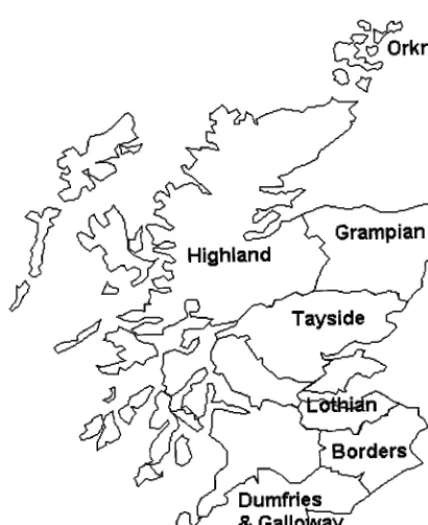
Two hundred and ten rabbits were killed between October and March 1996 on 22 farms located throughout Scotland. The regions sampled in the survey are shown in Fig. 1. Fourteen of the farms (A through N) had a history of paratu-berculosis among livestock, and eight of the farms (O through V) had no ob-served or recorded cases of paratuberculosis. Paratuberculosis was diagnosed on the affected farms by microscopic examination of feces and serum antibody tests. The rabbit carcasses were weighed, sexed, and examined at necropsy for visible lesions. Representative samples of the small and large intestine and mesenteric lymph nodes were removed aseptically and transferred to sterile containers. Separate instruments were used for each animal. Small portions of tissue were fixed in 10% formal saline for histopathological analysis.
Fecal samples were taken from cattle and sheep on farms A and C to culture
M. aviumsubsp.paratuberculosisisolates from the livestock for typing. Samples were processed as outlined below.
* Corresponding author. Mailing address: Moredun Research
Insti-tute, International Research Centre, Pentland Science Park, Bush Loan,
Penicuik, Midlothian EH26 0PZ, Scotland, United Kingdom. Phone:
44-131-445-5111. Fax: 44-131-445-6111. E-mail: [email protected].
† Present address: Histologia y Anatomia Patologica, Facultad de
Veterinaria, Universidad de Leon, Leon, Spain.
1746
on May 15, 2020 by guest
http://jcm.asm.org/
Histopathology.Fixed tissue samples were embedded in paraffin wax, and
4-mm sections were stained with hematoxylin and eosin and by the Ziehl-Neelsen
procedure for acid-fast bacilli.
Primary culture.Tissue and fecal homogenates were prepared as described
previously (7). Briefly, 1 g of feces or 0.5 cm3of finely chopped tissues were
homogenized for 30 s in 10 ml of sterile distilled water with a Colworth Stom-acher 80 (Seward Medical, London, United Kingdom). The homogenates were decontaminated by adding 10 ml of 1.5% hexadecyl pyridinium chloride and left overnight at room temperature to allow particulate materials to settle. The
supernatants were centrifuged at 3,8003gfor 30 min at 4°C, and each pellet was
resuspended in 10 ml of sterile distilled water. The centrifugation step was repeated, and each pellet was resuspended in 1 ml of sterile distilled water. The
suspension was transferred to a microfuge tube and centrifuged at 6,5003gfor
5 min. The pellet was resuspended finally in 0.5 ml of sterile distilled water. Two slants of Middlebrook 7H11 agar supplemented with Selectatabs (amphotericin B, polymixin B, carbenicillin, and trimethoprim; Code MS 24; MAST Labora-tories, Ltd., Merseyside, United Kingdom), 10% Middlebrook oleic acid-albu-min-dextrose-catalase (OADC) enrichment medium (Difco, Surrey, United
Kingdom) and 2mg of mycobactin J (Allied Monitor, Fayette, Mo.) per ml were
inoculated with 0.1 ml of the prepared suspension. The cultures were incubated at 37°C for up to 16 weeks and examined regularly for bacterial growth.
Analysis by PCR.The identity of the mycobacterium isolates was confirmed by
PCR. Two-hundred microliters of sterile distilled water was inoculated with a single bacterial colony from each positive culture. The mycobacteria were lysed by beating with 0.1-mm silica-zirconium beads (Biospec Products, Bartlesville, Okla.) in an ESPE capmix (Cottrell & Co.) twice for 40 s, with cooling on ice between each treatment. The DNA was extracted by using guanidine hydrochlo-ride as described by Challans et al. (2). Five-microliter aliquots of DNA were
analyzed by PCR assays directed against the IS900 sequence ofM. aviumsubsp.
paratuberculosis(33). PCR-amplified product was detected by continuous poly-acrylamide gel electrophoresis followed by silver staining (2).
Statistical analysis.The data were analyzed by using Genstat 5 software
(release 3.2) with the generalized linear modelling facilities with a logit link function. Excel 97 was used to manage the dataset, to examine the residuals, and to carry out simple hypothesis testing on the models generated by Genstat.
Molecular typing ofM. aviumsubsp.paratuberculosisisolates.Four leporine
isolates (R1, R7, R8, and R10) and four bovine isolates (F13, F14, F16, and F17) from farm A and seven leporine isolates (R186, R191, R193, R194, R195, R197, and R198) and four bovine isolates (JD1, JD2, JD3, and JD4) from farm C were selected for molecular typing. The primary cultures were subcultured on Middle-brook 7H11 agar supplemented with Selectatabs, OADC, and mycobactin J as described above and were sent to the various laboratories for typing. Due to a shortage of cultures at the time, it was not possible to analyze isolate F16 by IS900 RFLP or to chemotype isolate F17.
PFGE.Ten milliliters of Middlebrook 7H9 broth supplemented with Tween 80
(4% [wt/vol]), OADC (10% [vol/vol]), glycerol (2.5% [vol/vol]), and 2 mg of
mycobactin J per ml was inoculated with a single colony and incubated at 37°C until the cell density was at least McFarland standard 2, as assessed by using a densimat (BioMerieux, Lyon, France). Cultures were stirred during incubation with a magnetic stirrer bar to prevent clumping of the cells.
Cells were centrifuged at 3,5003gfor 20 min at 4°C. The cells were resuspended
in spheroplasting buffer (citrate phosphate buffer, pH 5.6 [0.2 M citrate, 0.5 M
phosphate]; 50 mM EDTA; 0.1% [wt/vol] Tween 80) to give 1.831010cells/ml.
One-half milliliter of the cell suspension was warmed to 45°C, mixed with 0.5 ml of prewarmed molten 1% (wt/vol) low-melting-point agarose (InCert Agarose; FMC Bioproducts, Flowgen, Staffordshire, United Kingdom), and poured into precooled moulds. Agarose plugs were allowed to set at 4°C for 15 min. Each plug was transferred to a plastic bijou bottle and incubated in 0.5 ml of lysis solution (10 mM Tris-HCl, pH 8.0; 1 mM EDTA, pH 8.0; 1 to 2 mg of lysozyme per ml) overnight at 37°C. After incubation, the lysis solution was discarded and replaced with 0.5 ml of ESP (0.5 M EDTA, pH 8.0; 1% [wt/vol] lauroyl sarcosine; 1 to 2 mg of proteinase K per ml) and then incubated with gentle agitation for at least 7 days at 55°C. The plugs were then transferred to fresh bijoux, and 5 ml of TE (10 mM Tris-HCl; 1 mM EDTA, pH 8.0) plus 1 mM phenylmethylsulfonyl fluoride was added; the mixture was then shaken gently for 30 min at room temperature. The samples were washed three times for 30 min in TE at room temperature with gentle agitation. Samples were stored in 0.5 M EDTA (pH 8.0) at 4°C until required for restriction analysis. Plugs were washed three times for 15 min with 5 ml of TE at room temperature, with shaking, prior to restriction analysis.
After equilibration in the appropriate restriction buffer for 1 h, the DNA was
restricted with 20 U ofHindIII orSpeI overnight at 37°C. Samples were
electropho-resed on a 1% (wt/vol) pulsed-field certified agarose gel in 0.53TBE (89 mM
Tris-borate; 89 mM boric acid, pH 8.3) by using a CHEF Mapper (Bio-Rad Labo-ratories, Ltd., Hertfordshire, United Kingdom) with parameters designed to give optimum separation in the 20- to 110-kb size range (gradient, 6 V/cm; included angle, 120°; linear ramping with an initial switch time of 2.98 s and a final switch time of 9.39 s; overall time, 26.56 h). PFGE profiles were analyzed with Phoretix PC software (Pharmacia Biotech, Ltd., Hertfordshire, United Kingdom).
IS900 RFLP analysis.DNA was prepared as previously described (17).
My-cobacteria were treated with lysozyme, immobilized in low-melting-point aga-rose, and lysed in a solution containing EDTA, sodium dodecyl sulfate, and
proteinase K. Agarose blocks, each containing approximately 4mg of DNA, were
digested withPstI andBstEII (New England Biolabs, Hertfordshire, United
Kingdom) according to the manufacturer’s instructions. The resulting restriction fragments were separated by field inversion gel electrophoresis at 80 V (4 V/cm) with linear ramping from 0.3 to 10 s for 16 h. The restriction fragments were vacuum blotted onto Hybond-N nylon membrane (Amersham Life Science, Ltd.) and hybridized to an IS900 probe as described previously (27, 28). The IS900 probe was prepared by PCR amplification of a 453-bp fragment with the primers
59-TGGACAATGACGGTTACGGAGGTGG-39and 59-GATCGGAACGTCG
GCTGGTCAGGCT-39according to the procedure described by Kunze et al.
(14). The product was electrophoresed on a 2% agarose gel, purified by using a Wizard PCR purification kit (Promega), and labelled by using the ECL Direct Labelling Kit (Amersham Life Science, Ltd.) according to the manufacturer’s instructions. DNA fingerprints were scanned with a CCD camera and processed with the software Gel Manager (Biosystematika, Tavistock, United Kingdom). RFLP types were defined as previously described (27, 28). There are currently
nine RFLP types generated by digestion withPstI which have been labelled A to
K (27, 28). RFLP types generated byBstEII can be divided into three groups
designated C, S, and I (27, 28). These three groups are further divided and designated with a new numerical series (C1–17, S1–3, and I1–2) (27, 28). Indi-vidual RFLP strain types were designated as an RFLP type after digestion with
both restriction endonucleasesPstI andBstEII (for example, B-C1).
Chemotyping.Isolates ofM. aviumsubsp.paratuberculosiswere grown on
Middlebrook 7H10 agar plates containing 100ml of Middlebrook OADC
en-richment medium per ml and 2mg of mycobactin J per ml added at a pH of 7.9
for 13 weeks at 37°C and 99% humidity.
Matrix solid-phase dispersion (MSPD) was performed as previously described by Hines et al. (11) and as modified by Hines and Frazier (10). Briefly, 60 mg of
mycobacteria was scraped from the plates and blended with 400 mg of C18
reversed-phase high-pressure liquid chromatography (HPLC) packing resin (Bakerbond; catalog no. 702500; J. T. Baker Co., Phillipsburg, N.J.). The blended material was transferred to a 10-ml syringe column, where it was eluted with 3 ml of each of the five solvents (hexane, methylene chloride, acetonitrile, methanol, and HPLC-grade water) in sequential order. An additional 7 ml of 100% meth-anol was mixed with the water-derived fraction to enhance evaporation. All five fractions from each bacterial sample were then evaporated to dryness under
nitrogen gas at 40°C. Once dried, the samples were resolubilized in 60ml of
chloroform-methanol-water (5:4:1) and vortexed briefly. Thin-layer chromatog-raphy (TLC) was performed by prescribed methods (38), with the following modifications. Five microliters of each of the five MSPD fractions from each organism were spotted onto separate lanes of a silica gel TLC plate (Baker S1250-PA, 19C; catalog no. 7009-04; J. T. Baker Co.). This was repeated three
times for a total of 15ml of each fraction per lane. The plates were allowed to dry
[image:2.612.65.279.67.329.2]at room temperature and then developed in a TLC chamber containing 100 ml of mobile phase comprised of chloroform-methanol-water (5:4:1). Plates were removed when the mobile phase had risen approximately 10 cm. After being dried in a chemical fume hood, the plates were sprayed with Bial’s reagent
FIG. 1. Map of Scotland showing regions sampled in the survey.
on May 15, 2020 by guest
http://jcm.asm.org/
(Sigma Chemical, St. Louis, Mo.), air dried, and then heated at 110 to 120°C for 15 to 20 min and photographed.
The Rx values were determined for each band by dividing the distance from the origin of each band by the distance from the origin of a common reference band in lane 4 (34). For standardization, band measurements were taken from the leading edge to prevent problems associated with thick bands. For crescent-or arrow-shaped bands, measurements were taken at the apex of the arc.
RESULTS
The results from the farm survey are detailed in Table 1 and
summarized in Table 2. A total of 130 rabbits were collected
from 14 farms with a history of Johne’s disease, and 80 rabbits
were collected from 8 farms with no history of Johne’s disease.
M. avium
subsp.
paratuberculosis
was cultured from rabbits
from 3 of the 14 paratuberculosis-affected farms which were
located all in the Tayside region. On these farms, between 8
and 53% of wild rabbits investigated were found to be infected.
M. avium
subsp.
paratuberculosis
also was cultured from a
rabbit on one farm in the Borders region with no known history
of Johne’s disease. In addition, an acid-fast organism was
cul-tured from a rabbit on a paratuberculosis-affected farm in
Orkney, although it was not possible to confirm the identity of
the isolate due to desiccation of the media and, for this reason,
this result was excluded from the statistical analysis. The
rab-bits were generally in good condition, as judged by their fat
reserves. No visible lesions were observed. Histopathological
changes were apparent in 28 (22%) rabbits on nine farms with
a history of Johne’s disease and in 6 (8%) rabbits on two farms
with no known history of Johne’s disease. Acid-fast bacilli
were associated with lesions in 10 (8%) of the rabbits on
paratuberculosis-affected farms and 2 (3%) of the rabbits on
the farms with no known history of Johne’s disease.
Statistical analysis.
For the statistical evaluation, the farms
were divided into two nonexclusive groups: farms with cattle
and farms with sheep. The species of ruminant on the farm was
considered important, since ovine
M. avium
subsp.
paratuber-culosis
isolates are notoriously difficult to grow in vitro, a factor
which could result in a lower number of isolates from sheep
farms. Two sets of variates were examined: rabbits which were
culture positive for
M. avium
subsp.
paratuberculosis
(a highly
specific, minimalist definition of presence) and rabbits which
were either culture positive or exhibited lesions on autopsy (a
general, maximalist definition of presence, since the lesions
could have been caused by other agents). Four situations were
evaluated as follows.
(i) Cattle farms, culture-positive rabbits.
Initial analysis
showed that the prevalence of
M. avium
subsp.
paratuberculosis
[image:3.612.49.551.83.325.2]infection in rabbits was significantly higher in Tayside than
elsewhere in Scotland. There were no discernible differences
between samples from any of the other regions included in the
study and, therefore, these regions were consolidated into one
TABLE 2. Relationship between farms with or without a known
history of Johne’s disease and the presence of rabbits from
which
M. avium
subsp.
paratuberculosis
was cultured
or for which lesions were observed with
or without acid-fast bacilli
Rabbit typea
No. of farms with (1) or without (2)
history of Johne’s disease
1 2 Total
Culture
1and/or lesion
19
2
11
Culture
2and lesion
25
6
11
Culture
13
1
4
Culture
211
7
18
Lesion
1and AFB
15
1
6
Lesion
1and AFB
29
2
11
aCulture1, positive forM. aviumsubsp.paratuberculosis; AFB, acid-fast
ba-cilli.
TABLE 1. Rabbit survey data
aFarm Location(region) Livestock held andJD historyb No. of rabbitsinvestigated No. of culture
1
rabbits
Histopathology (no. of rabbits)
Lesion1, AFB1 Lesion1, AFB2 Culture1and/or lesion1
A Tayside Cattle 10 5 3 2 8c
B Tayside Sheep 6 0 0 1 1
C Tayside Cattle,sheep 15 8 3 2 9
D Tayside Cattle 12 1 2 2 4
E Lothian Sheep, cattle 10 0 0 1 1
F Borders Cattle, sheep 5 0 0 0 0
G Borders Cattle, sheep,goats 4 0 0 0 0
H Dumfries and Galloway Cattle 9 0 0 3 3
I Dumfries and Galloway Sheep 10 0 0 0 0
J Dumfries and Galloway Cattle 9 0 0 0 0
K Grampian Cattle 10 0 1 4 5
L Highland Cattle,sheep 10 0 0 0 0
M Orkney Cattle, sheep 10 1d 1 2 3d
N Orkney Cattle, sheep 10 0 0 1 1
O Tayside Cattle, sheep 10 0 0 0 0
P Lothian Cattle, sheep 10 0 0 2 2
Q Borders Cattle, sheep 10 1 2 2 4
R Dumfries and Galloway Cattle 9 0 0 0 0
S Grampian Cattle, sheep 10 0 0 0 0
T Highland Cattle, sheep 10 0 0 0 0
U Highland Cattle, sheep 11 0 0 0 0
V Highland Cattle, sheep 10 0 0 0 0
Total 210 15 12 22 41
aAFB, acid-fast bacilli; culture1, positive forM. aviumsubsp.paratuberculosis.
bLivestock with a known history of Johne’s disease (JD) are indicated in boldface type.
cIncludes two rabbits which had lesions and from which a mycobacterium was cultured that was not IS900 positive or IS901 positive by PCR.
dCultured organism not identified due to dessication of media.
on May 15, 2020 by guest
http://jcm.asm.org/
overall “baseline” region. The effects of region, history of Johne’s
disease in cattle, presence of sheep, farm size (acreage), and
number of cattle on the farm were evaluated relative to a
con-stant which was deliberately chosen to be equivalent to a farm
located outside Tayside that had no history of Johne’s disease
in cattle and no sheep. The significance of each term in the
analysis was evaluated by using a chi-squared approximation.
The analysis indicated that the probability of finding
M. avium
subsp.
paratuberculosis
-infected rabbits was greater on farms in
Tayside with a previous history of Johne’s disease (
P
,
0.001).
Cattle farms with sheep were more likely to exhibit
M. avium
subsp.
paratuberculosis
-infected rabbits (
P
5
0.008), as were
small acreage farms (
P
5
0.008). Farms with larger numbers of
cattle were less likely to exhibit positive rabbits (
P
5
0.03).
(ii) Cattle farms, culture-positive and/or lesion-positive
rab-bits.
The same regional divisions and factors were used as for
situation i. Once again, there was a significantly greater
prev-alence of
M. avium
subsp.
paratuberculosis
-infected rabbits on
cattle farms in Tayside with a past history of Johne’s disease
(
P
5
0.013). The acreage of farm was marginally significant
(
P
5
0.05). The presence of sheep and the number of cattle
were not statistically significant. There is the possibility that
some of the observed lesions in the rabbits were caused by
agents other than
M. avium
subsp.
paratuberculosis
and that
these agents may be found on certain farms only. This results
in clustering of the data and the statistical model having a large
dispersion factor.
(iii) Sheep farms, culture-positive rabbits.
Initial examination
of the data suggested that a different division of Scotland was
appropriate in the case of sheep farms. Lothian, Dumfries,
High-land, and Grampian remain grouped together as a control
base-line, while Tayside and the Borders were grouped together as a
“hot” region. The analysis indicated that sheep farms in the hot
region that also maintained cattle had an enhanced probability of
exhibiting
M. avium
subsp.
paratuberculosis
-infected rabbits, with
a highly significant region by cattle interaction (
P
5
0.013). In the
sample of sheep farms studied, it was impossible to identify
whether positive rabbits were associated with the presence of
cattle or with a history of Johne’s disease in sheep. However, the
analysis described above, which assumed that the presence of
cattle is the important factor, gave more consistent and more
stable results and, therefore, was preferred.
(iv) Sheep farms, all culture-positive and/or lesion-positive
rabbits.
With the same regional division and factors as in
situation iii, the optimal analysis exhibits similar relationships.
A clear regional effect was still apparent (
P
5
0.003), and
rabbit infection was found to be associated with larger cattle
numbers (
P
5
0.036).
As part of the survey, farmers were asked to estimate the
size of the wild-rabbit population on their farms (high,
me-dium, or low). The size of the rabbit population was not found
to be a risk factor in any of the analyses evaluated.
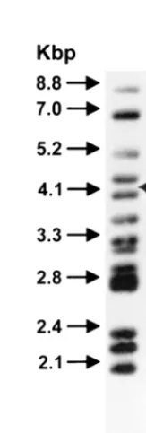
[image:4.612.56.290.69.277.2]Molecular typing.
The molecular genetic typing technique
PFGE did not detect any differences between the rabbit and
cattle isolates from the same farm or between farms. A typical
PFGE result with
Hin
dIII is shown in Fig. 2. There are
cur-rently no published PFGE profiles of
M. avium
subsp.
paratu-berculosis
restricted with
Hin
dIII. The
Spe
I profiles obtained
were identical to the S2 PFGE profile described previously (5).
The results of the IS900 RFLP analysis are shown in Fig. 3.
IS900 RFLP analysis identified all of the rabbit and cattle
isolates, except R8, as RFLP type B-C17, which is the
predom-inant RFLP type found in cattle and sheep strains in the
United Kingdom (26). The isolate R8 was found to be RFLP
type B-C16. The chemotype profiles of the rabbit and cattle
strains revealed minor differences which allowed division of
the isolates into six groups as follows (the farm identity is given
in parentheses): group I, rabbit isolates R1(A) and R186(C);
group II, rabbit isolate R196(C) and cattle isolate F16(A);
group III, rabbit isolates R7(A), R8(A), R10(A), R191(C), and
R194(C) and cattle isolates JD3(C) and JD4(C); group IV,
rabbit isolates R193(C) and R198(C) and cattle isolates JD1
(C), JD2(C), and F14(A); group V, cattle isolate F13(A); and
group VI, rabbit isolate R197(C). Groups III and IV are so
similar that they probably represent the same strain. These two
chemotype profiles were represented by both cattle and rabbit
FIG. 2. Long restriction fragments ofM. aviumsubsp.paratuberculosis
iso-lates from rabbits (R prefix) and cattle (F prefix) from farm A. Chromosomal
DNA was digested withHindIII and subjected to PFGE as described in Materials
and Methods. MW, PFGE molecular weight markers (Mid-Range II; New En-gland Biolabs); values are indicated in kilobases.
FIG. 3. DNA fingerprinting of isolates from rabbits and cattle (DNA types C16 and C17).
on May 15, 2020 by guest
http://jcm.asm.org/
[image:4.612.389.471.467.710.2]isolates and were the most common, comprising 39% (group
III) and 28% (group IV) of the isolates. Representative
che-motype profiles for the six groups are shown in the composite
photograph in Fig. 4. The distribution of chemotype profiles
was different on the two farms, as shown in Table 3. There
appeared to be no overlap between the chemotype profiles of
cattle and rabbit isolates on farm A, whereas on farm C there
is an overlap of chemotype groups 3 and 4.
DISCUSSION
This study has corroborated the findings of the pilot study
(7) and confirmed the presence of paratuberculosis in wild
rabbits in Scotland. The survey revealed that the Tayside
re-gion was a definite hot spot for
M. avium
subsp.
paratubercu-losis
infection of rabbits. Up to 53% of wild rabbits were found
to be infected on farms in this area, where paratuberculosis
was a problem in livestock.
M. avium
subsp.
paratuberculosis
also was cultured from a rabbit on a farm in the Borders
region, which is geographically distant from Tayside.
Analysis of the data showed a statistically significant
rela-tionship between a past or current problem of paratuberculosis
in cattle and in the wild rabbit population on the Tayside
farms. This could be interpreted as evidence for interspecies
transmission or it may be simply a further reflection of the
regional prevalence of
M. avium
subsp.
paratuberculosis
in both
host species. No evidence was found of a positive relationship
between a history of Johne’s disease in sheep and the
occur-rence of paratuberculosis in rabbits. However, this should not
be taken as evidence that no such relationship exists, since the
result may be a consequence of having cultured fewer isolates
from rabbits on sheep farms due to the difficulties in isolating
and growing ovine
M. avium
subsp.
paratuberculosis
strains and
the relatively small number of farms in the survey.
From the disease control perspective, the important
ques-tion is whether
M. avium
subsp.
paratuberculosis
can be
trans-mitted from rabbits to farm ruminants. Various typing
tech-niques were employed to determine whether there were two
distinct, noninteractive populations of
M. avium
subsp.
para-tuberculosis
in the livestock and the rabbits or whether one
population existed. The latter finding would support the
con-cept of interspecies transmission. The rabbit and cattle isolates
of
M. avium
subsp.
paratuberculosis
were morphologically
indistinguishable and had comparable growth rates in vitro on
primary isolation. Molecular genetic typing could not
discrim-inate between rabbit and cattle isolates from the same or
different farms, suggesting that a single strain may be
respon-sible for the disease in either host and that interspecies
trans-mission could have occurred. Unfortunately, it was not possible
to compare sheep isolates with cattle and rabbit isolates from
farm C due to difficulties encountered in culturing these
iso-lates in vitro. IS900 RFLP analysis is currently the most
suc-cessful technique for typing
M. avium
subsp.
paratuberculosis
and is considered to be the only technique with sufficient
dis-criminatory power to be used in epidemiological studies (27,
28). However, there appears to be little variation in the
geno-mic fingerprints of these isolates, which may indicate that
M. avium
subsp.
paratuberculosis
exhibits little diversity or
sim-ply that current techniques are inadequate for detecting
ge-netic variation in populations of this organism.
It was possible to discriminate between isolates according to
their chemotype profile. Six chemotype profiles were
identi-fied, and a difference in their distribution between cattle and
rabbit isolates on the two farms was observed. On farm C there
was overlap in the distribution of chemotypes III and IV
be-tween cattle and rabbit isolates, thus indicating that
interspe-cies transmission could have occurred. On farm A, the
chemo-TABLE 3. Distribution of chemotypes of cattle and
rabbit isolates from two positive farms
aFarm Chemotype profile in (host):
Cattle Rabbits
A
II, IV, V
I, III
C
III, IV
I, II, III, IV, VI
[image:5.612.56.547.70.286.2]aValues represent the chemotype profile of isolates from each farm.
FIG. 4. Composite photograph showing the six representative chemotypes ofM. aviumsubsp.paratuberculosisisolates from rabbits and cattle on farms A and C,
as determined by the MSPD-TLC method. Lanes: 1, hexane extract; 2, methylene chloride extract; 3, acetonitrile extract; 4, methanol extract; and 5, H2O extract. Note
the close similarity of groups III and IV.
on May 15, 2020 by guest
http://jcm.asm.org/
[image:5.612.311.553.665.719.2]type profiles of the cattle and rabbit isolates were different,
suggesting the existence of two independent populations of
M. avium
subsp.
paratuberculosis
associated with the two host
species. However, if chemotype groups III and IV represent
the same strain, interspecies transmission could have occurred.
It will be necessary to phenotype a larger number of isolates to
evaluate the epidemiological relationship between the cattle
and rabbit isolates on these farms.
Since molecular typing per se cannot prove that
interspe-cies transmission occurs, it will be necessary to demonstrate
interspecies transmission experimentally under controlled
conditions. Preliminary experiments have shown that lambs
ex-perimentally infected with a rabbit isolate develop lesions
(un-published observations). Further experiments are in progress.
This study highlights the fact that
M. avium
subsp.
paratu-berculosis
has a broad host range. The organism principally
affects ruminants, but the infection of lagomorphs may be
more common than was first thought. There have been
previ-ous reports of the isolation of a mycobactin-dependent
myco-bacterium from the European hare (19) and lesions containing
acid-fast bacilli in a wild rabbit in Scotland (1). More recently,
culture and PCR analysis have confirmed the presence of
M. avium
subsp.
paratuberculosis
in a single wild rabbit in Spain
(29). Other monogastrics known to have become infected
in-clude macaques (20) and humans (8). A variety of laboratory
animals, such as mice (25, 37), rats (12), hamsters (6), gerbils
(16), guinea pigs (21), and chickens (15), have been infected
experimentally with
M. avium
subsp.
paratuberculosis
.
How-ever, although the mycobacterium replicates in some of these
hosts, it does not produce the pathology and clinical signs
characteristic of paratuberculosis in ruminants.
ACKNOWLEDGMENTS
We thank Kathleen Connor, Amanda Pirie, and Karen Rudge for
maintaining the strain collection, Lenka Dvorska for the DNA
finger-printing, and Lisa Whittington for assistance with the chemotype profiles.
This work was funded by the Scottish Office Agriculture,
Environ-ment and Fisheries DepartEnviron-ment; the Animal Health Trust; the
Minis-try of Agriculture of the Czech Republic (grant number EP0960006087);
and the Veterinary Medical Experiment Station, University of
Geor-gia, Tifton, Ga.
REFERENCES
1.Angus, K. W.1990. Intestinal lesions resembling paratuberculosis in a wild
rabbit (Oryctolagus cuniculus). J. Comp. Pathol.103:101–105.
2.Challans, J. A., K. Stevenson, H. W. Reid, and J. M. Sharp.1994. A rapid
method for the extraction and amplification ofMycobacterium
paratubercu-losisDNA from clinical samples. Vet. Rec.134:95–96.
3.Chiodini, R. J., H. J. van Kruiningen, and R. S. Merkal.1984. Ruminant
paratuberculosis (Johne’s disease): the current status and future prospects.
Cornell Vet.74:218–262.
4.Chiodini, R. J., and H. J. Van Kruiningen.1983. Eastern white-tailed deer as
a reservoir of ruminant paratuberculosis. J. Am. Vet. Med. Assoc.182:168–169.
5.Feizabadi, M. M., I. D. Robertson, A. Hope, D. V. Cousins, and D. J.
Hamp-son.1997. Differentiation of Australian isolates ofMycobacterium
paratuber-culosisusing pulsed-field gel electrophoresis. Aust. Vet. J.75:887–889.
6.Gilmour, N. L., J. Campbell, and J. G. Brotherstone.1963. The pathogenesis
ofMycobacterium johneiin orally dosed hamsters. J. Comp. Pathol. Ther.73: 98–106.
7.Greig, A., K. Stevenson, V. Perez, A. A. Pirie, J. M. Grant, and J. M. Sharp.
1997. Paratuberculosis in wild rabbits (Oryctolagus cuniculus). Vet. Rec.140:
141–143.
8.Hermon-Taylor, J., M. Moss, M. Tizard, Z. Malik, and J. Sanderson.1990.
Molecular biology of Crohn’s disease mycobacteria. Balliere’s Clin.
Gastro-enterol.4:23–42.
9.Hillermark, K.1966. A disease resembling paratuberculosis (Johne’s
dis-ease) in roe deer (Capreolus capreolusL.). An aetiological and pathological
anatomical study. Acta Vet. Scand.7:330–363.
10. Hines, M. E., II, and K. S. Frazier.1993. Differentiation of mycobacteria on
the basis of chemotype profiles by using matrix solid-phase dispersion and
thin-layer chromatography. J. Clin. Microbiol.31:610–614.
11. Hines, M. E., II, A. R. Long, T. G. Snider III, and S. A. Barker.1991. Lysis
and fractionation ofMycobacterium paratuberculosisandEscherichia coliby
matrix solid-phase dispersion. Anal. Biochem.195:197–206.
12. Hole, N. H.1958. Johne’s disease. Adv. Vet. Sci.4:341–387.
13. Jessup, D. A., B. Abbas, D. Behymer, and P. Gogan.1981. Paratuberculosis
in tule elk in California. J. Am. Vet. Med. Assoc.179:1252–1254.
14. Kunze, Z. M., F. Portaels, and J. J. McFadden.1992. Biologically distinct
subtypes ofMycobacterium aviumdiffer in possession of insertion sequence
IS901. J. Clin. Microbiol.30:2366–2372.
15. Larsen, A. B., and H. W. Moon.1972. ExperimentalMycobacterium
paratu-berculosisinfection in chickens. Am. J. Vet. Res.33:1231–1235.
16. Larsen, A. B., J. M. Millar, and K. E. Kopecky.1976. Susceptibility of the
Mongolian gerbil (Meriones unguiculatus) toMycobacterium paratuberculosis.
Am. J. Vet. Res.37:1113–1114.
17. Levy-Frebault, V. V., M. F. Thorel, A. Varnerot, and B. Gicquel.1989. DNA
polymorphism inMycobacterium paratuberculosis, wood pigeon mycobacteria
and related mycobacteria analyzed by field-inversion gel electrophoresis.
J. Clin. Microbiol.27:2823–2826.
18. Libke, K. G., and A. M. Walton.1975. Presumptive paratuberculosis in a
Virginia white-tailed deer. J. Wildl. Dis.11:552–553.
19. Matthews, P. R., and A. Sargent.1977. The isolation of mycobacteria from
the brown hare (Lepus europaeus). Br. Vet. J.133:399–404.
20. McClure, H. M., R. J. Chiodini, D. C. Anderson, R. B. Swenson, W. R.
Thayer, and J. A. Coutu.1987.Mycobacterium paratuberculosisinfection in a
colony of stumptail macaques (Macaca arctoides). J. Infect. Dis.155:1011–1019.
21. Mohler, W. M.1939. Johne’s disease infection of laboratory animals with aid
of paraffin oil. J. Am. Vet. Med. Assoc.94:590–594.
22. Mokresh, A. H., and D. G. Butler.1990. Granulomatous enteritis following
oral inoculation of newborn rabbits withMycobacterium paratuberculosisof
bovine origin. Can. J. Vet. Res.54:313–319.
23. Mokresh, A. H., C. J. Czuprynski, and D. G. Butler.1989. A rabbit model for
study ofMycobacterium paratuberculosisinfection. Infect. Immun.57:3798–3807.
24. Mondal, D., and R. P. Sinha.1992. Pathogenicity of caprine strain of
My-cobacterium paratuberculosisin rabbit. Ind. J. Anim. Sci.62:1117–1120.
25. Mutwiri, G. K., D. G. Butler, S. Rosendal, and J. Yager.1992. Experimental
infection of severe combined immunodeficient beige mice with
Mycobacte-rium paratuberculosisof bovine origin. Infect. Immun.60:4074–4079.
26. Pavlik, I., A. Horvathova, L. Dvorska, P. Svastova, R. du Maine, B. Fixa, and
I. Rychlik.Unpublished results.
27. Pavlik, I., A. Horvathova, L. Dvorska, J. Bartl, P. Svastova, R. du Maine, and I.
Rychlik.Restriction-fragment-length polymorphisms with the probe IS900 in
My-cobacterium aviumsubspeciesparatuberculosis. J. Microbiol. Methods, in press.
28. Pavlik, I., L. Bejckova, M. Pavlas, Z. Rozsypalova, and S. Koskova.1995.
Characterisation by restriction endonuclease analysis and DNA hybridisa-tion using IS900 of bovine, ovine, caprine and human mycobactin-dependent
strains ofMycobacterium paratuberculosisisolated in various localities. Vet.
Microbiol.45:311–318.
29. Perez-Perez, V., and F. Garcia-Marin.Personal communication.
30. Riemann, H., M. R. Zaman, R. Ruppanner, O. Aalund, J. B. Jorgensen, H.
Worsaae, and D. Behymer.1979. Paratuberculosis in cattle and free-living
exotic deer. J. Am. Vet. Med. Assoc.174:841–843.
31. Ris, D. R., K. L. Hamel, and A. M. Weaver.1988. Natural transmission of
Johne’s disease to feral goats. N. Z. Vet. J.36:98–99.
32. Ris, D. R., K. L. Hamel, and J. M. Ayling.1987. Can sheep become infected
by grazing pasture contaminated by cattle with Johne’s disease? N. Z. Vet. J. 35:137.
33. Sanderson, J., M. T. Moss, M. L. V. Tizard, and J. Hermon-Taylor.1992.
My-cobacterium paratuberculosisDNA in Crohn’s disease tissue. Gut33:890–896.
34. Schaefer, W. B.1965. Serologic identification and classification. Am. Rev.
Respir. Dis.92(Suppl.):85–93.
35. Sharp, J. M., K. Stevenson, J. A. Challans, C. Ramage, D. Hitchcock, and
H. W. Reid.1996. Mycobacterial infections of free-living deer in Scotland,
p. 180–182.InProceedings of the Fifth International Colloquium on
Para-tuberculosis. International Association for Paratuberculosis, Inc., East Prov-idence, R.I.
36. Sweeney, R. W. (ed.).1996. Paratuberculosis (Johne’s disease). Vet. Clin.
North Am. Food Anim. Pract.12:No. 2.
37. Tanaka, S., M. Sato, T. Taniguch, and Y. Yokomizo.1994. Histopathological
and morphometrical comparison of granulomatous lesions in BALB/c and
C3H/HeJ mice inoculated withMycobacterium paratuberculosis. J. Comp.
Pathol.110:381–388.
38. Touchstone, J. C., and M. F. Dobbins.1983. Practice of thin-layer
chroma-tography, 2nd ed., p. 9–15. Wiley Interscience, New York, N.Y.
38a.Veterinary Laboratories Agency.1993. Veterinary Investigation Diagnostic
Analysis report, 1991–1993. Veterinary Laboratories Agency, Addlestone, United Kingdom.
39. Williams, E. S., and T. R. Spraker.1979. Paratuberculosis in free-ranging
big-horn sheep and a Rocky Mountain goat with a brief review of the disease in wild
species, p. 122–124.InAnnual Proceedings of the AAZV. American
Associa-tion of Zoo Veterinarians, Denver, Colo.
40. Williams, E. S., S. P. Snyder, and K. L. Martin.1983. Pathology of
sponta-neous and experimental infection of North American wild ruminants with
Mycobacterium paratuberculosis. Vet. Pathol.20:274–290.