ORIGINAL RESEARCH ARTICLE
CELL THERAPY WITH MESENQUIMIAL STEM CELLS OF BONE MARROW IMPROVES RENAL
FUNCTION IN PRE-CLINICAL MODULE
*1,2
Peterson Vieira de Assis,
1,2Silvia Cordeiro das Neves,
1,2Luiz Carlos Takita,
1,2Bethânia
Borges Tura,
1,2,3Rodrigo Juliano Oliveira and
1,2Andréia Conceição Milan
Brochado Antoniolli-Silva
1
Centro de Estudos em Células-Tronco, Terapia Celular e Genética Toxicológica - CeTroGen, Hospital
Universitário Maria Aparecida Pedrossian - HUMAP, Universidade Federal de Mato Grosso do Sul - UFMS,
Campo Grande, Mato Grosso do Sul, Brasil
2
Programa de Pós-graduação em Saúde e Desenvolvimento na Região Centro-Oeste, Faculdade de Medicina Dr.
Hélio Mandetta - FAMED, Universidade Federal de Mato Grosso do Sul - UFMS, Campo Grande, Mato Grosso do
Sul, Brasil
3
Programa de Pós-graduação em Genética e Biologia Molecular, Centro de Ciências Biológicas - CCB,
Universidade Estadual de Londrina - UEL, Londrina, Paraná, Brasil
ARTICLE INFO ABSTRACT
Acute kidney injury (AKI) is a disease characterized by a drop in glomerular filtration rate and is considered a public health problem because it causes morbidity and mortality. In some cases, cell therapy with mesenchymal stem cells (MSC) may be an option for resumption of kidney function and resting. Thus, the present study evaluated the effects of cell therapy with bone marrow-derived MSC on acute renal injury in male Wistar rats exposed to the chemotherapeutic agent cisplatin. The animals were divided into 4 experimental groups (n = 06): Bone marrow donor group (DG); Control Group (CG) - animals treated with PBS and not receiving MSC transplantation; Nephrotoxicity Group (NG) - animals treated with the chemotherapeutic cisplatin (4mg/kg) and that did not receive MSC transplantation; and the Nephrotoxicity Group + Mesenchymal Stem Cell (NG + MSC) – animals treated with cisplatin and who received MSC transplantation (1,0 x 10⁶ cells). The results demonstrated that the cellular therapy caused reduction of plasma levels of urea and creatinine. Histopathological analysis showed a reduction (p <0.05) in the frequency of NG + MSC damage in relation to NG. In view of the results presented, it is inferred that cell therapy with mesenchymal stem cells is a possibility of choice for the treatment of acute renal injury.
Copyright © 2019,Peterson Vieira de Assis et al. This is an open access article distributed under the Creative Commons Attribution License, which permits unrestricted use, distribution, and reproduction in any medium, provided the original work is properly cited.
INTRODUCTION
Acute renal injury (AKI) is a disease characterized by a drop in the glomerular filtration rate (Santos and Mendonça, 2015) and increased serum creatinine and urea levels (Yamaki et al., 2012). According to KDIGO's Clinical Practice Guidelines for Acute Renal Injury, creatinine clearance is considered a strong biochemical indicator of this disease (Khwaja, 2012). Soon this is a common examination in hospital beds (Peres et al., 2013).
*Corresponding author: Andréia Conceição Milan Brochado Antoniolli-Silva
During hospitalization, AKI is considered a substantial problem, since one in five adults and one in three children present renal damage or renal insufficiency determined in particular by drug treatments (Nargesi et al., 2017). In this context, mortality rates vary from 3 to 5%, and this risk is increased to 30 to 50% in patients admitted to Intensive Care Units (Patschan et al., 2018). Thus, the treatment of AKI, which can lead to morbidity and mortality in a short time, is a public health challenge. Added to this is the fact that AKI can further determine the gradual loss of renal function and progress in the long term to chronic kidney disease (Bellomo
et al., 2012; Kellum et al., 2012; Schrezenmei et al., 2017).
ISSN: 2230-9926
International Journal of Development Research
Vol. 09, Issue, 03, pp.26613-26619, March, 2019
Article History:
Received 24th December, 2018
Received in revised form 17th January, 2019
Accepted 19th February, 2019
Published online 31st March, 2019
Key Words:
Cisplatin, Kidney, Immunomodulation, Transplantation
Citation: Peterson Vieira de Assis, Silvia Cordeiro das Neves, Luiz Carlos Takita, Bethânia Borges Tura, Rodrigo Juliano Oliveira and Andréia
Conceição Milan Brochado Antoniolli-Silva. 2019. “A scientometric study of the journal of philosophy of education”, International Journal of
Development Research, 09, (03), 26613-26619.
because, in general, chronic kidney treatments require high investments (Nargesi et al., 2017). Treatment of AKI may require the identification and treatment of the cause, simple hydration, suspension of medications, removal of kidney stones and/or treatment of diseases, for example, autoimmune diseases affecting the kidneys (Marini e Wheeler, 1999; Siqueria et al., 2008; Brunner e Suddarth, 2011; Santos et al., 2013). However, in some cases even using these therapeutic resources the kidney does not resume its function and this demands the development of new therapies. In recent years cell therapy with mesenchymal stem cells (MSC) has emerged as an alternative for the treatment of different diseases (Bareeqa et al., 2018; Hong et al., 2018; Mancuso et al., 2019; Svistushkin et al., 2019), including renal (Hu e Zou, 2017;
Nargesiet al., 2017; Zhang et al., 2018; Zhuang et al., 2019)
through the resumption of organ/tissue function and homeostasis. This healing process can take place: (I) by the transdifferentiation that is when the stem cells are lodged in the lesion and, by stimuli of the tissue itself, they become healthy cells of that organ (Lee et al., 2018; Los et al., 2018); e/ou (II) for paracrine and autocrine effects (Gnecchi et al., 2008; Souza et al., 2010) which is when the cell produces a variety of endogenous cytokines and/or growth factors that exert function on the MSCs (autocrine effect) or adjacent cells (paracrine effect) and determine cell division, differentiation, anti- inflammatory, antiapoptotic, pro-angiogenic and/or endogenous repairing effect (Gnecchi et al., 2008; Souza et al., 2010) which facilitators of the organ recovery process in search of homeostasis and the adequate functioning. The present study evaluated the effects of mesenchymal bone marrow stem cell therapy in promoting improvement and reestablishment of renal function in a preclinical model.
MATERIAL AND METHODS
Chemical Agents: For the induction of nephrotoxicity, the
chemotherapeutic cisplatin (Fauldcispla LIBBS®, Lot: 12GO702) at the dose of 4mg/kg (b.w., i.p.) was used. The applications were performed once a week for 4 consecutive weeks (Canta et al., 2011).
Animals: Twenty-four male and sexually mature Wistar rats
from the Central Biotherm of the Institute of Biosciences of the Federal University of Mato Grosso do Sul (INBIO/UFMS) were used. The animals were kept in trios in propylene boxes covered by brush and fed with commercial feed (Nuvital®) and filtered water ad libitum. The boxes were maintained on a ventilated shelf (Alesco®) under standard conditions of 12-hour photoperiod (12 12-hours clear: 12 12-hours dark), temperature around 22 ± 2ºC and relative humidity 55 ± 10%. The experiment was conducted in accordance with the guidelines of the Universal Declaration of the Rights of the Animals and with the approval of the Committee of Ethics in the Use of Animals of UFMS (Protocol # 532/2013).
Experimental Design: The animals were divided into four
groups: (I) Donors group - DG group (n = 6) - used as donors of mesenchymal stem cells of bone marrow; (II) Control Group - CG (n = 6) - the animals were treated with phosphate buffered saline (PBS) at a ratio of 1 ml/100g (b.w.; i.p.) once weekly for 4 consecutive weeks, and a new administration of PBS was done intravenously (i.v.), through the caudal vein , 24 hours the last administration i.p.; (III) Nephrotoxicity Group -
concentration of 4 mg/kg (b.w., i.p.) once weekly for 4 consecutive weeks, and administration of PBS (i.v.) was done by the caudal vein, the last administration i.p., and (IV) Nephrotoxicity Group + Mesenchymal Stem Cells - NG + MSC (n = 6) - the animals were treated with cisplatin at the concentration of 4 mg/kg (b.w., i.p.) once a week for 4 consecutive weeks, and MSC (i.v.) administration was done via the caudal vein, 24 hours after the last administration i.p.. For each animal 1x10⁶ cells were transplanted (Urt-Filho et al., 2016). The euthanasia of the animals occurred 30 days after the administration of MSC
Obtaining, Cultivating and Expansion of Mesenchymal Stem Cells from the Bone Marrow: DG animals, called donors, were submitted to euthanasia by excessive anesthetic dose (Tiopental®). Then the femurs were collected and each had their intraosseous lumen washed with 5mL of phosphate buffered saline (PBS) supplemented with 1% antibiotic (Penicillin/Streptomycin, LGB Biotechnology®) under sterile conditions. Sample processing and culturing of the cells was performed as described by Urt-Filho et al. (2016).
Osteogenic, Adipogenic and Chondrogenic Differentiation:
The flasks destined for the differentiation experiments, when they reached 80% confluency, were again trypsinized and the cells were seeded in 6 flasks of 25cm2, at the concentration of 2x10⁵ cells (Urt-Filho et al., 2016). Three flasks of 25cm2, used as controls, were maintained in supplemented culture medium. For adipogenic, osteogenic and chondrogenic differentiation, cells were maintained for 24 hours in supplemented culture medium. Then, this was replaced by culture medium STEMPRO Adipogenic, Osteogenesis and
Chondrogenesis Differentiation Kit (Invitrogen®) and
maintained in culture for 14 days for adipogenic differentiation and for 21 days for osteogenic and chondrogenic cultures, with changes twice a week (Urt-Filho et al., 2016). For the confirmation of the adipogenic differentiation, after discarding the differentiating medium, the cells were fixed for 60 minutes at room temperature with 10% formaldehyde. The cells were then washed with 60% isopropanol and then incubated with
Oil Red O (Sigma®) for 20 minutes at room temperature. The
excess dye was removed by washing with distilled water.
The differentiation was confirmed by accumulation of intracellular lipids on the 14th day (Pesarini et al., 2017; Schweich et al., 2017; Pesarini et al., 2018). For osteogenic differentiation, after discarding of the differentiating medium, the cells were fixed for 10 minutes at room temperature with 10% formaldehyde. The cells were then washed 2 times with PBS and stained with Alizarin Red (Sigma®) for 5 minutes at room temperature. Excess dye was removed by distilled water washes. Osteogenic differentiation was demonstrated by the visualization of calcium deposits on day 21 (Pesarini et al., 2017; Schweich et al., 2017; Pesarini et al., 2018). During the cell culture process of the chondrogenic differentiation the cells were grouped forming a spheroid. At the end of the process this was collected by aspiration with a Pasteur pipette. The spheroid was fixed in 10% buffered formalin at room temperature and subsequently subjected to the histological routine in the automated tissue processor TP09 TS Lupetec® according to manufacturer's instructions. Subsequently, the spheroid was cut into a microtome Leica® RM2235 in cuts with thickness 3 µm. The slides were stained with Alcian Blue
specifications. The differentiation was confirmed by the presence of rich extracellular matrix of glycosaminoglycans on day 21 (Pesarini et al., 2017; Schweich et al., 2017; Pesarini et al., 2018).
Preparation of Mesenchymal Stem Cells for
Transplantation: After the confluence of the third passage, the
culture flasks were trypsinized. The cell suspension was centrifuged at 2000 rpm for 5 minutes and the pellet
resuspended in PBS to obtain the cells used in cell therapy. The animals from the NG + MSC groups received MSC transplantation (i.v.) 24 hours after the administration of the last dose of cisplatin at the concentration of 1.0x10⁶ célls (Urt-Filho et al., 2016).
Biochemical Evaluations of Renal Function: Biochemical
evaluations of renal function, urea and creatinine were performed 30 days after administration of the last dose of cisplatin. Blood collection was performed through the retro-orbital plexus after anesthesia (Ketamina – 50mg/kg b.w., i.p.; Xilasina – 10mg/kg p.c., i.p.). After collection, the sedimentation of the blood was awaited at 4° C and the serum was frozen at -20° C until analysis. The analysis was performed on automated equipment COBAS 6000 (Roche Diagnostics®) according to the manufacturer's recommendations.
Histopathological Analysis: After the 30-day experimental period, the animals were submitted to euthanasia by excessive anesthetic dose as described. The kidneys were then collected, sectioned, fixed in 10% buffered formalin and prepared according to routine histopathological procedures according to Urt-Filho et al. (2016). For histopathological analysis, the double-blind model and the criteria of the classification Banff 97 were used, according to Racusen et al. (1999) with Urt-Filho et al. (2016) modifications where it is reported: 1) Tubulitis: intratubular lymphocytic infiltrate (necrosis and fibrosis replacement not assessed); 2) Tubular hydropic degeneration: cytoplasmic balanonization of the cells of the proximal contorted tubule (1+<25%; 2+25–75%; 3+>75%); 3) Intratubular protein: presence of eosinophilic material within the tubules (proximal contortion, distal, loop of Henle) – glomerular filtration failure, tubular reabsorption failure (1+<25%; 2+25–75%; 3+>75%); 4) Glomerulitis: lymphocytic infiltrate in the glomerulus; 5) Arteritis: lymphocytic infiltrate in arterioles; 6) Interstitial infiltrate: lymphocytes in the
interstitium (1+0–25%; 2+25–75%; 3+>75%); 7) Interstitial fibrosis: deposition of scarring collagen; 8) Glomerular sclerosis: glomerular fibrosis (failure of the nephron); 9) Calcifications; 10) Retraction/Cortical atrophy: partial replacement of the renal parenchyma by fibrosis and/or inflammatory infiltrate; 11) Apoptosis / tubular necrosis: total or partial degeneration of the tubules (1+<25%; 2+25–75%; 3+>75%); 12) Global necrosis: glomeruli and tubules involved.
Statistical analysis: Results were expressed as average ±
standard error of the average. Statistical analysis was performed on the Software GraphPad InStat 5 according to the data distribution and for that purpose the ANOVA/Tukey tests were applied for parametric data and Kruskal-Wallis/Dunn for non-parametric data. Differences were considered significant when p≤0.05.
RESULTS
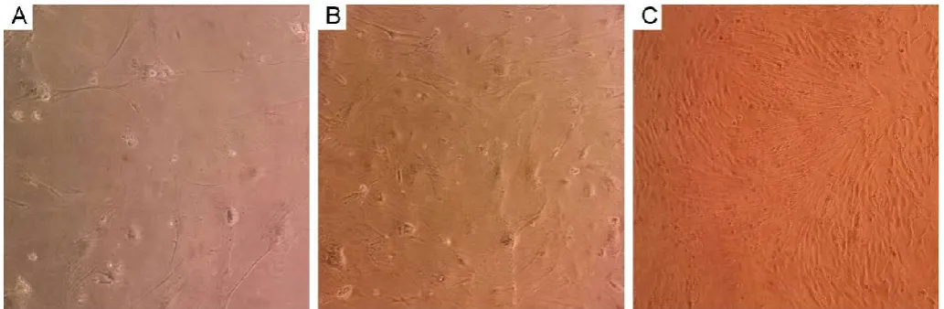
Expansion of Mesenchymal Stem Cells:After isolation of the
MSC, they were expanded in culture flasks with successive tests. Viability was observed in all passages and only cultures with viability greater than 95% were maintained. In Figure 1 the aspects of the culture used in the experiments can be observed.
Mesenchymal Stem Cells Differentiate in Osteogenic,
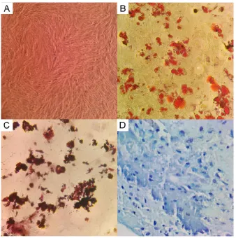
Adipogenic and Chondrogenic Cells in vitro: The
confirmation that the cells in culture really were MSC was through the adipogenic, osteogenic and chondrogenic differentiations where it was possible to identify the accumulation of lipids stained with Oil red O, calcium deposits stained with Alizarin Red and the rich extracellular matrix of glycosaminoglycans stained with Alcian Blue, respectively (Figure 2).
Evaluation of the Effects of Mesenchymal Stem Cell Transplantation on Cisplatin Induced Nephrotoxicity:
[image:3.595.43.560.54.223.2]Administration of cisplatin caused an increase (p <0.05) in plasma urea concentration in the nephrotoxicity group compared to the negative control (Figure 3A). Likewise, it was observed increased (p <0.05) significantly creatinine (Figure 3B). These same parameters were evaluated in animals that received treatment with cisplatin and subsequently received MSC transplantation.
The results indicated that there were no significant differences between this group (NG + MSC) and the negative control group (Figure 3). However, significant differences were observed, with a reduction (p <0.05) in urea and creatinine levels when compared to NG and NG + MSC groups (Figure 3).
Histopathological Analysis: Histopathological analysis
revealed that in CG the changes were tubular hydropic degeneration, intratubular protein accumulation, tubular necrosis/apoptosis with medians of 0.5, 0.25, 0.75, respectively (Figure 4A).
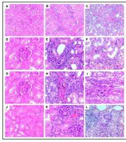
[image:4.595.129.468.56.397.2]In the NG and NG + MSC groups the observed changes were tubulitis, tubular hydropic degeneration, tubular protein accumulation, glomerulitis, interstitial infiltrate, interstitial fibrosis, glomerular sclerosis and apoptosis / tubular necrosis with medians 0.5 e 1; 1.25 e 0.75; 0.75 e 0.75; 0.5 e 0.5; 1.25 e 0.75; 0.75 e 0.25; 0.5 e 0.25; 0.5 and 0.5 respectively (Figure 4A, Figure 5). There was no statistically significant difference between groups. However, there is a tendency to reduce tubular hydropic degeneration, interstitial infiltrate, interstitial fibrosis and glomerular sclerosis in the group that received MSC transplantation (Figure 4A).
Figure 2. MSC photomicrographs: (A) undifferentiated culture, (B) culture in adipogenic differentiation with lipid vacuoles stained
by Oil red O, (C) culture in osteogenic differentiation with calcium deposits stained by Alizarin Red, (D) Culture in chondrogenic
differentiation with rich extracellular matrix of glycosaminoglycans stained by Alcian Blue. 400x magnification
Figure 3. Plasma concentrations of urea and creatinine in the different experimental groups. Administration of 4 mg/kg cisplatin once weekly for four consecutive weeks was able to increase the plasma concentrations of these two biochemical markers of renal function.
[image:4.595.113.483.450.603.2]Figure 4. Median and standard deviation of the score of histopathological changes of the cross-sectional kidneys according to classification Banff 97 (A) and evaluation of global damage (sum of all damages classified according to Banff 97) (B). CG - Control Group - evaluation 30 days after treatment with PBS 1ml/100g (b.w.; i.p.). NG –Nephrotoxicity Group - evaluation 30 days after treatment with cisplatin (4mg/Kg, b.w., i.p.); NG + MSC – Nephrotoxicity Group + Mesenchymal StemCell - evaluation 30 days after treatment with cisplatin at the concentration of 4mg/Kg (b.w., i.p.) and cell therapy with MSC (i.v.) performed by the caudal vein, 24 hours after the last i.p. of cisplatin (Statistical Analysis: Kruskal-Wallis/Dunn Statistical Test, p> 0.05)
However, when a global analysis of the histological damages was performed, a significant increase (p <0.05) of 6.13x NG was observed in relation to the CG and a statistically significant (p <0.05) reduction of 4.25x NG + MSC compared to NG (Figure 4B).
DISCUSSION
Acute renal injury can determine the gradual loss of renal function triggering mortality or the development of a chronic disease that correlates directly with morbidities (Sarnak et al., 2003) and high costs for the health systems since the chronic renal patient needs dialysis and/or renal transplantation
(Nargesi et al., 2017). Thus new strategies of prevention of
[image:5.595.42.289.49.452.2]renal damage are necessary to prevent the progression of AKI to chronic renal damage. In this context, MSC cell therapy is inserted.
Figure 5. Histological sections of the renal tissue of the animals of the experimental groups CG (A, D, G and J), NG (B, E, H and K) and NG + MSC (C, F, I and L). Figure A, B and C overview of tissue from CG, NG and NG + MSC groups. Figures E and F indicate interstitial infiltrate (*). Figures H and I demonstrate interstitial fibrosis (#) and Figures K and L indicate glomerular sclerosis (+). Increase of 100x in Figures A, B and C and 400x in Figures D-L.
In the present study to evaluate the effects of this cellular therapy was used the model of acute renal damage caused by medicines. One of the drugs described as capable of causing these renal lesions is the chemotherapeutic cisplatin (Nargesi
et al., 2017; Patschan et al., 2018). Cisplatin is a
[image:5.595.302.558.51.341.2]Patschan et al., 2018). In addition to improving renal function, cell therapy was able to reduce the overall frequency of histological damage. This reduction was statistically significant and was also reported in other preclinical models. According to Lin et al. (2016); Nargesi et al. (2017); Patschan
et al. (2018). These results demonstrate that cell therapy with
mesenchymal stem cells may be a good alternative for the treatment of AKI within 30 days. It is believed that the MSC can act by two main routes that are (I) regeneration and the (II) paracrine effect. According to Little et al., (2018), regeneration occurs when the MSCs go to the site of the lesion adhere to the tissue and are transdifferentiated. This fact can occur because the injured tissue itself produces growth factors and differentiating factors that induce MSC to transform into the tissue of the organ that requires regeneration. Paracrine action occurs when MSCs migrate to the lesion site and produce endogenous repair factors, anti-inflammatory and antiapoptotic factors that aid in the recovery of the injured organ/tissue. However, without integrating MSC into the tissue matrix (Souza et al., 2010). Although there is still no consensus in the literature about the main action of bone marrow MSC in renal tissue recovery, it is believed that these two forms of action may justify the findings described by this study. Thus, it is inferred from the results presented that cell therapy with mesenchymal stem cells is a possibility of choice for the treatment of acute renal injury. It is also worth noting that this therapy may bring benefits to clinical practice, including for patients oncological treatment with cisplatin, since the preclinical model reinforces the possibility of use in humans in the near future.
REFERENCES
Albertoni G, Schor N. 2015. Resveratrol plays important role in protective mechanisms in renal disease-mini-review.
Brazilian Journal of Nephrology. 37:106-114.
Bareeqa SB, Bibi F, Ahmed SI, Samar SS. 2018. Advancement in Stem Cell Therapy for Ischemic Myocardial Cell: A systematic Review. International Journal of Hematology-Oncology and Stem Cell Research.
12:281-289.
Bellomo R, Kellum JA, Ronco C. 2012. Acute kidney injury. The Lancet. 380: 756-766.
Brunner LS, Suddarth DS. 2011. Tratado de enfermagem cirúrgica. In Tratado de enfermagem médico-cirúrgica, Guanabara.
Canta, A. Chiorazzi A, Carozzi V, Meregalli C, Oggioni N, Sala B, Crippa L, Avezza F, Forestieri D, Rotella G, Zucchetti M, Cavaletti G. 2011. In vivo comparative study of the cytotoxicity of a liposomal formulation of cisplatin (lipoplatin™). Cancer chemotherapy and pharmacology, pp 1001-1008.
Chen YT, Sun CK, Lin YC, Chang LT, Chen YL, Tsai TH, Chung SY, Chua S, Kao YH, Yen CH, Shao PL, Chang KC, Leu S, Yip HK. 2011. Adipose-derived mesenchymal stem cell protects kidneys against ischemia-reperfusion injury through suppressing oxidative stress and inflammatory reaction. Journal of translational medicine. 9:51.
Gnecchi M, Zhang Z, Ni A, Dzau VJ. 2008. Paracrine mechanisms in adult stem cell signaling and therapy. Circulation research. 103:1204-1219.
He L, Li J, Zhan J, Yi F, Fan X, Wei Y, Zhang W. 2019. The value of serum cystatin C in early evaluation of renal
systematic review and meta-analysis. Cancer chemotherapy and pharmacology. 1-11.
Hong B, Lee S, Shin N, Ko Y, Kim D, Lee J, Lee W. 2018. Bone regeneration with umbilical cord blood mesenchymal stem cells in femoral defects of ovariectomized rats. Osteoporosis and Sarcopenia. 4:95-101.
Hu H, Zou C. 2017. Mesenchymal stem cells in renal ischemia-reperfusion injury: Biological and therapeutic perspectives. Current stem cell research & therapy. 12: 183-187
Kellum JA, Bellomo R, Ronco C. 2012. Kidney attack. Jama. 307:2265-2266.
Khwaja A, Acute Kidney Injury Work Group. 2012. KDIGO clinical practice guideline for acute kidney injury. Kidney Int Suppl. 2:(sSuppl 1).
Ko SF, Chen YT, Wallace CG, Chen KH, Sung PH, Cheng BC, Huang TH, Chen YL, Chang HW, Lee MS, Yang CC, Yip HK. 2018. Inducible pluripotent stem cell-derived mesenchymal stem cell therapy effectively protected kidney from acute ischemia-reperfusion injury. American
journal of translational research. 10:3053.
Lee KW, Kim TM, Kim KS, Lee S, Cho J, Park JB, Kwon GY, Kim SJ. 2018. Renal Ischemia-Reperfusion Injury in a Diabetic Monkey Model and Therapeutic Testing of Human Bone Marrow-Derived Mesenchymal Stem Cells. Journal of diabetes research. 2018:1-9.
Lin KC, Yip HK, Shao PL, Wu SC, Chen KH, Chen YT, Yang CC, Sun CK, Kao GS, Chen SY, Chai HT, Chang CL, Chen CH, Lee MS. 2016. Combination of adipose-derived mesenchymal stem cells (ADMSC) and ADMSC-derived exosomes for protecting kidney from acute ischemia– reperfusion injury. International journal of cardiology.
216:173-185.
Little MH, Kumar SV, Forbes T. 2018. Recapitulating kidney development: Progress and challenges. In Seminars in cell & developmental biology. Academic Press.
Los MJ, Hudecki A, Wiechec E. 2018. Stem Cells and Biomaterials for Regenerative Medicine. Academic Press. Mancuso P, Raman S, Glynn A, Barry F, Murphy JM. 2019.
Mesenchymal Stem Cell Therapy for Osteoarthritis: The Critical Role of the Cell Secretome. Frontiers in
bioengineering and biotechnology. 7.
Marini JJ, Wheeler AP. 1999. Terapia intensiva: o essencial. Editora Manole.
Nargesi AA, Lerman LO, Eirin A. 2017. Mesenchymal stem cell-derived extracellular vesicles for kidney repair: current status and looming challenges. Stem cell research & therapy. 8:273.
Patschan D, Buschmann I, Ritter O, Kribben A. 2018. Cell-Based Therapies in Acute Kidney Injury (AKI). Kidney
and Blood Pressure Research. 43:673-681.
Patschan D, Buschmann I, Ritter O, Kribben A. 2018. Cell-Based Therapies in Acute Kidney Injury (AKI). Kidney
and Blood Pressure Research. 43:673-681.
Peres LAB, Cunha Júnior ADD, Schäfer AJ, Silva ALD, Gaspar AD, Scarpari DF, Alves JBF, Neto RG, Oliveir, TFTD. 2013. Biomarkers of acute kidney injury. Brazilian
Journal of Nephrology.35:229-236.
adipogenic differentiation. Biomedicine &
Pharmacotherapy.108:914-924.
Pesarini JR, Oliveira RJ, Pessatto LR, Antoniolli-Silva ACMB, Felicidade I, Nardi NB, Camassola M, Mantovani MS, Ribeiro LR. 2017. Vitamin D: Correlation with biochemical and body composition changes in a southern Brazilian population and induction of cytotoxicity in mesenchymal stem cells derived from human adipose tissue. Biomedicine & Pharmacotherapy. 91:861-871. Rahyussalim AJ, Saleh I, Kurniawati T, Lutfi, APWY. 2017.
Improvement of renal function after human umbilical cord mesenchymal stem cell treatment on chronic renal failure and thoracic spinal cord entrapment: a case report. Journal
of medical case reports. 11:334.
Santos ES, Marinho CM. 2013. Main causes of acute renal insufficiency in intensive care units: nursing intervention. Referência. 3:181-189.
Santos, J. C. O., Mendonça, M. A. O. 2015. Factors predisposing for acute kidney injury in patients in critical condition: Integrative review. Revista da Sociedade
Brasileira de Clínica Médica, 13(1), 69-74.
Sarnak MJ, Levey AS, Schoolwerth AC, Coresh J, Culleton B, Hamm LL, McCullought PA, Kasiske BL, Kelepouris E, Klag MJ, Parfrey P, Pfeffer M, Raij L, Spinosa DJ, Wilson PW. 2003. American heart association councils on kidney in cardiovascular disease, high blood pressure research, clinical cardiology, and epidemiology and prevention. Kidney disease as a risk factor for development of cardiovascular disease: a statement from the American Heart Association Councils on Kidney in cardiovascular disease, high blood pressure research, clinical cardiology, and epidemiology and prevention. Hypertension, 42:1050-1065.
Schrezenmeier EV, Barasch J, Budde K, Westhoff T, Schmidt Ott KM 2017. Biomarkers in acute kidney injury–pathophysiological basis and clinical performance. Acta physiologica. 219:556-574.
Schweich LC, Oliveira EJT, Pesarini JR, Hermeto LC, Camassola M, Nardi NB, Brochado TM, Antoniolli-Silva ACMB, Oliveira RJ. 2017. All-trans retinoic acid induces
mitochondria-mediated apoptosis of human adipose-derived stem cells and affects the balance of the adipogenic differentiation. Biomedicine & Pharmacotherapy. 96:1267-1274.
Siqueira MEMD, Francalacci LC, Perreira CG. 2008. Insuficiência renal aguda e rabdomiólise induzida pelo uso de estatina. Relato de caso. Rev Bras Clin Med. 6:273-275. Souza CFD, Napoli PD, Han SW, Lima VCD, Carvalho AC
2010. Células-tronco mesenquimais: células ideais para a regeneração cardíaca?. Revista Brasileira de Cardiologia Invasiva.
Svistushkin MV, Kotova SL, Shekhter AB, Svistushkin VM, Akovantseva AA, Frolova AA, Fayzullin AL, Starostina SV, Bezrukov EA, Sukhanov RB, Timashev SF, Butnaru DV, Timashev PS 2019. Collagen fibrillar structures in vocal fold scarring and repair using stem cell therapy: a detailed histological, immunohistochemical and atomic force microscopy study. Journal of microscopy. 2019:1-14. Urt-Filho A, Oliveira RJ, Hermeto LC, Pesarini JR, David ND,
Cantero WDB, Falcão G, Marks G, Antoniolli-Silva ACMB (2016). Mesenchymal stem cell therapy promotes the improvement and recovery of renal function in a preclinical model. Genetics and molecular biology. 39:290-299.
Yamaki VN, Gonçalves TB, Coelho JVB, Pontes RVS, Costa FLDS, Brito MVH 2012. Protective effect of remote ischemic per-conditioning in the ischemia and reperfusion-induce renal injury in rats. Revista do Colégio Brasileiro de Cirurgiões. 39:529-533.
Zhang Y, Zhou S, Hu JM, Chen H, Liu D, Li M, Guo Y, Fan LP, Li LY, Liu YG, Zhao M. 2018. Preliminary Study of Bone Marrow-Derived Mesenchymal Stem Cells Pretreatment With Erythropoietin in Preventing Acute Rejection After Rat Renal Transplantation. Transplantation Proceedings. 50:3873-3880.
Zhuang Q, Ma R, Yin Y, Lan T, Yu M, Ming Y. 2019. Mesenchymal Stem Cells in Renal Fibrosis: The Flame of Cytotherapy. Stem Cells International. 2019:1-18.