Received 8 May 2014|Accepted 24 Sep 2014|Published 4 Nov 2014
Structural basis for ion selectivity revealed
by high-resolution crystal structure of Mg
2
þ
channel MgtE
Hironori Takeda
1,2, Motoyuki Hattori
1,2,3,4, Tomohiro Nishizawa
1,2, Keitaro Yamashita
5, Syed T.A. Shah
6,
Martin Caffrey
6, Andre
´s D. Maturana
7, Ryuichiro Ishitani
1,2& Osamu Nureki
1,2Magnesium is the most abundant divalent cation in living cells and is crucial to several biological processes. MgtE is a Mg2þ channel distributed in all domains of life that contributes to the maintenance of cellular Mg2þ homeostasis. Here we report the high-resolution crystal structures of the transmembrane domain of MgtE, bound to Mg2þ, Mn2þ and Ca2þ. The high-resolution Mg2þ-bound crystal structure clearly visualized the hydrated Mg2þ ion within its selectivity filter. Based on those structures and biochemical analyses, we propose a cation selectivity mechanism for MgtE in which the geometry of the hydration shell of the fully hydrated Mg2þ ion is recognized by the side-chain carboxylate groups in the selectivity filter. This is in contrast to the Kþ-selective filter of KcsA, which recognizes a dehydrated Kþion. Our results further revealed a cation-binding site on the periplasmic side, which regulate channel opening and prevents conduction of near-cognate cations.
DOI: 10.1038/ncomms6374 OPEN
1Department of Biological Sciences, Graduate School of Science, University of Tokyo, 2-11-16 Yayoi, Bunkyo-ku, Tokyo 113-0032, Japan.2Global Research
Cluster, RIKEN, 2-1 Hirosawa, Wako-shi, Saitama 351-0198, Japan.3Precursory Research for Embryonic Science and Technology (PRESTO), Japan Science
and Technology Agency, 4-1-8 Honcho, Kawaguchi, Saitama 332-0012, Japan.4School of Life Sciences, Fudan University, 220 Handan Road, Yangpu District,
Shanghai 200433, China.5SR Life Science Instrumentation Unit, RIKEN SPring-8 Center, 1-1-1 Kouto, Sayo-cho, Sayo-gun, Hyogo 679-5148, Japan.
6Membrane Structural and Functional Biology Group, School of Medicine, and School of Biochemistry and Immunology, Trinity College Dublin, Dublin 2,
Ireland.7Department of Bioengineering Sciences, Graduate School of Bioagricultural Sciences, Nagoya University, Furo-cho, Chikusa-ku, Nagoya 464-8601,
T
he magnesium ion, Mg2þ, is involved in many cellular functions, such as the formation of RNA and protein structures, genome stability and enzymatic catalysis1,2, and thus is an essential cation for life. The intracellular homeostasis of the Mg2þ concentration is important for these functions, and thus is maintained by Mg2þ transporters and channels that translocate Mg2þ across biological membranes. The cellular concentrations of other biological cations (that is, Naþ, Kþ and Ca2þ) are strictly controlled, and thus the selective transport of Mg2þ by these transporters and channels is of particular importance. The selective transport mechanism of monovalent cations has been well investigated, based on the crystal structures of Naþ and Kþ channels. The atomic resolution crystal structures of KcsA3,4 and MthK5 revealed that Kþ is dehydrated and directly recognized by the protein atoms in the selectivity filter. In contrast, the selective transport mechanism for divalent cations, such as Ca2þ and Mg2þ, has remained elusive. The dehydration energies of divalent cations are much higher than those of monovalent cations, and thus their recognition mechanisms are attracting significant interest. The crystal structures of divalent cation channels, including CorA6–9, Orai10 and Ca2þ conductive Naþ channel11, suggested that their selectivity filters consist of multiple acidic residues, with carboxylate groups that recognize the divalent cation. However, the medium-resolution structures of these channels limited the clear understanding of the ion recognition mechanism by the selective filters for divalent cations.MgtE is a Mg2þchannel widely conserved in archaea, bacteria and eukaryotes12. The bacterial MgtE is a high-conductance Mg2þ-selective channel gated by the cytosolic Mg2þ concentration and is involved in the cellular homeostasis of Mg2þ. The vertebrate homologues of MgtE are known as the SLC41 family transporters, and the SLC41A1, SLC41A2 and SLC41A3 members of this family were also shown to mediate Mg2þtransport across the plasma or organellar membranes13,14. Several mutations related to Parkinson’s disease exist in the
SLC41A1gene15. The crystal structures ofThermus thermophilus
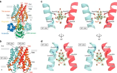
MgtE revealed that MgtE forms a homodimer, consisting of the cytosolic region and the transmembrane (TM) domain16,17. The cytosolic region comprises the N domain, the CBS domain and the plug helix. These domains contain several Mg2þ-binding sites and were suggested to function as sensors for the cytosolic Mg2þ concentration. The ion-conducting pore is formed at the homodimeric interface of the TM domain, which may selectively recognize Mg2þ to allow its conduction across the membrane. The cytosolic side of the pore is sealed by the plug helix, while its periplasmic side is closed by the hydrophobic gate, suggesting that the structures represent a closed form.
In the ion-conducting pore, Asp432 is the sole acidic residue that is widely conserved in the MgtE family members, including the vertebrate SLC41 transporters. A strong electron density peak was observed around the Asp432 carboxylate groups, suggesting the binding of Mg2þ. These observations strongly suggested that the site at the middle of the pore including Asp432 functions as the selectivity filter of MgtE16,17. However, the detailed mechanism of Mg2þ recognition by the selectivity filter has remained elusive, because the medium resolution of the crystal structures could not distinguish the water molecules coordinated to Mg2þ.
In this study, we determined the crystal structure of the MgtE TM domain (MgtE-TMD) at 2.2 Å resolution. The high-resolution crystal structure clearly revealed the metal ion and its coordinated water molecules bound to the selectivity filter. To our knowledge, this is the first visualization of the fully hydrated divalent cation recognized by the selectivity filter of ion channels. In combination with biochemical analyses,
we proposed the divalent cation selectivity mechanism by MgtE.
Results
Crystal structure of MgtE-TMD. In the previously determined full-length structure of MgtE, the ion-conducting pore is formed at the dimer interface, which harbours two conserved Asp residues (Asp432 in TM5) in the middle of the TM segment (M1 site, Fig. 1a). This M1 site is mainly composed of TM2 and TM5, and is expected to function as a Mg2þ-specific selectivity filter16. To obtain high-resolution structural information about the MgtE selectivity filter, we crystallized the TM domain of MgtE (278–450; MgtE-TMD), using the lipidic cubic phase method18,19. We finally determined the crystal structures of MgtE-TMD, bound to Mg2þ, Mn2þ and Ca2þ, at 2.3, 2.2 and 3.2 Å resolutions, respectively (Table 1). The overall structures of MgtE-TMD are essentially the same as the full-length structure determined at 2.94 Å resolution16, with a root mean squared deviation of 1.30 Å for all Caatoms (Fig. 1a,b). The hydrophobic gate located just above the M1 site is closed similar to that in the full-length structure. In contrast, the M1 site is accessible from the cytoplasmic side, as MgtE-TMD lacks the plug helix and the cytosolic domain (Fig. 1b).
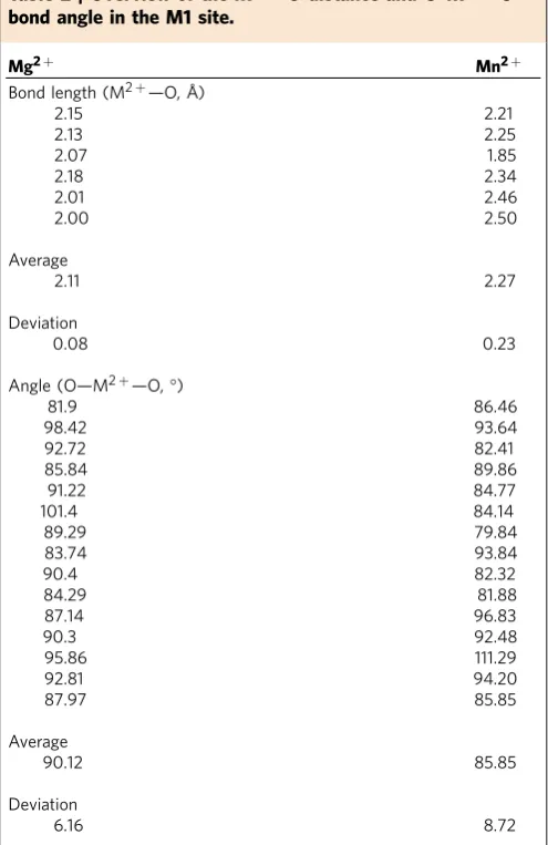
Structure of the selectivity filter. The Mg2þ-bound structure revealed a strong electron density peak surrounded by six weaker satellite peaks in the M1 site (Fig. 1c and Supplementary Fig. 1). Given that the Mg2þ ion adopts an octahedral geometry coor-dinating six water molecules20, the present crystal structure indicates that the fully hydrated Mg2þ ion is bound to the M1 site. The distances between the Mg2þ ion and the coordinated water molecules are 1.97–2.20 Å (Table 2) and are comparable to the typical Mg-O distance (2.1 Å) in an aqueous environment20. This assignment is also consistent with the fact that the crystallization conditions contained 100 mM Mg2þ. The four carboxylate oxygen atoms of Asp432 hydrogen bond with four of the six water molecules in the first hydration shell, thereby anchoring the fully hydrated Mg2þ ion to the M1 site (Fig. 1c). Besides the first hydration shell, the water molecules in the second hydration shell are also recognized in the M1 site. In addition to the carboxyl oxygen atoms of Asp432, the backbone carbonyl oxygen atoms of Pro321 and Leu324 in TM2, and Ala428 in TM5 are within hydrogen-bonding distance of the water molecules in the second hydration shell (Fig. 1c,d). The Gly and Pro residues clustered in the centre of TM2 would confer flexibility to the helix (Supplementary Fig. 2), which might allow the backbone carbonyl groups in the a-helix region to form bifurcated hydrogen bonds with both the water molecules and backbone amide groups. These structural elements involved in the Mg2þ recognition are also conserved in the human SLC41 transporters (Supplementary Fig. 2), suggesting that they also recognize divalent cations in a similar manner.
In the Ca2þ-bound structure, a strong density peak was observed in the M1 site (Fig. 2b), which probably arises from the Ca2þ ion contained in the crystallization conditions. The distances between the centre of the density peak and the carboxylate oxygen atoms of Asp432 are B4 Å, suggesting that the Ca2þ ion is bound to this site in a fully hydrated state, as in the cases of the Mg2þand Mn2þions. We observed broad peaks around Ca2þin themFo–DFcdensity map (Fig. 2b); however, we
could not model water molecules into these density peaks, because of the medium resolution. The coordination geometry of the Ca2þ ion is dynamic and exhibits a broad range of coordination numbers, that is, six to eight water molecules, in an aqueous environment21–23. This is quite different from those of the Mg2þ and Mn2þ ions, which assume rather stable first hydration shells with six water molecules. This difference may result in an unstable interaction of Ca2þ with the M1 site, and thus explain the lower resolution of the Ca2þ-bound structure. Overall, these structures suggested that the M1 site specifically recognizes divalent cations that can assume an octahedral geometry, such as Mg2þ and Mn2þ.
Structure of the periplasmic metal-binding sites. In the crystal structure of the Mn2þ-bound form, we observed three strong peaks around the periplasmic side of MgtE-TMD in the electron density map (M2, M20and M3 sites, Fig. 2c). We also observed strong peaks at the M2 and M3 sites in the anomalous difference Fourier map (4.8 and 4.0s, respectively; Supplementary Fig. 3b,e). These observations suggested that the Mn2þ ions are bound to these periplasmic sites.
The M2 and M20 sites are located on the surface of the
periplasmic side and are related by the twofold non-crystal-lographic symmetry (NCS) axis between the MgtE-TMD
protomers (Fig. 2c). The M2 site is not involved in any crystal-packing interactions, while the Mn2þ ion in the M20 site forms interactions with an adjacent molecule in
the crystalline lattice (Fig. 2c). Consequently, the ligand atoms of the M2 site are different from those of the M20site. The M2 site consists of the side chains of Gln304 and Glu307 in one protomer and His383 in another protomer (Supplementary Fig. 3b). The Mn2þ ion bound to the M2 site is directly coordinated by the imidazole nitrogen of His383 and the side-chain carbonyl oxygen of Gln304. The Mn2þ ion is further surrounded by four satellite peaks, which probably arise from the coordinated water molecules (Supplementary Fig. 3b). In contrast, the M20 site
consists of the side chains of Glu307 and Glu311, as well as the side chain of Asp348 and main chains of Arg345 and Asp346 in the symmetry-related molecule (Fig. 2c). The M3 site is located at the periplasmic entrance of the pore and is situated nearly on the twofold NCS axis. The Mn2þ ion of this site is directly coordinated by the side-chain carboxylate group of Glu311 in one protomer and the main-chain carbonyl group of Glu311 of another protomer (Supplementary Fig. 3e). The M3 site is involved in crystal-packing interactions: the Mn2þ ion is coordinated by the side-chain carboxylate group of Glu275 in the symmetry-related molecule (Supplementary Fig. 3e).
In the crystal structure of the Ca2þ-bound form, we observed a strong density peak only in the M3 site (2.9s, Supplementary Fig. 2f), which may arise from the Ca2þ ion. The Ca2þ ion is directly coordinated by the side-chain carboxylate groups of Glu275 in the symmetry-related molecule and Glu311 (Supplementary Fig. 3f); however, the coordination manner is slightly different from that in the Mn2þ-bound form (Supplementary Fig. 3e). By contrast, no strong density peaks were observed in the M2, M20and M3 sites in the Mg2þ-bound
structure (Supplementary Fig. 3a,d). Therefore, these observations
TM2 TM2
TM2
TM5 TM5
TM5 TM5 TM5
M1 site
Asp432 Asp432
Mg2+
Mg2+ M1 site
M1 site
M1 site
M2 site M3 site
Mg2+ M1 site
Out
In
CBS domain TM domain
N domain
Plug helix
Pro321
Leu324
Pro321
Leu324
TM1
L1 L1
In
Out TM5
TM2
a c
d b
TM4 TM4
TM3 L1 L1 TM5
TM4 TM4
TM2
TM5
TM2
Pro321
Leu324
Pro321
Leu324
TM2 TM2
90° 90°
Ala428 Ala428
Asp432 Asp432
Mg2+
TM5 TM5
Ala428 Ala428
TM1
TM2 TM3
TM1
[image:3.595.91.502.51.306.2]TM2
suggested that these periplasmic cation-binding sites can discriminate divalent cations by directly coordinating them: the M3 site can bind both Mn2þand Ca2þ, while the M2 (and M20)
site can bind Mn2þ, but not Ca2þ.
Mg2þ transport activity of MgtE and its mutants. To investi-gate the role of the metal-binding sites in the TM domain, we analysed the ion-transport activity of wild-type (WT) MgtE and its mutants in vitro. We quantified the amounts of divalent cations accumulated within proteoliposomes reconstituted with MgtE or MgtE mutants, using the divalent cation-specific fluor-escent indicator KMG-20. Although KMG-20 was developed as a Mg2þindicator24, it is also useful for the quantification of Ca2þ and Mn2þ (Supplementary Fig. 4a,b).
At first, we measured the Mg2þtransport activity of WT MgtE and the M1 site mutants (D432A and D432N). The results showed that the D432A and D432N mutants have no activity, as compared with the WT MgtE (Fig. 3a), consistent with the previous patch-clamp analysis results16. Next, we measured the activity of the M2 and M3 site mutants. In the M2A and M3A mutants, the residues involved in the cation binding (that is, Gln304, Glu307 and His383 for M2 and Glu311 for M3) are replaced by Ala, while all of these residues are replaced by Ala in the M2M3A mutant. The results showed that all of the M2A, M3A and M2M3A mutants possess comparable Mg2þ transport activity to that of WT MgtE, suggesting that the M2 and M3 sites do not affect the Mg2þ transport mechanism (Fig. 3b and
Supplementary Fig. 5). This is consistent with the crystal structure, in which Mg2þ binding was disrupted at the M2 and M3 sites (Supplementary Fig. 3a,d). Therefore, these results indicated that the M1 site, but not the M2 and M3 sites, is directly involved in the Mg2þ transport mechanism.
Cation selectivity of MgtE and its mutants. To explore the ion selectivity of MgtE, we examined its Mn2þ and Ca2þ transport activities, using the liposome-based assay. WT MgtE exhibited almost no Mn2þ transport activity, whereas the M2A and M3A mutants exhibited a 3.2-fold increase in Mn2þ permeability, as compared with that of WT MgtE (Fig. 3c). The M2M3A mutant exhibited as much as a 5.2-fold increase over that of the WT MgtE (Fig. 3c). However, it should be noted that the Mn2þ transport activity of the M2M3A mutant is stillB20% of that of Mg2þ (Fig. 3b,c). The M2 and M3 sites may partly contribute to the low Mn2þ transport by MgtE, and thus their disruption resulted in the increased permeability for Mn2þ. It should be noted here that the disruption of the M3 site affected the Mn2þ transport activity (Fig. 3c). This suggested that the M3 site also interacts with Mn2þ in the lipid bilayer environment, although the Mn2þ ion in the M3 site is coordinated by the Asp275 side chain in the symmetry-related molecule in the crystal (Fig. 2c).
As for the Ca2þ ion, the M3A and M2M3A mutants showed
[image:4.595.45.549.71.428.2]B1.3-fold higher Ca2þ transport activity than those of the WT and the M2A mutant (Fig. 3d). These results suggested that the M3 site, but not the M2 site, has a slight effect on the Ca2þ
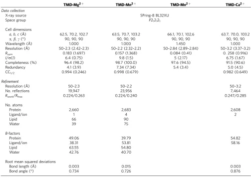
Table 1 | Data collection and refinement statistics.
TMD-Mg2þ TMD-Mn2þ TMD-Mn2þ TMD-Ca2þ
Data collection
X-ray source SPring-8 BL32XU
Space group P212121
Cell dimensions
a,b,c(Å) 62.5, 70.2, 102.7 63.5, 70.7, 103.2 66.1, 70.1, 102,6 63.7, 70.0, 103.2
a,b,g(°) 90, 90, 90 90, 90, 90 90, 90, 90 90, 90, 90
Wavelength (Å) 1.000 1.000 1.450 1.000
Resolution (Å) 50–2.3 (2.42–2.3) 50–2.2 (2.32–2.2) 50–2.84 (2.89–2.84) 50–3.2 (3.37–3.2)
Rsym 0.183 (1.697) 0.157 (1.368) 0.084 (0.41) 0. 258 (0.916)
I/s(I) 6.4 (0.75) 9.8 (1.5) 5 (2.17) 6.75 (1.67)
Completeness (%) 96.4 (98.2) 98.7 (100.0) 97.6 (94.5) 91.5 (90.6)
Redundancy 4.1 (3.9) 7.34 (7.34) 5.4 (3.4) 5.0 (4.5)
CC1/2 0.994 (0.246) 0.998 (0.679) 0.982 (0.649)
Refinement
Resolution (Å) 50–2.3 50–2.2 50–3.2
No. reflections 19,947 23,956 7,464
Rwork/Rfree 0.224/0.263 0.224/0.240 0.247/0.285
No. atoms
Protein 2,660 2,683 2,608
Ligand/ion 1 4 2
Lipid 66 90
Water 39 75
B-factors
Protein 49.06 39.79 54.82
Ligand/ion 38.31 53.81 58.16
Lipid 63.55 54.80
Water 42.76 40.70
Root mean squared deviations
Bond length (Å) 0.003 0.015 0.003
Bond angle (°) 0.734 0.726 0.876
transport activity. This is consistent with the observation in the crystal structure, in which Ca2þ is bound to the M3 site (Supplementary Fig. 3h). Furthermore, we showed that both the WT and M2A mutant have almost no transport activity for Naþ and Kþ, using Sodium Green and PBFI, respectively (Supplementary Fig. 6a,b). These results indicated that the M1 site strictly discriminates the monovalent and divalent cations. Taken together, the M1 site is mainly involved in the higher permeability of Mg2þ over Mn2þ and Ca2þ by MgtE, which is further enhanced by the periplasmic cation-binding sites.
Competitive inhibition of Mg2þ transport activity by cations. Next, to investigate the ion-selectivity mechanism by the M2 and M3 sites, we further examined the effects of Mn2þ, Ca2þ, Naþ and Kþon the Mg2þtransport activity. The results revealed that Mn2þ has a strong inhibitory effect on the transport activity of WT MgtE: the Mg2þ transport activity decreased toB33% in the presence of 1mM Mn2þ (Fig. 4a). A similar result was obtained with a fluorescent microscopy-based method, using MgtE-expressing giant spheroplasts (Supplementary Fig. 7). This inhibitory effect is reduced in the M2A, M3A and M2M3A mutants, while a weak inhibitory effect still remains in the M2M3A mutant (Fig. 4a). These results suggested that the binding of Mn2þ to the M2 and M3 sites strongly inhibits the Mg2þ transport activity of MgtE.
In contrast, Ca2þ has only a small effect on the Mg2þ transport activity of WT MgtE, as compared with Mn2þ (Fig. 4b). Moreover, similar results were obtained with all of the M2A, M3A and M2M3A mutants. Thus, these results suggested that the presence of Ca2þ has almost no inhibitory effect on the Mg2þ transport activity, and that Ca2þ binding to the M3 sites does not inhibit the Mg2þ transport activity.
In the competition assay using Naþ and Kþ, the Mg2þ transport activity was not affected by Naþ and Kþ, even at concentrations over 1 M (Fig. 4c). These monovalent cations may have much lower affinity than Mg2þ to the M1 site, and thus are not transported by MgtE. These observations indicated that the periplasmic cation-binding sites are mainly involved in the selectivity of divalent cations.
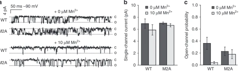
Gating mechanism by the periplasmic cation-binding site. Given that the periplasmic cation-binding sites are formed across the dimer interface (Fig. 2c), the binding of divalent cations can fix the arrangement of the MgtE protomers in the closed form, which may prevent the ion conduction. To assess this hypothesis, we performed the single-channel analysis using the patch-clamp technique. The results showed that the presence of Mn2þ on the periplasmic side significantly decreases the open-channel probability of the WT MgtE channel, while such a change in the open-channel probability was not observed in the M2A mutant (Fig. 5). Thus, the low permeability of MgtE in the presence of Mn2þ (Fig. 4a) is mainly ascribed to the change in the open probability of the channel. The Mn2þbinding to the M2 site may stabilize the closed form of MgtE, thereby preventing the opening of the cation-conducting pore.
Periplasmic cation-binding sites of MgtEs from other species. The MgtEs from several species also have acidic and/or hydro-philic residues around the M2 and M3 sites, as suggested from the amino-acid sequence alignment and the homology modelling (Supplementary Figs 2 and 8a). Thus, it is likely to be that the acidic and/or hydrophilic residues of these MgtEs also form cation-binding sites and thereby function as periplasmic gating sites. To test this hypothesis, we analysed the Mg2þ and Mn2þ transport activities of the WT MgtE and its mutants (M2A and M3A mutants) from Methanosarcina mazei (MmMgtE), which seems to have structurally conserved M2 and M3 sites (Supplementary Figs 2 and 8a). In the M2A and M3A mutants, the acidic and/or hydrophilic residues around the M2 and M3 sites (that is, Glu300 and Glu378 for M2 and Gln304 for M3) are replaced by Ala, while all of these residues are replaced by Ala in the M2M3A mutant. The results showed that the WT MmMgtE has similar Mg2þ transport activity as that of T. thermophilus
[image:5.595.44.292.61.443.2]MgtE (TtMgtE), while it has almost no Mn2þ transport activity (Supplementary Fig. 8b,c). Furthermore, the M2A and M3A mutants of MmMgtE showed about two- and threefold higher Mn2þ transport activities than that of the WT, respectively, as observed in the case of TtMgtE (Supplementary Fig. 8b,c). Thus, these results support our proposal that the M2 and M3 sites of MmMgtE also function as periplasmic gating sites. Next, to further verify our hypothesis, we analysed the effects of Mn2þon the Mg2þ transport activity of MmMgtE and its mutants. The results showed that the Mg2þ transport activity of the WT MmMgtE decreased to B42% in the presence of 1mM Mn2þ (Supplementary Fig. 8d), and this inhibitory effect was reduced in the M2A and M3A mutants (Supplementary Fig. 8d). Therefore, these observations strongly support our proposal that Mn2þ binding to the M2 and M3 sites inhibits the Mg2þ transport activity of MmMgtE, as observed in the case of TtMgtE. Taken together, the M2 and M3 sites are structurally and functionally
Table 2 | Overview of the M2þ–O distance and O–M2þ–O bond angle in the M1 site.
Mg2þ Mn2þ
Bond length (M2þ—O, Å)
2.15 2.21
2.13 2.25
2.07 1.85
2.18 2.34
2.01 2.46
2.00 2.50
Average
2.11 2.27
Deviation
0.08 0.23
Angle (O—M2þ—O,°)
81.9 86.46
98.42 93.64
92.72 82.41
85.84 89.86
91.22 84.77
101.4 84.14
89.29 79.84
83.74 93.84
90.4 82.32
84.29 81.88
87.14 96.83
90.3 92.48
95.86 111.29
92.81 94.20
87.97 85.85
Average
90.12 85.85
Deviation
conserved among the MgtEs, while they are not strictly conserved at the amino-acid sequence level.
Discussion
In this work, we determined the high-resolution crystal structures of MgtE-TMD bound to Mg2þ, Mn2þ and Ca2þ, and performed biochemical analyses based on the structures. The crystal structure of the Mg2þ-bound form revealed that the M1 site recognizes the Mg2þ ion in a fully hydrated state. The structure suggested that the M1 site, including the carboxylate
groups of the Asp432 side chains, strictly recognizes the size and geometry of the Mg2þ hydration shells, which may be important for the selective transport of Mg2þ over other cations, such as Naþ, Kþ and Ca2þ.
The present crystal structure of MgtE-TMD is essentially the same as that of the full-length MgtE in the closed conformation. The hydrophobic gate located just above the M1 site is closed in the structure of MgtE-TMD, whereas the M1 site is accessible from the cytosolic side (Fig. 1b). Thus, in the open-form of MgtE, this hydrophobic gate will be open to permeate ions and Asp432 will become accessible from the extracellular side. Our previous
a
b
c
Asp432 Asp432
Mn2+
M1 site TM5TM5 TM5TM5 M1 site TM5TM5 TM5TM5
Asp432 Asp432
Mn2+
M1 site M1 site
Ca2+
Asp432 Asp432
TM5
TM5 TM5TM5
Ala428 Ala428 Ala428 Ala428
Ala428 Ala428
90° Glu311
Glu311 L1 L1 L1 L1
His383 His383
Gln304 Gln304 Glu307 Glu307 Glu311 Glu311 Asp348
Asp348
Asp346
Asp346 Glu275Glu275 Arg345
Arg345
Mn2+
TM2 TM2
TM2 TM2
TM2 TM2
TM2 TM2 TM3
TM3
TM3 TM3
TM3 TM3 TM3
TM3 Symmetry molecule
L0 L0
L1 L1 L1 L1
His383 His383
Gln304 Gln304 Glu307
Glu307
Glu307 Glu307Glu307Glu307 Glu311
Glu311
Glu311 Glu311
Mn2+
Mn2+
Mn2+
Mn2+
Mn2+
Mn2+
L1 L1 L1 L1 Ca2+
Asp432 Asp432
TM5
TM5 TM5TM5
Ala428 Ala428
Mn2+
M2 site M2’ site
M3 site M2 site
M2’ site
M3 site
TM1 TM1
TM1 TM1 TM1
TM1
TM5 TM5
TM5 TM5
TM5 TM5 TM4
TM4
TM4 TM4
TM4 TM4 Mn2+
Mn2+
Mn2+
Glu307 Glu307
[image:6.595.112.479.46.501.2]Mn2+
Figure 2 | Mn2þ and Ca2þbinding sites in MgtE-TMD.(a) Stereo view of TM5 in the M1 site of the Mn2þ-bound structure. The anomalous difference Fourier map derived from the Mn-peak data set contoured at 5.0sand the 2mFoDFcelectron density map contoured at 3.5sare shown in magenta and green, respectively. Water molecules and Mn2þions are represented by red and purple spheres, respectively. (b) Stereo view of TM5 in the M1 site of the Ca2þ-bound structure, with the 2mFoDFcandmFo–DFcelectron density maps contoured at 2.5s(green) and 3.0s(blue), respectively. Ca2þ ions are represented by green spheres. (c) Mn2þ binding to the M2, M20and M3 sites. Water molecules and Mn2þ ions are represented by red and purple
patch-clamp analysis results demonstrated the high Mg2þ conductivity of MgtE (96 pS), which is similar to that of the prokaryotic potassium channel KcsA (20B100 pS)16,25,26. Therefore, the opening of the hydrophobic gate may be large enough to accommodate the fully hydrated Mg2þ ion. Here we hypothesized that the structure of the selectivity filter (that is, the M1 site) in the present structure is also conserved in this open-form MgtE, as in the cases of other channels, including KcsA27 (Fig. 6a). Based on this hypothesis, the present crystal structure excellently explains the cation selectivities of the M1 site of MgtE for the biological cations, Naþ, Kþ, Mg2þ and Ca2þ, observed
in the present biochemical analysis using the M2M3A mutant. The size of the fully hydrated Mg2þ ion (B6 Å) is slightly larger than the constriction between the Asp432 residues (B5 Å) in the MgtE pore. The electrostatic interaction between Mg2þ and the
2 charge of Asp432 in the M1 site is strong enough to decrease the steric barrier of this constriction, thereby enabling the high Mg2þ conductivity. The hydrogen bonds between the M1 site and the water molecules in the first- and second-hydration shells may further reduce this steric barrier (Fig. 6a). In contrast, the radius of the first hydration shell of the Ca2þ ion is larger than that of Mg2þ, which may prevent its efficient transport. In
c b
a
0 0.2 0.4 0.6 0.8 1.0 1.2 1.4
Normalized cation uptake
Normalized cation uptake
0 0.2 0.4 0.6 0.8 1.0 1.2 1.4
WT
M3A M2A
M2M3A
WT
M3A M2A
M2M3A
10–6 10–3 1
CaCl2 (mM) Monocation (mM)
10–6 10–3 1
MnCl2 (mM)
[Mg2+]
out = 25 mM
[Mg2+]
out = 25 mM Normalized cation uptake [Mg2+]out = 25 mM
M2A + NaCl M2A + KCl WT + NaCl WT + KCl
0 0.2 0.4 0.6 0.8 1.0 1.2
[image:7.595.57.547.50.192.2]0 1 10 100 1,000
Figure 4 | Competition assay with Mn2þ, Ca2þ, Naþ and Kþ.(a,b) Inhibition of the transport activity of WT MgtE and its mutants in the presence of 25 mM MgCl2and the indicated concentrations of Mn2þ(a) and Ca2þ (b). (c) Competition assay with Naþ and Kþof WT MgtE and the M2A mutant in the presence of 25 mM MgCl2and the indicated concentrations of monocations. All data points are mean±s.e.m. (n¼3).
0 2 4 6 8 10
0.0 0.2 0.4 0.6 0.8 1.0
Open-channel probability
50 ms –90 mV
WT
M2A WT
M2A
10 µM Mn2+
0 µM Mn2+ 10 µM Mn2+
0 µM Mn2+
a b c
+ 10 µM Mn2+
+ 0 µM Mn2+
c o
c o
c o
c o
Single-channel current (pA)
WT WT
4 pA
M2A M2A
Figure 5 | Single-channel analysis of WT MgtE and the M2A mutant by the patch-clamp technique.(a) Current traces of WT MgtE and the M2A mutant in the inside-out configuration at –90 mV, in the presence and absence of 10mM Mn2þ on the periplasmic side. C and O indicate the closed and open states of MgtE, respectively. (b,c) Single-channel current (b) and open-channel probability (c) of WT MgtE and the M2A mutant in the presence and absence of 10mM Mn2þ on the periplasmic side.
[Mg2+]out = 100 mM
Mg
2+
uptake
(
µ
mol per mg
protein per min)
Mg
2+
uptake
(
µ
mol per mg
protein per min)
Mn
2+
uptake
(
µ
mol per mg
protein per min)
Ca
2+
uptake
(
µ
mol per mg
protein per min)
WT
No protein
D432A D432N
a b c d
No protein
WT M2A M3A M2M3A
No protein
WT M2A M3A M2M3A
No protein
WT M2A M3A M2M3A 0
5 10 15 20
0 1 2 3 4
0 1 2 3 4
[image:7.595.53.547.253.386.2]0 5 10 15 20
[image:7.595.102.497.450.567.2]addition, the formation of specific hydrogen bonds between the M1 site and the water molecules coordinated to Ca2þ is geometrically impossible, which may also increase the barrier for Ca2þto pass through the M1 site. Furthermore, in the case of the monovalent cations, the electrostatic interaction between the monovalent cations and the –2 charge of Asp432 in the M1 site is weaker than that of the divalent cations, which may prevent the interaction of the monovalent cations with the M1 site.
The present results also suggested that the M3 site is involved in the low Ca2þ permeability: the M3A mutant exhibited higher Ca2þ transport activity than that of the WT MgtE (Fig. 3d). However, in the competition assay, Ca2þ had almost no inhibitory effect on the Mg2þ transport activities of both the WT MgtE and its mutants (Fig. 4b). This result suggested that unlike Mn2þ, Ca2þ does not strongly bind to the TM pore of MgtE to fix the conformation of MgtE in the closed state (Fig. 6b). The M3 site may contribute to the low Ca2þ permeability, transiently trapping Ca2þ at the entrance of the cation-conducting pore of MgtE (Fig. 6b).
Numerous crystal structures of ion channels have revealed the structural bases for their ion selectivity mechanisms. Particularly, the atomic-resolution crystal structures and functional analyses of the voltage-gated Kþ channel superfamily, including KcsA3,4, MthK5 and NaK28, provided insights into the ion selectivity mechanism of Kþ: the backbone carbonyl oxygen atoms recognize the cations in the dehydrated forms. In striking contrast, the present crystal structure revealed that the fully hydrated Mg2þ ion is recognized by the Asp432 side chains in the M1 site of MgtE, which is quite different from the cases of the monovalent cations. The proper arrangement of the two carboxylate groups in the M1 site may strictly recognize the specific hydration-shell structure of the Mg2þ ion, thereby enabling the selective transport of Mg2þ over other biological cations. This MgtE mechanism seems to be reasonable for the fast and selective conduction of Mg2þ, as the energy required for the dehydration of divalent cations is four to five times higher than that of monovalent cations29. Moreover, it was proposed that Mg2þchannel CorA also transport Mg2þion in a fully hydrated form, based on its inhibition by the Co2þ hexammin compound8. Thus, selectivity based on the hydrated Mg2þ may be a common feature of Mg2þ channels.
Transition metal cations, such as Fe2þ, Co2þ, Ni2þ and Mn2þ, are important as essential trace elements, but over-exposure to them is toxic for living organisms. The present crystal structure revealed that Mn2þbound to the M2 and M3 sites. The direct interaction between the cation and the protein ligand atoms, including the carbonyl oxygens of Glu307 and Gln304, and the imidazole nitrogen atoms of His383, may enable the discrimination between Mn2þ and Mg2þ. The functional analysis revealed that Mn2þ binding to the M2 and M3 sites strongly inhibits the cation transport activity of MgtE. The patch-clamp analysis further showed that the presence of Mn2þ on the periplasmic side decreases the open probability of the channel (Fig. 5). Therefore, the binding of transient-metal cations, including Mn2þ, to these periplasmic gating sites (that is, M2 and M3 sites) may fix the arrangement of the MgtE protomers in the closed form, thereby preventing the excess uptake of these toxic cations (Fig. 6c). The present work further suggested the
Closed state Open state
Closed state Open state
Mg2+
In Out
Ca2+
D432 D432 D432 D432
E311 E311 E311 E311
Mn2+ a
c b
D432 D432 D432 D432
M2 site M3 site
Ion-conduction pore M2’ site
M1 site
In Out
M1 site M3 site M3 site
Closed state Open state
E311 E311 E311 E311
TMD
TMD
TMD N domain
CBS domain
Plug helix
Hydrophobic gate
[image:8.595.46.290.276.743.2]Hydrophobic gate
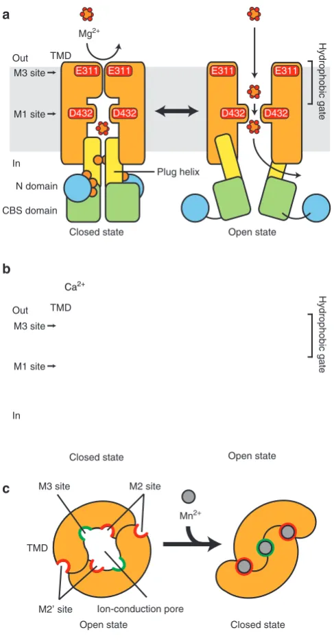
Figure 6 | Proposed model for the ion-conduction mechanism of MgtE. (a) Model for the Mg2þtransport mechanism. A schematic representation
of MgtE, viewed from the plane in the membrane, is shown. The Mg2þ binding to the cytosolic domain stabilizes the MgtE in the closed state (left panel), while the M2 and M3 sites do not bind to Mg2þ, and thus the
periplasmic Mg2þdoes not affect the gating of MgtE. After the dissociation
of Mg2þfrom the cytosolic domain, MgtE adopts the open form (right panel). In the open form, the electrostatic interaction between Asp432 and Mg2þ, and the hydrogen bonds between Asp432 and the Mg2þhydration
shells enable the precise recognition and high-speed conduction of Mg2þ. (b) Model for the Ca2þconduction mechanism. Solid and dashed lines represent the major and minor ion pathways for Ca2þ, respectively.
Although periplasmic Ca2þ can interact with the M3 site, its weak affinity does not stabilize MgtE in the closed form (left panel). In the open form (right panel), Ca2þ cannot pass through the selectivity filter (that is,
Asp432) efficiently, because the interaction between Asp432 and its hydration shell is not optimal. The interaction between the M3 site and Ca2þalso contributes to the slowing of its transport rate. The major
portion of Ca2þ diffuses back to the periplasmic side through the
hydrophobic gate, while a small portion overcomes the barrier in the selectivity filter, which results in the weak Ca2þ transport activity of MgtE. (c) Gating model of the ion-conducting pore by the periplasmic gating sites. A schematic representation of MgtE, viewed from the periplasmic side, is shown. Periplasmic Mn2þ binds to the M2, M20and M3 sites with high
presence of these periplasmic gating sites in MgtEs from other species, including M. mazei(Supplementary Fig. 8).
In addition, we found that the M2M3A mutant, which disrupts all of the periplasmic gating sites, still preferably transports Mg2þ over Mn2þ (Fig. 3b,c). The results of the competitive inhibition analysis further showed that Mn2þ has an inhibitory effect on the cation transport activity of the M2M3A mutant (Fig. 4a). These results suggested that the M1 site has higher affinity for Mn2þ than Mg2þ. The higher affinity may trap Mn2þ in the M1 site, which may result in the decreased Mg2þpermeability in the presence of Mn2þ, as well as the Mn2þ conductance itself. However, it seems to be difficult to explain this difference in the affinity from only the present crystal structure: both Mg2þ and Mn2þ bind to the M1 site in similar manners, in which the Asp432 carboxylate groups indirectly recognize these cations through the water molecules in their first hydration shell (Figs 1c and 2a). Thus, the question arises as to what causes this difference in the affinity of the M1 site for the fully hydrated Mg2þ and Mn2þ ions. Previous quantum chemical calculations of Mg2þ and Mn2þ in complex with water molecules revealed that the hydration geometry of the Mn2þ ion deviates from the ideal octahedral arrangement, due to the excessive polarization and charge-transfer effects of Mn2þ with open d orbitals30,31. This difference between Mg2þ and Mn2þ was suggested to affect the substrate specificity of restriction enzymes32. We expect that this difference in the electronic states and polarization would also contribute to the difference in the affinities of the M1 site for Mg2þ and Mn2þ. Further understanding of the discrimination mechanism of Mg2þ and transition metal cations will require structural and computational analyses, including ab initio
quantum chemical calculations. Overall, our study provides structural insights into the specific recognition of Mg2þ over other cations by the MgtE Mg2þ channel, to achieve fast and selective ion permeation in combination with the selectivity filter, M1, and the periplasmic gating sites, M2 and M3.
Methods
Expression and purification.Full-lengthT. thermophilusMgtE (TtMgtE) was subcloned into a pET-modified vector, including an amino-terminal His6tag16,17. The tobacco etch virus (TEV) protease cleavage site was inserted into the L0 loop, which connects the cytosolic and TM domains of TtMgtE, by PCR with the primers 50-ATTTTCAAGGAGTGCCCGACCTCGTCTACAGCGAGG-30and 50-TAT TTAAAAGCAGGTGCCGCGGCTCGAACACCTAC-30. The protein was overexpressed inEscherichia coliC41 (DE3) cells, induced with 0.5 mM isopropyl D-thiogalactoside for 18 h at 20°C. The cells were disrupted and the membrane fraction was collected by ultracentrifugation. This fraction was solubilized by 1.5% (w/v) n-dodecyl-beta-D-maltopyranoside (DDM) in buffer A (50 mM HEPES (pH 7.0), 300 mM NaCl and 20 mM imidazole). The solubilized fraction was applied to a Ni-NTA column equilibrated with buffer A containing 0.1% (w/v) DDM and mixed for 30 min. The column was washed with buffer A containing 0.1% (w/v) DDM and 50 mM imidazole, and TtMgtE was eluted in the same buffer containing 250 mM imidazole. To cleave the TEV protease cleavage site, the purified MgtE was treated with His6-tagged TEV protease and dialysed against buffer B (25 mM HEPES (pH 7.0), 150 mM NaCl, 0.1% (w/v) DDM). The sample was reloaded on a Ni-NTA column equilibrated with buffer B, to remove the cytosolic domain and the TEV protease. The flow-through fraction containing the TM domain of MgtE (MgtE-TMD) was collected and further purified by chromatography on a Superdex200 10/300 column in buffer B. The purified MgtE-TMD was concentrated toB10 mg ml1, using an Amicon Ultra 50K filter.
Crystallization.For the lipidic cubic phase (LCP) crystallization trials19, we used monoolein (Sigma) or 7.7MAG33as the host lipid. MgtE-TMD was mixed with the host lipid in a 2:3 protein to lipid ratio (w/w), using the twin-syringe mixing method. Aliquots (100 nl) of the protein–LCP mixture were spotted on a 96-well sandwich plate and overlaid by 800 nl of precipitation solution by the crystallization robot, mosquito LCP (TTP LabTech). Crystals for data collection were grown at 20°C in monoolein, with a reservoir containing 30–35% (w/v) PEG600, 100 mM Tris (pH 8.0), 120 mM Mg-formate and 100 mM NaCl. For crystallization in the presence of Mn2þand Ca2þ, the purified protein solution was diluted to a DDM concentration of 0.05% and was concentrated to
B10 mg ml–1. Crystals were obtained as described above, in a reservoir containing 30–32% (w/v) PEG300, 100 mM HEPES-NaOH (pH 7.5) and 80–120 mM MnCl2,
and a reservoir containing 20–28% (w/v) PEG400, 100 mM HEPES (pH 7.5), 120–160 mM CaCl2and 100 mM NaSCN.
Data collection and structure determination.All diffraction data sets were collected at the SPring-8 BL32XU (Hyogo, Japan). The data sets were processed with the programmes HKL2000 (HKL Research, Inc.) and XDS34. Phases were obtained by molecular replacement with the programme MOLREP35, using the TM domain of the previous MgtE coordinates (PDB 2ZY9) as a search model. The model was subsequently improved through iterative cycles of manual building with COOT and refinement with the programme PHENIX36. The structural refinement statistics are summarized in Table 1. Molecular graphics were illustrated with CueMol (http://www.cuemol.org/).
Divalent cation uptake assay.The divalent cation uptake assay was performed as previously described37, with some modifications. Purified WT and mutant TtMgtE proteins were reconstituted into liposomes at a 50:1E. colipolar lipid to protein ratio (w/w), by the freeze–thaw method. The proteoliposomes were loaded with buffer C (25 mM HEPES (pH 7.0), 100 mM NaCl) and the reaction was initiated by adding MgCl2. After an incubation for 20 min at room temperature, 2 mM Co(III)hexamine was added to stop the cation uptake through the MgtE protein16. The reaction mixture was centrifuged at 210,000gfor 10 min. The pellet was washed three times in buffer C containing 2 mM Co(III)hexamine and was resuspended in buffer C containing 0.5% SDS. The suspension was mixed with 10mM divalent cation indicator, KMG-20 (ref. 24) (Wako), and incubated for 2 h at 37°C. The KMG-20 fluorescence was excited at 440 nm and detected at 500–530 nm, using an F-7000 fluorescence spectrophotometer (Hitachi). Divalent cation uptake was calculated from a Benesi–Hildebrand plot24, obtained from the known concentration of each indicated divalent cation in buffer C containing 0.5% SDS. NaCl and KCl have no effect on the fluorescence intensity of KMG-20 (ref. 24). In the previous report16, divalent cation binding to the cytosolic domain strongly stabilized the closed state of the ion-conducting pore at concentrations over 10 mM. Thus, in this experiment, the MgtE with the cytosolic domain facing towards the extraliposomal side does not contribute to the Mg2þtransport
activity, because we measured the activity under conditions with an extraliposomal Mg2þconcentration higher than 10 mM.
Naþand Kþuptake assays.The Naþand Kþuptake assays were performed as previously described38–40. Briefly,E. colipolar lipids and ergosterol, at a 50:1 ratio (w/w), were suspended in buffer D (25 mM Tris (pH 7.5), 100 mM n-methyl-D-glucamin) with 10mM Sodium Green and 60mM PBFI, respectively, and sonicated with a large tip for 30 s. The liposome solution was centrifuged at 210,000gfor 10 min and the pellet was resuspended in buffer D. The purified WT MgtE, the M2A mutant and the Naþionophore, a nystatin–ergosterol complex (10 nmol)41, were reconstituted into liposomes by the freeze–thaw method. The liposomes were centrifuged at 210,000gfor 10 min and the pellet was resuspended in buffer D. The Naþ and Kþuptake reactions were initiated by the addition of NaCl and KCl, respectively, and were monitored by the fluorescence of Sodium Green (lex/lem¼488/540) and PBFI (lex/lem¼493/515), respectively.
Patch-clamp analysis.E. coligiant spheroplasts were prepared as described previously16. Spheroplasts expressing WT MgtE and the M2A mutant were plated on glass coverslips in a bath medium (250 mM N-methyl-D-glucamine, 40 mM MgCl2, 300 mM glucose and 10 mM HEPES, pH 7.2). Borosilicate pipettes (Harvard Apparatus, Kent, UK), with about 5–8 MOresistance, were filled with a pipette solution (250 mM N-methyl-D-glucamine, 40 mM MgCl2, 300 mM glucose and 10 mM HEPES, pH 7.2). In some experiments, 10mM MnCl2was added to the pipette solution. After the formation of a gigaseal, a membrane patch was excised and the bath solution was exchanged using a perfusion system with a batch solution: 250 mM N-methyl-D-glucamine, 0.2 mM MgCl2, 300 mM glucose and 10 mM HEPES, pH 7.2. The membrane patch voltage was clamped and currents were recorded using an Axopatch 200B amplifier (Axon CNS, Molecular Devices), coupled to an A/D converter (Axon CNS, Molecular Devices) and controlled by the pclamp10 software (Axon CNS, Molecular Devices). Currents were filtered at 2 kHz and sampled at 5 kHz.
References
1. Cowan, J. A. Structural and catalytic chemistry of magnesium-dependent enzymes.Biometals15,225–235 (2002).
2. Hartwig, A. Role of magnesium in genomic stability.Mutat. Res.475,113–121 (2001).
3. Zhou, Y., Morais-Cabral, J. H., Kaufman, A. & MacKinnon, R. Chemistry of ion coordination and hydration revealed by a Kþchannel-Fab complex at 2.0A resolution.Nature414,43–48 (2001).
4. Morais-Cabral, J. H., Zhou, Y. & MacKinnon, R. Energetic optimization of ion conduction rate by the Kþselectivity filter.Nature414,37–42 (2001). 5. Ye, S., Li, Y. & Jiang, Y. Novel insights into Kþselectivity from
high-resolution structures of an open Kþchannel pore.Nat. Struct. Mol. Biol.17, 1019–1023 (2010).
6. Lunin, V. V.et al.Crystal structure of the CorA Mg2þtransporter.Nature
440,833–837 (2006).
7. Eshaghi, S.et al.Crystal structure of a divalent metal ion transporter CorA at 2.9 angstrom resolution.Science313,354–357 (2006).
8. Guskov, A.et al.Structural insights into the mechanisms of Mg2þuptake, transport, and gating by CorA.Proc. Natl Acad. Sci. USA109,18459–18464 (2012).
9. Pfoh, R.et al.Structural asymmetry in the magnesium channel CorA points to sequential allosteric regulation.Proc. Natl Acad. Sci. USA109,18809–18814 (2012). 10. Hou, X., Pedi, L., Diver, M. M. & Long, S. B. Crystal structure of the
calcium release-activated calcium channel Orai.Science338,1308–1313 (2012).
11. Tang, L.et al.Structural basis for Ca2þselectivity of a voltage-gated calcium channel.Nature505,56–61 (2014).
12. Moomaw, A. S. & Maguire, M. E. The unique nature of Mg2þchannel.
Physiology (Bethesda)275–285 (2009).
13. Alexander, R. T., Hoenderop, J. G. & Bindels, R. J. Molecular determinants of magnesium homeostasis: insights from human disease.J. Am. Soc. Nephrol.19, 1451–1458 (2008).
14. Sahni, J. & Scharenberg, A. M. The SLC41 family of MgtE-like magnesium transporters.Mol. Aspects Med.34,620–628 (2013).
15. Kolisek, M.et al.Substitution p.A350V in Naþ/Mg2þexchanger SLC41A1, potentially associated with Parkinson’s disease, is a gain-of-function mutation.
PLoS ONE8,e71096 (2013).
16. Hattori, M.et al.Mg2þ-dependent gating of bacterial MgtE channel underlies Mg2þhomeostasis.EMBO J.28,3602–3612 (2009).
17. Hattori, M., Tanaka, Y., Fukai, S., Ishitani, R. & Nureki, O. Crystal structure of the MgtE Mg2þ transporter.Nature448,1072–1075 (2007).
18. Cherezov, V. Lipidic cubic phase technologies for membrane protein structural studies.Curr. Opin. Struct. Biol.21,559–566 (2011).
19. Caffrey, M. & Cherezov, V. Crystallizing membrane proteins using lipidic mesophases.Nat. Protoc.4,706–731 (2009).
20. Persson, I. Hydrated metal ions in aqueous solution: How regular are their structures?Pure Appl. Chem.82,1901–1917 (2010).
21. Katz, A. K., Glusker, J. P., Beebe, S. A. & Bock, C. W. Calcium ion coordination: a comparison with that of beryllium, magnesium, and zinc.J. Am. Chem. Soc.
118,5752–5763 (1996).
22. Pavlov, M., Siegbahn, P. E. M. & Sandstro¨m, M. Hydration of beryllium, magnesium, calcium, and zinc ions using density functional theory.J. Phys. Chem. A102,219–228 (1998).
23. Jalilehvand, F.et al.Hydration of the calcium ion. An EXAFS, large-angle X-ray scattering, and molecular dynamics simulation study.J. Am. Chem. Soc.123, 431–441 (2001).
24. Suzuki, Y.et al.Design and synthesis of Mg2þ-selective fluoroionophores based on a coumarin derivative and application for Mg2þmeasurement in a living cell.Anal. Chem.74,1423–1428 (2002).
25. Meuser, D., Splitt, H., Wagner, R. & Schrempf, H. Exploring the open pore of the potassium channel fromStreptomyces lividans.FEBS Lett.462,447–452 (1999). 26. Schrempf, H.et al.A prokaryotic potassium ion channel with two predicted
transmembrane segments fromStreptomyces lividans.EMBO J.14,5170–5178 (1995).
27. Cuello, L. G., Jogini, V., Cortes, D. M. & Perozo, E. Structural mechanism of C-type inactivation in Kþchannels.Nature466,203–208 (2010).
28. Alam, A. & Jiang, Y. Structural analysis of ion selectivity in the NaK channel.
Nat. Struct. Mol. Biol.16,35–41 (2009).
29. Yatsimirskii, K. Electronic structure, energy of hydration, and stability of metal aquo ions.Theor. Exp. Chem.30,1–9 (1994).
30. Marino, T., Toscano, M., Russo, N. & Grand, A. Structural and electronic characterization of the complexes obtained by the interaction between bare and hydrated first-row transition-metal ions (Mn2þ, Fe2þ, Co2þ, Ni2þ, Cu2þ, Zn2þ) and glycine.J. Phys. Chem. B110,24666–24673 (2006). 31. Aakesson, R., Pettersson, L. G. M., Sandstroem, M. & Wahlgren, U. Ligand field
effects in the hydrated divalent and trivalent metal ions of the first and second transition periods.J. Am. Chem. Soc.116,8691–8704 (1994).
32. Solt, I., Simon, I., Csa´sza´r, A. G. & Fuxreiter, M. Electrostatic versus nonelectrostatic effects in DNA sequence discrimination by divalent ions Mg2þand Mn2þ.J. Phys. Chem. B111,6272–6279 (2007).
33. Misquitta, L. V.et al.Membrane protein crystallization in lipidic mesophases with tailored bilayers.Structure12,2113–2124 (2004).
34. Kabsch, W. Xds.Acta Crystallogr. D Biol. Crystallogr.66,125–132 (2010). 35. Vagin, A. & Teplyakov, A. MOLREP: an automated program for molecular
replacement.J. Appl. Crystallogr.30,1022–1025 (1997).
36. Adams, P. D.et al.PHENIX: building new software for automated crystallographic structure determination.Acta Crystallogr. D Biol. Crystallogr.
58,1948–1954 (2002).
37. Ishijima, S., Shigemi, Z., Adachi, H., Makinouchi, N. & Sagami, I. Functional reconstitution and characterization of theArabidopsisMg2þtransporter AtMRS2-10 in proteoliposomes.Biochim. Biophys. Acta1818,2202–2208 (2012).
38. Suzuki, T., Ozaki, Y., Sone, N., Feniouk, B. A. & Yoshida, M. The product of uncI gene in F1Fo-ATP synthase operon plays a chaperone-like role to assist c-ring assembly.Proc. Natl Acad. Sci. USA104,20776–20781 (2007). 39. Garlid, K. D., Paucek, P., Yarov-Yarovoy, V., Sun, X. & Schindler, P. A. The
mitochondrial KATP channel as a receptor for potassium channel openers.
J. Biol. Chem.271,8796–8799 (1996).
40. Bajgar, R., Seetharaman, S., Kowaltowski, A. J., Garlid, K. D. & Paucek, P. Identification and properties of a novel intracellular (mitochondrial) ATP-sensitive potassium channel in brain.J. Biol. Chem.276,33369–33374 (2001).
41. Kleinberg, M. E. & Finkelstein, A. Single-length and double-length channels formed by nystatin in lipid bilayer membranes.J. Membr. Biol.80,257–269 (1984).
Acknowledgements
We thank H. Nishimasu (University of Tokyo) for helpful discussions, A. Kurabayashi for technical support, the RIKEN Integrated Cluster of Clusters (RICC) for providing computational resources and the beamline staff members at BL32XU of SPring-8 (Hyogo, Japan) for technical assistance during data collection. The synchrotron radiation experiments were performed at BL32XU of SPring-8, Japan, with approval from RIKEN (Proposal Nos. 2012B1146, 2012B1162, 2013A1128, 2013A1168 and 2013A1128), and at I03 of the Diamond Light Source, UK. This work was supported by the Platform for Drug Discovery, Informatics and Structural Life Science from the Ministry of Education, Culture, Sports, Science and Technology (MEXT), by JSPS KAKENHI (grant numbers 22117007, 24227004, 25291011 and 26711003), by the FIRST programme, by PRESTO, JST, by a Grant-in-Aid for JSPS Fellows, by a grant for the HPCI STRATEGIC PRO-GRAM Computational Life Science and Application in Drug Discovery and Medical Development from MEXT, and by grants from the Private University Strategic Research Foundation Support Program (MEXT).
Author contributions
H.T. expressed and purified MgtE and its mutants for crystallization, collected the dif-fraction data, solved the structures and performed liposome-based assay. K.Y. assisted with the structure refinement. S.T.A.S. and M.C. synthesized lipids for crystallization. A.D.M. performed patch-clamp analysis. M.H. and T.N. contributed to the early stage of the project. H.T., R.I. and O.N. wrote the manuscript. O.N. directed and supervised all of the research.
Additional information
Accession codes:Atomic coordinates and structure factors for theT. therMgtE bound to Mg2þ, Mn2þand Ca2þ have been deposited in the Protein Data Bank, under the
accession codes 4U9L, 4U9N and 4WIB, respectively.
Supplementary Informationaccompanies this paper at http://www.nature.com/ naturecommunications
Competing financial interests:The authors declare no competing financial interest.
Reprints and permissioninformation is available online at http://npg.nature.com/ reprintsandpermissions/
How to cite this article:Takeda, H.et al.Structural basis for ion selectivity revealed by high-resolution crystal structure of Mg2þchannel MgtE.Nat. Commun.5:5374 doi: 10.1038/ncomms6374 (2014).