Series Introduction: Intrinsic and innate
defenses in the lung: intersection of pathways
regulating lung morphogenesis, host defense,
and repair
Jeffrey A. Whitsett
J Clin Invest.
2002;
109(5)
:565-569.
https://doi.org/10.1172/JCI15209
.
This Perspective series seeks to summarize the present concepts regarding the biological
processes that mediate intrinsic and innate host defense against microbial invasion of the
lung. From an evolutionary standpoint, the requirements for an extensive gas exchange
region that comes in direct contact with inhaled particles and pathogens present a
formidable challenge that has been countered by the layering and intersection of several
intrinsic defense systems. These systems, detailed in the accompanying articles, maintain
pulmonary form and function required for ventilation by means of structural, mechanical,
chemical, and cellular strategies, as well as innate and acquired host defenses. The
epithelial barrier itself represents a first line of defense against pathogens, and its
effectiveness is greatly enhanced by fluid homeostasis and mucociliary clearance. As
discussed by Knowles and Boucher in this series, these mechanisms protect the epithelium
and physically remove inhaled pathogens from the lung. Additional protection comes from
polypeptide mediators of the innate host defense, such as the defensins and other antibiotic
peptides reviewed by Ganz in this series, and the collectin family, considered by
McCormack and Whitsett. Professional phagocytes also play their part in host defense in
this tissue, in part by responding to molecules found on the surface of common pathogenic
microbes. Finally, the cytokine and chemokine pathways that cooperate with host defense
polypeptides to mediate initial host […]
Perspective
Find the latest version:
PERSPECTIVE SERIES
Jeffrey A. Whitsett, Series Editor
Innate defenses in the lung
This Perspective series seeks to summarize the present concepts regarding the biological processes that medi-ate intrinsic and innmedi-ate host defense against microbial invasion of the lung. From an evolutionary standpoint, the requirements for an extensive gas exchange region that comes in direct contact with inhaled particles and pathogens present a formidable challenge that has been countered by the layering and intersection of sev-eral intrinsic defense systems.
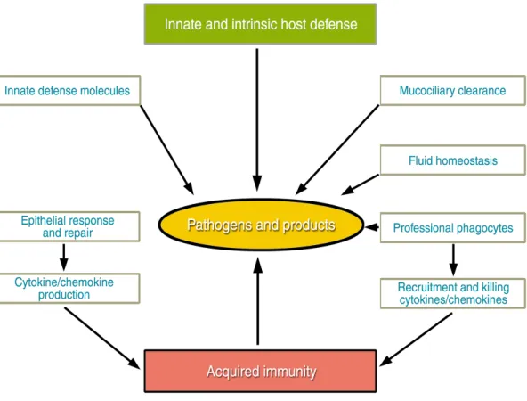
These systems, detailed in the accompanying arti-cles, maintain pulmonary form and function required for ventilation by means of structural, mechanical, chemical, and cellular strategies, as well as innate and acquired host defenses. The epithelial barrier itself represents a first line of defense against pathogens, and its effectiveness is greatly enhanced by fluid homeostasis and mucociliary clearance. As discussed by Knowles and Boucher in this series, these mechanisms protect the
epithe-lium and physically remove inhaled pathogens from the lung. Additional protection comes from polypeptide mediators of the innate host defense, such as the defensins and other antibiotic peptides reviewed by Ganz in this series, and the collectin fami-ly, considered by McCormack and Whitsett. Professional phagocytes also play their part in host defense in this tissue, in part by responding to molecules found on the surface of common pathogenic microbes. Finally, the cytokine and chemokine
pathways that cooperate with host defense polypep-tides to mediate initial host defenses by phagocytes also orchestrate tissue repair and subsequent acquired immune responses following infection, as discussed by Strieter et al. The interplay of these var-ious mechanisms is depicted in Figure 1.
The concerted effects of mechanical, innate, and acquired host defense systems serve to recognize, localize, kill, and remove pathogens to maintain sterility of pulmonary tissues and the host. In gener-al, pathogens are cleared from the lung without per-sistent or robust inflammation, thus protecting its inherent structure and maintaining its function fol-lowing exposure to microbes. It is the failure of vari-ous arms of the host defense systems that allows local or systemic infection and destruction of lung tissue that are frequently the basis of morbidity and mor-tality from common lung diseases, whether related to
SERIES INTRODUCTION
Intrinsic and innate defenses in the lung:
intersection of pathways regulating lung morphogenesis,
host defense, and repair
Jeffrey A. Whitsett
Children’s Hospital Medical Center, Divisions of Neonatology and Pulmonary Biology, 3333 Burnet Avenue, Cincinnati, Ohio 45229-3039, USA.
Phone: (513) 636-4830; Fax: (513) 636-7868; E-mail: [email protected].
[image:2.576.242.538.509.734.2]J. Clin. Invest.109:565–569 (2002). DOI:10.1172/JCI200215209.
Figure 1
genetic or environmental disease processes. Deficient or uncontrolled host defenses and inflammation underlie some of the most common pulmonary dis-orders — chronic obstructive pulmonary disease, cys-tic fibrosis, acute and chronic pneumonia, asthma, emphysema, and bronchopulmonary dysplasia.
Structure and function of the respiratory epithelium: form dictates function
The lung is a complex organ consisting of a series of branching tubules and alveoli that are highly vascu-larized to provide a large gas exchange surface. The respiratory tract is lined by endodermally derived epithelial cells that differentiate from the foregut endoderm. Commitment and proliferation of respi-ratory epithelial cells are dependent upon mesenchy-mal-epithelial interactions, mediated by a number of distinct and intersecting autocrine-paracrine path-ways (e.g., bone morphogenetic protein–4 [BMP-4], FGF, sonic hedgehog [SHH]; see ref. 1 for review), which, in turn, regulate gene transcription to influ-ence cell fate, proliferation, and function. In the lung, commitment and differentiation of the respiratory epithelium are mediated, at least in part, by the homeodomain protein NKx2.1 (also known as thy-roid transcription factor-1 [TTF-1]), as well as tran-scription factors of the forkhead family (hepatocyte nuclear factor-3β[HNF-3β], HNF-3α, and hepato-cyte factor homologue-4 [HFH-4]) and the zinc fin-ger family (GATA-6) (reviewed in refs. 2–4). Lung tubules undergo stereotypic, dichotomous branching to form bronchi, bronchioles, and the peripheral air-ways and alveoli.
Initially, the lung is lined by an undifferentiated columnar epithelium, but as lung morphogenesis proceeds, the tubules are lined by an increasingly diverse population of respiratory epithelial cells that vary both spatially — along the cephalocaudal axis — and temporally. In the adult human lung, conducting airways are lined by a pseudostratified epithelium
consisting of at least a dozen morphologically distinct epithelial cells, including squamous, ciliated, noncil-iated bronchiolar (Clara cells), basal, intermediate, serous, goblet, and neuroepithelial cells, as well as alveolar type II and type I epithelial cells. This diversi-ty of cell diversi-type accomplishes the various physiologic tasks that optimize mucociliary clearance, precise reg-ulation of fluid homeostasis, and the synthesis and secretion of a myriad of such host defense proteins as lysozyme, defensins, surfactant proteins (SPs), and lactoferrin. The abundance and activity of the various cell types, which are strongly influenced by infection and other inflammatory stimuli, can themselves influence host defense function and respiratory cell proliferation and differentiation during repair. In the normal lung, the diverse repertoire of respiratory epithelial cells is maintained in their proper places and activation states, and they interact with profes-sional phagocytes and the lymphoid components of the acquired immune system. In addition, an exten-sive system of tracheal-bronchial glands is lined by distinct epithelial cell types that produce mucous, fluid, and other host defense proteins critical for mucociliary activity that clears particles and pathogens from the lung. The generation and main-tenance of this panoply of epithelial cell types provide structural framework required for host defense func-tions. Acute injury and chronic injury lead to epithe-lial cell dysplasia and metaplasia, as in the conducting airways in smokers, where squamous cell metaplasia and goblet cell hyperplasia are common. Epithelial defenses and mucociliary clearance are thus compro-mised, rendering the lung susceptible to injury by par-ticles and pathogens.
Transcriptional control of lung formation and repair
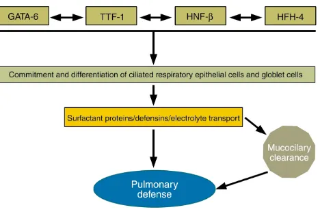
The formation, maintenance, and repair of the post-natal lung depend upon precisely regulated expres-sion of many of the same transcription factors involved in formation of the lung earlier in lung mor-phogenesis. Three distinct families of transcription factors, including the homeodomain protein TTF-1, the zinc finger protein GATA-6, and a forkhead fami-ly member, HNF-3β(also known as Foxβ2), play crit-ical roles in the differentiation of foregut endoderm to form the progenitors of the lung parenchyma (Fig-ure 2). GATA-6 and HNF-3βare involved in formation of the endoderm per se (5–8), while TTF-1 is required for the generation of epithelial cells that form the lung periphery (9). Later in development, these same transcription factors regulate the transcription of host defense molecules, such as SP-A, SP-B, SP-C, and Clara cell secretory protein, modifiers of lung inflam-mation following infection or injury (10).
The precise temporal/spatial formation and func-tion of the respiratory epithelium are controlled at the level of transcription, mediated by the con-certed actions of both tissue-specific and ubiqui-tous nuclear proteins. These nuclear transcription proteins influence differentiation of specific
epithe-Figure 2
[image:3.576.60.294.58.216.2]lial cell types and regulate the synthesis of the dis-tinct host defense proteins elaborated by subsets of respiratory epithelial cells. For example, during development, HFH-4 (also known as Foxj1) is required for ciliated cell differentiation, expression of
β-tubulin, and formation of cilia in conducting air-ways (11). Defects in ciliated cell function, as seen in ciliary dyskinesia and Kartagener syndrome, cause situs inversus and disturb mucociliary clearance, leading to chronic infection and destruction of lung tissues. While initially derived from common pro-genitors, the heterogeneous epithelial cell types lin-ing the respiratory tract are generated under the direction of cell-cell, cell-matrix, autocrine-paracrine, and transcriptional pathways that are influenced by their microenvironments. Distinct respiratory epithelial cells synthesize diverse classes of host defense molecules that maintain pulmonary sterility. There is increasing evidence that the genetic path-ways that initially direct lung morphogenesis during development are later employed to regulate the syn-thesis of host defense molecules and cellular respons-es to pathogens that, in turn, influence cell prolifer-ation and tissue remodeling. Repair following injury, at least in part, recapitulates molecular and cellular processes mediating lung formation.
Shared pathways mediating lung morphogenesis, innate host defenses, and repair
The vertebrate lung is a relatively late evolutionary adaptation required for terrestrial survival, first appearing approximately 350–400 million years ago in the mid-Devonian fossil record. Of the estimated 40,000 distinct genes expressed by the human genome, relatively few are uniquely expressed in the lung, many of the molecular pathways controlling lung morphogenesis being borrowed from similar processes in other organs conserved among diverse phyla that were established long before the advent of the lung. Unique combinations of molecules mediat-ing signalmediat-ing and transcriptional pathways in many organs are used to form and maintain lung structure and function.
Signaling pathways involved in lung morphogene-sis, lung defense, and repair are highly conserved. FGF receptor–, transcription factor–, cytokine-, and NF-κB–dependent pathways shared among diverse phyla participate in lung morphogenesis and repair. It is perhaps the unique combinatorial use of ancient signaling mechanisms that provides the molecular framework underlying lung formation and function. Indeed, the genes involved in generation and func-tion of the lung have ancient antecedents, for exam-ple in Caenorhabditis elegansandDrosophila. FGF sig-naling via the Drosophilagenes branchlessand breathless
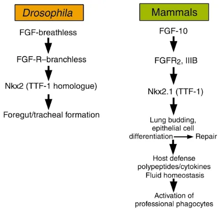
(an FGF receptor [FGFR] and ligand, respectively) is required for formation of the tracheal system in the fly (12, 13), much as it is for lung formation in verte-brates (Figure 3). Likewise, FGF signaling medi-ates diverse functions of the respiratory epithelium. Deletion of either FGF-10 or FGFR signaling results
in nearly complete lack of lung formation in the mouse (14, 15). Surprisingly, mutation in FGF-10 (11) or the FGFR2IIIb (16) also blocks formation of the limbs, FGF signaling mutants being both “lung-less” and “limbless.” It may be significant that the FGF pathway determines the formation of two organs that have been critical for adaptation of ver-tebrates to terrestrial habitats; namely, breathing and tetrapod ambulation.
While FGF signaling is critical for lung formation, FGF family members also enhance epithelial cell pro-liferation and differentiation in the postnatal lung (17). Both FGF-7 and FGF-10 increased respiratory epithelial cell proliferation and enhanced expression of TTF-1 and its downstream targets, e.g., SP-A, -B, and -C, that are involved in innate defense and surfac-tant activity in the lung (18, 19). Furthermore, FGF stimulated production of cytokines and chemokines, recruiting and activating professional phagocytes. Likewise, FGF increased the expression of aquaporin V and C1-dependent fluid secretion to regulate fluid transport by the respiratory epithelium (18–20).
NF-κB: another intersection between lung morphogenesis and innate host defense
[image:4.576.305.520.452.658.2]Transcriptional control of both acquired and innate host defense systems in the lung and other organs depends upon the nuclear translocation and activa-tion of NF-κB in target cells (21). NF-κB family mem-bers include at least three polypeptides, P50, P65, and Rel-A. Like the FGF signaling pathway, NF-κB plays a dual role in lung morphogenesis and inflammatory responses to various pathogens. NF-κB is regulated by cell surface receptors of the Toll-like receptor (TLR)
Figure 3
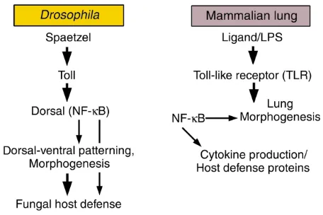
family. Activation of TLR/NF-κB is regulated, at least in part, by binding of pattern recognition molecules that present carbohydrate peptide or lipid compo-nents of pathogens (for example, endotoxin and pro-teoglycan) to professional phagocytes or epithelial cell surfaces (reviewed in ref. 22). In the lung, both SP-A and SP-D, members of the collectin family of polypep-tides, bind promiscuously to complex carbohydrates, serving as nonself pattern recognition molecules, binding the surfaces and products of respiratory pathogens, presenting them for phagocytosis, and modulating subsequent inflammatory responses dur-ing infection (23). Furthermore, SP-A and SP-D inter-act with CD14/TLR to influence cellular responses to pathogens via transcriptional pathways activated by NF-κB translocation to the nucleus of responding cells (24, 25). Members of the TLR family and associ-ated pathways were first recognized for their involve-ment in dorsal-ventral patterning during Drosophila
development and in fungal host defense (26). TLRs and their homologues are critical mediators of innate defenses in plants and animals, activating gene tran-scription via NF-κB to enhance killing of microor-ganisms and the inflammatory response (Table 1; see also ref. 22). Thus, the ancient NF-κB pathway serves complex and critical roles in both morphogenesis and antimicrobial host defense.
NF-κB and gene expression in lung morphogenesis and repair
Increased activity of NF-κB in embryonic lung mes-enchyme inhibits growth and budding of the respirato-ry epithelium, while inhibition of NF-κB signaling enhances proliferation and budding of the respiratory epithelial cells in a process likely mediated by autocrine-paracrine mechanisms (27). While the mechanisms of action of NF-κB in the lung mesenchyme are not fully clarified, enhanced NF-κB activity blocks FGF-10 expression in the embryonic lung, suggesting that NF-κB and FGF signaling are linked during both lung and limb morphogenesis. NF-κB also regulates the expression of the water transport protein aquaporin V (28), various innate host defense proteins (defensins and mucins), and cytokines, and it activates professional phagocytes following pulmonary injury or infection.
Events in lung morphogenesis, at least in part, are reca-pitulated during inflammation and repair. As shown in Figure 4, the multicomponent innate defense response mollifies inflammation and initiates morphogenetic events that result in the orderly repair of the respiratory epithelium and underlying stroma. Indeed, FGF and NF-κB pathways are critically involved in lung repair. TTF-1 and its downstream targets are also increased dur-ing repair followdur-ing severe lung injury (29). FGF protects respiratory epithelial cells during lung injury in vivo (30) and enhances expression of TTF-1 and its demonstrat-ed targets, SP-A, SP-B, and SP-C, as well as fluid trans-port proteins (18–20). TTF-1 regulates epithelial cell dif-ferentiation and proliferation and influences the expression of innate host defense molecules. In turn, these molecules influence the activation state, oxidant production, and apoptotic activity of professional phagocytes that are involved in clearance of apopto-tic cells and pathogens from the lung (23).
[image:5.576.61.289.59.211.2]The multiplicity and interaction of structural and cel-lular components of the intrinsic and innate host defenses of the lung are orchestrated temporally and spatially and are graded stochastically to prevent initial invasion, to limit the extent of injury, and to achieve rapid repair following pulmonary infection by respira-tory pathogens. Detailed knowledge of these pathways should improve our understanding of the pathogene-sis of common lung diseases and lead to new therapies based on knowledge of lung morphogenesis, repair, and innate host defense systems.
Table 1
Conservation of NF-κB–dependent pathways in morphogenesis, lung inflammation, and host defense
Drosophila Mammalian lung
Ligand Spaetzel LPS
Receptor Toll TLR
Transcription factor induced Dorsal (NF-κB) NF-κB Developmental role Dorsal-ventral patterning, morphogenesis Lung morphogenesis
Role in host defense Antifungal responses Cytokine production/host defense proteins
A proteolytic process originating in the dorsal region of the fly embryo produces Spaetzel that binds Toll on the cell surface, ultimately translocating Dorsal (an NF-κB homologue) to the nucleus of target cells to influence transcription of genes critical for morphogenesis or host defense. In vertebrates, homologous signal-ing systems recognize bacterial components, activatsignal-ing TLRs, in turn activatsignal-ing NF-κB to regulate host defense responses. NF-κB also plays a critical role in lung morphogenesis in vertebrates.
Figure 4
[image:5.576.57.522.628.695.2]1. Hogan, B.L.M. 1999. Morphogenesis. Cell.96:225–233.
2. Perl, A.K.T., and Whitsett, J.A. 1999. Molecular mechanisms controlling lung morphogenesis. Clin. Genet.56:14–27.
3. Costa, R.H., Kalinichenko, V.V., and Lim, L. 2001. Transcription factors in mouse lung development and function. Am. J. Physiol.280:L823–L838. 4. Warburton, D., et al. 2000. The molecular basis of lung morphogenesis.
Mech. Dev.92:55–81.
5. Ang, S.L., and Rossant, J. 1994. HNF-3βis essential for node and noto-chord formation in mouse development. Cell.78:561–574.
6. Morrisey, E.E., Ip, H.S., Lu, M.M., and Parmacek, M.S. 1996. GATA-6: a zinc finger transcription factor that is expressed in multiple cell lineag-es derived from lateral mlineag-esoderm. Dev. Biol.177:309–322.
7. Koutsourakis, M., Langeveld, A., Patient, R., Beddington, R., and Grosveld, F. 1999. The transcription factor GATA6 is essential for early extraembryonic development. Development.126:723–732.
8. Keijzer, R., et al. 2001. The transcription factor GATA6 is essential for branching morphogenesis and epithelial cell differentiation during fetal pulmonary development. Development.128:503–511.
9. Kimura, S., et al. 1996. The T/ebp null mouse: thyroid-specific enhancer-binding protein is essential for the organogenesis of the thyroid, lung, ventral forebrain, and pituitary. Genes Dev.10:60–69.
10. Bohinski, R.J., DiLauro, R., and Whitsett, J.A. 1994. The lung-specific surfactant protein B gene promoter is a target for thyroid transcription factor 1 and hepatocyte nuclear factor 3, indicating common factors for organ-specific gene expression along the foregut axis. Mol. Cell. Biol.
14:5671–5681.
11. Brody, S.L., Yan, X.H., Wuerffel, M.K., Song, S.-K., and Shapiro, S.D. 2000. Ciliogenesis and left-right axis defects in forkhead factor HFH-4-null mice. Am. J. Respir. Cell Mol. Biol.23:45–51.
12. Sutherland, D., Samakoulis, C., and Krasnow, M.A. 1996. Branchless encodes a Drosophila Fgf homolog that controls tracheal migration and the pattern of branching. Cell.87:1091–1101.
13. Glazer, L., and Shilo, B.Z. 1991. The Drosophila FGF-R homolog is expressed in the embryonic tracheal system and appears to be required for directed tracheal cell extension. Genes Dev.5:697–705.
14. Peters, K., et al. 1994. Targeted expression of a dominant negative FGF receptor blocks branching morphogenesis and epithelial differentiation of the mouse lung. EMBO J.13:3296–3301.
15. Min, H., et al. 1998. Fgf-10 is required for both limb and lung develop-ment and exhibits striking functional similarity to Drosophila branch-less. Genes Dev.12:3156–3161.
16. Hajihosseini, M.K., Wilson, S., De Moerlooze, L., and Dickson, C. 2001.
A splicing switch and gain-of-function mutation in FgfR2-IIIc hemizy-gotes causes Apert/Pfeiffer-syndrome-like phenotypes. Proc. Natl. Acad. Sci. USA.98:3855–3860.
17. Ulich, T.R., et al. 1994. Keratinocyte growth factor is a growth factor for type II pneumocytes in vivo. J. Clin. Invest.93:1298–1306.
18. Tichelaar, J.W., Lu, W., and Whitsett, J.A. 2000. Conditional expression of fibroblast growth factor-7 in the developing and mature lung. J. Biol. Chem.275:11858–11864.
19. Clark, J.C., et al. 2001. FGF-10 disrupts lung morphogenesis and causes pulmonary adenomas in vivo. Am. J. Physiol.280:L705–L715. 20. Zhou, L., et al. 1996. Keratinocyte growth factor stimulates
CFTR-inde-pendent fluid secretion in the fetal lung in vitro. Am. J. Physiol.
271:L987–L994.
21. Kopp, E.B., and Medzhitov, R. 1999. The Toll-receptor family and con-trol of innate immunity. Curr. Opin. Immunol.11:13–18.
22. Hoffmann, J.A., Kafatos, F.C., Janeway, C.A., and Ezkowitz, R.A.B. 1999. Phylogenetic perspectives in innate immunity. Science.284:1313–1318. 23. LeVine, A.M., and Whitsett, J.A. 2001. Pulmonary collectins and innate
host defense of the lung. Microbes Infect.3:161–166.
24. Sano, H., et al. 2000. Surfactant proteins A and D bind CD14 by differ-ent mechanisms. J. Biol. Chem.275:22442–22451.
25. Yoshida, M., Korfhagen, T.R., and Whitsett, J.A. 2001. Surfactant protein D regulates NF-κB and matrix metalloproteinase production in alveo-lar macrophages via oxidant-sensitive pathways. J. Immunol.
166:7514–7519.
26. Lemaitre, B., Nicolas, E., Michaut, L., Reichhart, J.-M., and Hoffmann, J.A. 1996. The dorsoventral regulatory gene cassette spätzle/Toll/cactus
controls the potent antifungal response in Drosophila adults. Cell.
86:973–983.
27. Muraoka, R.S., Bushdid, P.B., Brantley, D.M., Yull, F.E., and Kerr, L.D. 2000. Mesenchymal expression of nuclear factor-kappaB inhibits epithe-lial growth and branching in the embryonic chick lung. Dev. Biol.
225:322–338.
28. Borok, Z., et al. 1998. Keratinocyte growth factor modulates alveolar epithelial cell phenotype in vitro: expression of aquaporin 5. Am. J. Respir. Cell Mol. Biol.18:554–561.
29. Stahlman, M.T., Gray, M.E., and Whitsett, J.A. 1996. Expression of thy-roid transcription factor-1 (TTF-1) in fetal and neonatal human lung.
J. Histochem. Cytochem.44:673–678.