© 2015, IRJET ISO 9001:2008 Certified Journal Page 2370
Developing an Image Fusion Algorithm Using Double Density Dual-tree
Complex Wavelet Transform
Pooja Chandrakar, Ravi Mishra
Master Of Engineering, Electronics And Communication Engineering, SSTC Bhilai, Chhattisgarh Senior Assistant Professor of Electrical and Electronics Dept, SSTC Bhilai, Chhattisgarh, India
Abstract - Double-density Dual-tree Complex Wavelet transform is introduced to image fusion based on multi resolution, images are decayed by double-density dual tree complex wavelet transform with multi-level, multi direction and shift-invariance and according to characters of low and high frequency coefficients correspondingly, different permutation rules are adopted to fuse images and combination coefficients are reconstructed by double-density dual-tree complex wavelet inverse transform. Some fusion experiments are completed by numerous sets of images with different modalities and objective performance assessments are satisfied to calculate fusion results. The tentative results indicate that the planned approach can appreciably outperform the traditional image fusion method based on Laplacian pyramid and discrete wavelet transform. For medical diagnosis, doctors usually monitor the images manually and fuse them in the mind. The aim of image fusion is to acquire useful complementary information from CT/MRI multimodality images. By this method we can get more balancing information and also satisfactory Entropy, Better correlation coefficient, PSNR (Peak- Signal-to-Noise Ratio) and less MSE (Mean square error).
Keywords: Double Density Dual Tree Complex Wavelet Transform, image fusion.
1. INTRODUCTION
Medical image fusion has been also a admired research topic. In general, medical image fusion means the matching and fusion between two or more images of the same lesion area from different medical imaging equipment, and aims to obtain harmonizing information and increase the amount of in succession. Medical image fusion procedure is to merge the information of a multiplicity of images with computer-based image processing technique. It is being used for medical image fusion so as to get a superior image which is clearer and contains more information. In the experimental diagnosis and treatment, the use of fused images can provide more useful information. It is key for lesion
location, diagnosis, making treatment and pathological study.
© 2015, IRJET ISO 9001:2008 Certified Journal Page 2371 particular image fusion sequence (where the
input data consists of image sequences). A
possible application is the fusion of forward
looking infrared (FLIR) and low light visible images (LLTV) obtained by an airborne sensor platform to aid a pilot navigates in poor weather conditions or darkness. In pixel-level image fusion, some generic necessities can be compulsory on the fusion result. The fusion
process should preserve all significant
information of the input imagery in the merged image (pattern conservation). The fusion act should not introduce any artifact inconsistency which would divert the human observer or subsequent processing stages. The fusion procedure should be shift and rotational invariant, i.e. the fusion result should not depend on the location or orientation of an object the input imagery. In case of image succession fusion arises the further crisis of temporal stability and reliability of the fused image sequence. The human visual scheme is primarily sensitive to moving light stimuli, so moving artifacts or time depended contrast changes introduced by the fusion process are highly disturbing to the human observer. So, in case of image succession fusion the two supplementary requirements apply. Temporal stability: The fused image progression should be temporal stable. Temporal consistency: Gray level changes going on the input sequences must be present in the fused progression without any interruption or contrast change.
II. METHODS OF FUSION
I. Existing method
Image averaging and maximization
method
Principal component analysis
Discrete wavelet transform
Thresholding and K means clustering
methods for segmentation.
Fig 1 Blocks Diagram Of Basic Image Fusion Process
Here we take four types of images which are as follows: multispectral images, medical images, merging out of focus images, Applications and trends navigation aid.
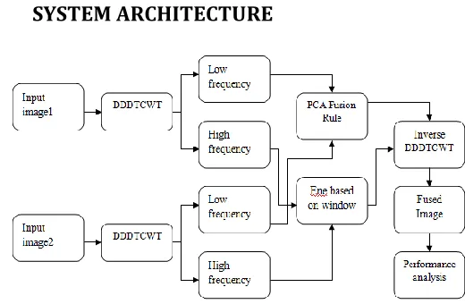
[image:2.612.324.563.200.322.2]II. SYSTEM ARCHITECTURE
Fig 2: Block Diagram of DDDTCWT
III. METHODOLOGIES
[image:2.612.324.584.454.624.2]© 2015, IRJET ISO 9001:2008 Certified Journal Page 2372
institutions to test image processing and image
compression algorithms. The images are in many
cases chosen to represent natural or typical images that a class of processing techniques would need to deal with. Other test images are chosen because they present a range of challenges to image reconstruction algorithms, such as the reproduction of fine detail and textures, sharp transitions and edges, and uniform regions. Preprocessing of any image is to rectify the inconsistencies that are inside in captured images for obtaining better object for further processing.
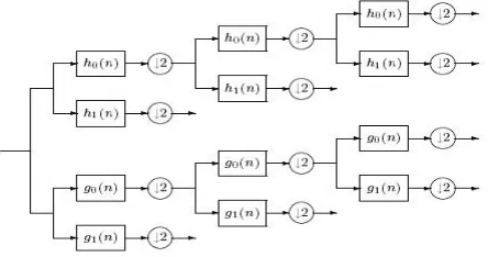
[image:3.612.47.269.560.677.2]2. Double-density Dual-tree Complex Wavelet transforms: - Multiscale image fusion includes three stages: decomposition, combination and reconstruction. At decomposition stage, the input images are decomposed by DD-DTCWT to low-frequency and high-frequency subbands representing different physical meanings. At the combination stage, because of their different physical meaning, the low-frequency and high-frequency subbands should be treated by different fusion rules to form different fused coefficients. Finally, Double density dual-tree complex wavelet inverse transform (DD-DTCWIT) is employed to reconstruct an image.
Fig 3: Iterated filter bank for the double-density dual-tree CWT
3. Fusion rules (PCA & Energy based on window)
3.1 PCA fusion rule of approximation images:
- Assuming that source approximation images
are I1L and I2L, combined approximate image is IL, PCA fusion steps are followed.
Step 1: The coefficient matrices of decomposed approximate images I1L and I2L are arranged by fore-row-post-column to create one dimension vectors, that is x, y respectively.
Step 2: Calculate the mean of vector x, y.
Step 3: Calculating the covariance of vector x , y
Step 4: Calculating the covariance matrix.
Step 5: Calculating eigen values and eigenvectors of covariance matrix.
Step 6: Confirming principal component Eigen value and calculating approximate combination image.
3.2 Selecting bigger Energy based on window region fusion rule of detail images
Step 1: In coefficient matrices of decomposed detail images.
Local energy (E) and local medium are local
characteristic of image. The local energy of any
area which center is Med (i,j) in image G has been
© 2015, IRJET ISO 9001:2008 Certified Journal Page 2373
IV. EXPERIMENTAL RESULTS
In this section, for test the validity of the planned approach, four sets of different modality images i.e. multifocus images, CT and MRI images, infrared and visual images, remote sensing images respectively are provided to be fused and experiment results are shown below.
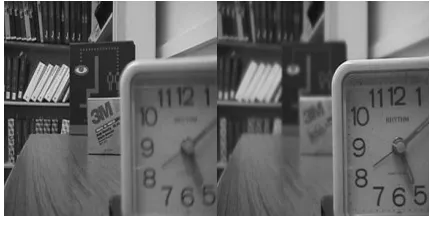
(a)Left focus image (b) right focus image
[image:4.612.33.552.50.735.2](c) DDDTCWT image
Fig 4 Multi Focus Images
(a)CT image (b) MRI image
[image:4.612.34.249.232.349.2](c) DDDTCWT image
Fig 5 Medical Images
(a)Saras 51 (b) Saras 52
(b)DDDTCWT image
© 2015, IRJET ISO 9001:2008 Certified Journal Page 2374
(a)Infrared image (b) visible image
(c)DDDTCWT image
Fig 7 Visible and Infrared Images
SSIM is used for measuring the similarity
between two images. The SSIM index is a full
reference metric; in other words, the
measurement or prediction of image quality is based on an initial uncompressed or distortion-free image as reference. SSIM is designed to
improve on traditional methods such as peak
signal-to-noise ratio (PSNR) and mean squared
error (MSE), which have proven to be
inconsistent with human visual perception. The SSIM index is calculated on various windows of an image. The measure between two windows
and of common size N×N is:
With the average of ; the average of ;
the variance of ; the variance of ; the
covariance of and ; , two
variables to stabilize the division with weak denominator;
Table 1: Fusion Performance Of Different Images Parameters Multi
focus images
Medical
images Remote sensing images
Visible & infrared images
E 7.23391 6.61546 4.6593 6.8008 MI 3.11522 2.80051 2.67055 1.3913 SSIM 0.896539 0.49839 0.931509 0.682633
From tab.1 data, fusion performance by planned approach is best, this is due to DDDTCWT that can decompose 16 main orders, each of the main orders includes two wavelets that is measured as the real and imaginary parts of complex wavelet which can describe characteristic more accurately, in addition, the trait of approximate shift-invariance can be more improve the accuracy of image decay and reform.
V. CONCLUSION
© 2015, IRJET ISO 9001:2008 Certified Journal Page 2375
References
[1]. Aniveni Mahesh, Ch. Sridhar, and Ahmed Zeeshan, Double Density Dual Tree Complex Wavelet Transform Based Satellite Image Resolution Enhancement, International Journal of Engineering & Science Research, IJESR, August
2014, Vol-4,Issue-8,595-601.
[2]. K.P.Indira , Dr.R.Rani Hemamalini, Analysis on Image Fusion Techniques for Medical
Applications, International Journal of Advanced Research in Electrical, Electronics and
Instrumentation Engineering, September 2014, Vol. 3, Issue 9.
[3]. Kusum Rani1, Reecha Sharma, Study of Different Image fusion Algorithm, International Journal of Emerging Technology and Advanced Engineering, May 2013 Volume 3, Issue 5.
[4]. G. Chen and Y. Gao, Multisource Image Fusion Based on Double Density Dual-tree Complex Wavelet Transform, International Conference on Fuzzy Systems and Knowledge Discovery (FSKD 2012 ) IEEE: pp 1864-1868.
[5]. Qingping Li, Junping Du, and Liang Xu ,
Multi-Focus Image Fusion Using the Local Fractal Dimension, International Journal Advnce Robotic System, IEEE 2013, Vol. 10: pp 251.
[6]. Yu Chen Lin and Tian Hua Chen, Infrared and Vsible Image Fusion Method Based on Wavelet Transform, International Conference on
Advanced Computer Science and Electronics Information (ICACSEI 2013): pp 632-635.
[7]. R. K. Sarawale andS.R. Chougul, Image Denoising using Dual-Tree Complex DWT and Double-Density Dual-Tree Complex DWT,
International Journal of Advanced Research in Computer Engineering & Technology (IJARCET) Vol 2(6), June 2013: pp 2148-2154.
[8]. K. R. Penmetsa, N.V. Rao and V.G.P.
Naraharisetti, An Image Fusion Technique For Colour Images Using Dual-Tree Complex Wavelet Transform. International Journal of Engineering Research & Technology (IJERT) Vol. 1 Issue 8, October – 2012.
[9]. D. S. Reddy and S. Varadarajan, 2D Dual-Tree Complex Wavelet Transform Based Image
Analysis, Contemporary Engineering Sciences, Vol. 5, 2012, no. 3: pp127 – 136.
[10]. Sandhya G, Kishore K. Denoising of Images corrupted by Random noise using Complex Double Density Dual Tree Discrete Wavelet Transform. International Journal of Engineering Research and Applications 2012; 2(3): 79-87.
[11]. Deepak Kumar Sahu, M.P.Parsai, Different Image Fusion Techniques –A Critical Review, International Journal of Modern Engineering Research (IJMER), Vol. 2, Issue. 5, Sep.-Oct. 2012 pp-4298-4301.