Single-Center Study
Yanjiao Zhou,a,b Carey-Ann D. Burnham,cTiffany Hink,dLei Chen,eNurmohammad Shaikh,aAye Wollam,bErica Sodergren,e George M. Weinstock,ePhillip I. Tarr,a,fErik R. Dubberked
Department of Pediatrics, Washington University School of Medicine, St. Louis, Missouri, USAa
; The Genome Institute, Washington University School of Medicine, St. Louis, Missouri, USAb
; Department of Pathology & Immunology and Pediatrics, Washington University School of Medicine, St. Louis, Missouri, USAc
; Department of Medicine, Washington University School of Medicine, St. Louis, Missouri, USAd
; The Jackson Laboratory for Genomic Medicine, Farmington, Connecticut, USAe
; Department of Molecular Microbiology, Washington University School of Medicine, St. Louis, Missouri, USAf
Clostridium difficileinfections (CDI) are a growing concern in North America, because of their increasing incidence and sever-ity. Using integrated approaches, we correlated pathogen genotypes and host clinical characteristics for 46C. difficileinfections in a tertiary care medical center during a 6-month interval from January to June 2010. Multilocus sequence typing (MLST) dem-onstrated 21 known and 2 novel sequence types (STs), suggesting that the institution’sC. difficilestrains are genetically diverse. ST-1 (which corresponds to pulsed-field gel electrophoresis strain type NAP1/ribotype 027) was the most prevalent (32.6%); 43.5% of the isolates were binary toxin gene positive, of which 75% were ST-1. All strains were ciprofloxacin resistant and metro-nidazole susceptible, and 8.3% and 13.0% of the isolates were resistant to clindamycin and tetracycline, respectively. The corre-sponding resistance loci, including potential novel mutations, were identified from the whole-genome sequencing (WGS) of the resistant strains. Core genome single nucleotide polymorphisms (SNPs) determining the phylogenetic relatedness of the 46 strains recapitulated MLST types and provided greater interstrain differentiation. The disease severity was greatest in patients infected with ST-1 and/or binary gene-positive strains, but genome-wide SNP analysis failed to provide additional associations with CDI severity within the same STs. We conclude that MLST and core genome SNP typing result in the same phylogenetic grouping of the 46C. difficilestrains collected in a single hospital. WGS also has the capacity to differentiate those strains within STs and allows the comparison of strains at the individual gene level and at the whole-genome level.
C
lostridium difficileinfections (CDI) are the most commonin-fectious antibiotic-associated gastrointestinal disorders. C.
difficilecolonization of the intestine results in a range of clinical
states, ranging from asymptomatic carriage to self-limited diar-rhea to life-threatening colitis. CDI was the leading cause of gas-troenteritis- and gastrointestinal tract infection-associated deaths between 1999 and 2007 in the United States (1). Risk factors for CDI include antibiotic exposures (especially fluoroquinolones [FQ] and cephalosporins), advanced age, and the severity of the underlying illness (2,3,4).
The most commonC. difficilestrain that has emerged in the past decade in North America and some areas in Europe has been classified as 027 by ribotyping, NAP1 by pulsed-field gel electro-phoresis (PFGE), BI by restriction endonuclease analysis, and ST-1 by multilocus sequence typing (MLST). ST-1 strains account for half of the sporadic hospital-associated CDI in some settings (5). Some studies have reported that ST-1 strains elaborateC.
difficiletoxins (TCDs) at high concentrations; its purported
hy-pervirulence is plausibly related to this trait. This strain has single and 18-bp deletions oftcdC, a negative regulator oftcdAandtcdB. These mutations cause premature stops, and this truncation is believed to cause toxin overproduction (6,7). However, this as-sumption was challenged by recent studies showing no significant difference in toxin production between hypervirulent and nonhy-pervirulentC. difficilestrains, and no association of thetcdC ge-notype and toxin production (8,9).
C. difficilestrains containingcdtAandcdtBbinary toxin genes
are associated with greater mortality in their hosts than strains in which these genes are absent (10). However, it is not clear if the binary toxin genes increase the virulence of ST-1 or if they are
simply epidemiologic markers ofC. difficilestrains with increased virulence (i.e., guilt by association). It is also notable that other ribotypes with binary toxin, such as 078 (ST-11), can also cause severe CDI, especially in young adults. TheseC. difficileribotype 078 strains were highly related to animals and food-borneC.
dif-ficilestrains (11). It is concerning that 078 strains have increased in
prevalence from 3% (2008) to 13% (2011) (1). CDI caused by both 027/ST-1 and 078/ST-11 are associated with an increased risk of death (12).
Our understanding of the pathogenesis ofC. difficileis based largely on studies in outbreak strains. While the epidemiology of CDI is changing, analysis ofC. difficile, especially the strains caus-ing severe CDI, in a nonoutbreak settcaus-ing might shed light on the mechanism of the pathogenicity of sporadicC. difficileand possi-bly produce more generalizable data. The objective of this study, therefore, was to characterize the phenotypes and genotypes of 46 nonoutbreakC. difficileisolates from a large academic medical center using conventional microbiological analyses and
whole-Received24 July 2014 Returned for modification2 September 2014 Accepted25 September 2014
Published ahead of print1 October 2014
Editor:D. J. Diekema
Address correspondence to Erik R. Dubberke, [email protected].
Supplemental material for this article may be found athttp://dx.doi.org/10.1128
/JCM.02115-14.
Copyright © 2014, American Society for Microbiology. All Rights Reserved.
doi:10.1128/JCM.02115-14
on May 16, 2020 by guest
http://jcm.asm.org/
genome sequencing and to investigate the associations between strain phenotypes and genotypes and clinical outcomes.
MATERIALS AND METHODS
CDI severity, bacterial strains, and ribotyping and binary toxin charac-terization.This study was approved by the Washington University Hu-man Research Protection Office. All subjects were prospectively inter-viewed and examined as part of aC. difficileassay comparison evaluation (13). The presence of clinically significant diarrhea and the severity of CDI were determined. Patients without clinically significant diarrhea or those who were colonized with a nontoxigenic strain ofC. difficilewere not classified as having CDI. Severe CDI was defined according to the clinical practice guidelines for CDI in adults (14): subjects with a white blood cell count ofⱖ15,000 cells/mm3and/or serum creatinine ofⱖ1.5 times the premorbid level at the time of CDI diagnosis. Specimens were collected, andC. difficilestrains were isolated and characterized as part of aC. diffi-cilelaboratory method study (13). Ribotyping (15) and detection of the binary toxin genes from the isolates were performed by PCR as previously described (16,17).
Antibiotic susceptibility testing.C. difficilestrains were tested for antibiotic susceptibility using a gradient diffusion method according to the manufacturer’s recommendations. Isolates ofC. difficilewere grown in an anaerobic environment on prereduced sheep blood agar (BBL; BD, Sparks, MD). A bacterial suspension was prepared in 0.9% saline to a 1 McFarland standard and then applied as a lawn of growth toBrucellaagar with hemin and vitamin K (Hardy Diagnostics, Santa Maria, CA). Etest strips for metronidazole, clindamycin, moxifloxacin, ciprofloxacin, and tetracycline (bioMérieux) were applied to the agar and incubated with quality control strains according to the manufacturer’s recommenda-tions. The resulting MIC values were interpreted according to the Clinical and Laboratory Standards Institute guidelines (18).
Whole-genome sequencing and analysis.Genomic DNA was ex-tracted from the 46 isolates by a BiOstic bacteremia DNA isolation kit (MO BIO Laboratories). A genome paired-end library was constructed with average insert lengths from 150 to 200 bp, following the Illumina library construction protocol. The libraries were sequenced at an Illumina 2⫻100 bp platform. The genome assembly was performed by Velvet (version 1.1.04-64) (19). All assemblies were subjected to host contami-nation screening and met the criterion for draft genomes used in the Human Microbiome Project (20). Gene annotation employed the online RAST annotation pipeline with manual inspection (http://www.nmpdr .org/FIG/wiki/view.cgi/FIG/RapidAnnotationServer). The open reading frames (ORFs) were at least 300 bp long. Core gene sets were determined by pan-genome analysis pipeline (PGAP) with default parameters using all of the 46C. difficilestrains and reference strain 630 (21). The contigs from the draft genomes were aligned to theC. difficileMLST database (http://pubmlst.org/cdifficile/) to determining the sequence type (ST) us-ing Mummer (22). The mutations from the novel ST types, regulatory genes in the pathogenicity locus (PaLoc), binary genes, and resistance genes were verified by manual inspection of the read alignment to refer-ence alleles. Targeted PCR was performed to close the gaps in specific genes such astcdEthat were not fully covered in a subset of isolates.
Single nucleotide polymorphisms (SNPs) were identified with the SNP detection pipeline developed at the Washington University Genome Institute (TGI) by aligning the reads to theC. difficilereference strain 630 using BWA aligner (version 0.5.9) and SAMtools (version 0.1.12). SNP calling was performed as previously described but with increased strin-gency (23) as follows: (i) the coverage of a SNP is at least 10 reads and (ii) the number of reads supporting a SNP calling/the number of reads not supporting a SNP calling isⱖ10 (3)ⱖ12 bp between two SNP sites. A phylogenetic tree based on the SNPs from the core gene sets was con-structed using the neighbor-joining algorithms in Phylip (http: //evolution.genetics.washington.edu/phylip.html).
Statistical analysis.Fisher’s exact test or the chi-square test was used to assess whether ST-1 and the presence of binary genes inC. difficile
isolates are associated with CDI severity.Pvalues⬍0.05 were considered statistically significant.
RESULTS
Isolate characterization.The 46C. difficileisolates represented 23 STs and 20 ribotypes. ST-1 (which corresponds to NAP1/027) accounted for 32.6% of the isolates. Other STs with at least two representatives in the collection were STs 2, 6, and 8 (Table 1). Three strains were not assignable to any STs in the current MLST database. One was a new allele profile (adk3,atpA7,dxr14,glyA
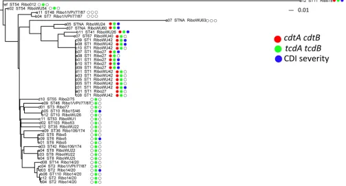
8,recA6,sodA25,tpi10), and the other two had an identical SNP at nucleotide position 198 insodA. The correlation of ribotypes and STs was observed: ST-1 corresponded with ribotypes 027 and WU42, ST-2 with ribotypes 001/VPI/77/87 and 014/020, ST-6 with ribotype 005, and ST-8 with ribotypes WU22 and WU25. In addition, ribotypes 027 and WU42 were exclusively found in iso-lates belonging to ST-1, whereas ribotypes 001/VPI/77/87 were found among four STs (Table 1,Fig. 1). Thus, the ribotypes and ST types did not have a 1:1 correlation.
We next studied the toxin-related genes, includingtcdA,tcdB, and binary genescdtAandctdB. tcdAandtcdBgenes were detected by conventional PCR and were further validated by sequencing. PCR results were perfectly correlated with the sequence data for detectingtcdAandtcdB. Binary toxin genes were also detected by PCR and successfully reconstructed from whole-genome se-quencing. Isolates were grouped into three categories based on the presence of the toxin genes: (i) positive fortcdA,tcdB,cdtA, and
cdtB, which comprised 43.5% (20 of 46) of theC. difficileisolates; (ii) positive fortcdAandtcdB⫹and negative forcdtAandcdtB, which comprised 50.0% of the strains (23 of 46); and (iii) neg-ative fortcdA,tcdB,cdtA, andcdtB, which accounted for only three isolates (Fig. 1). Of the 20tcdA-,tcdB-,cdtA-, andcdtB -positive strains, 15 belonged to ST-1. Other STs containing
tcdA, tcdBand binary toxin genes were ST-11, ST-41, ST-67, and the two novel STs.
All 46 isolates were susceptible to metronidazole (MICs from 0.032 to 4g/ml) and resistant to ciprofloxacin (MICs of⬎32
g/ml). The 46 strains had MICs between 2 and⬎32g/ml to moxifloxacin (overall MIC50and MIC90 of 8 and⬎32g/ml,
respectively), and 36, 8, and 2 isolates were resistant, intermediate, or susceptible to moxifloxacin, respectively. Of the isolates, 8.3% and 13.0% were resistant to clindamycin and tetracycline, respec-tively. Two (from ST35 and ST48) of the 46 isolates were resistant to all tested antibiotics except metronidazole; these two isolates were binary toxin gene negative and one wastcdAandtcdB nega-tive. Five isolates were resistant to at least three antibiotics, includ-ing ciprofloxacin, tetracycline, and moxifloxacin.
Phylogenetic concordance between ST and WGS SNP typing. WGS was performed on an Illumina HiSeq platform with 2⫻100 bp read lengths at 100⫻coverage on average. Read assemblies yielded 193⫾53 contigs per genome. The contigs were annotated to provide the gene calling for each isolate. The gene content ranged from 3,612 to 4,054 ORFs per genome, indicating signifi-cant genetic variations acrossC. difficilestrains. ST-1 isolates had between 3,700 and 3,768 genes, corresponding to 121 fewer genes on average than the other ST types in this study (see Table S1 in the supplemental material).
To determine the core genes used for phylogeny inference, we computed the shared genes from the 46C. difficilestrains and the
C. difficilereference strain 630 (an ST-54 strain first isolated from
on May 16, 2020 by guest
http://jcm.asm.org/
a patient with pseudomembranous colitis). TheC. difficilestrain 630 was chosen as the reference because its genome was well an-notated, and it has been widely used as the reference for SNP identification. We identified a total of 2,871 core genes across the isolates in this study, accounting for 64.0% of their gene content. Between 1,096 and 44,935 SNPs were identified from the whole
[image:3.585.43.543.86.594.2]genomes of these isolates, of which 60.4% to 80.5% were distrib-uted among the core genes. The median (interquartile range [IQR]) number of core genome SNPs was 6,926 per stain. No strains were identical at the SNP level, and 46 SNPs were identified between the two closest strains. The phylogenetic tree constructed using the SNPs from core genes from the 46 strains and theC.
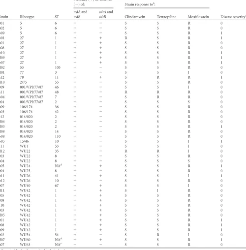
TABLE 1Phenotypic and genotypic characterization of 46C. difficilestrainsa
Strain Ribotype ST
Presence (⫹) or absence
(⫺) of: Strain response tob:
Disease severityc tcdAand
tcdB
cdtAand
cdtB Clindamycin Tetracycline Moxifloxacin
e01 5 6 ⫹ ⫺ S S R 0
a02 5 6 ⫹ ⫺ S S R 0
b09 5 6 ⫹ ⫺ S S R 1
b01 27 1 ⫹ ⫹ R S R 1
a01 27 1 ⫹ ⫹ S S R 0
a08 27 1 ⫹ ⫹ S S R 0
b10 27 1 ⫹ ⫹ S S R 1
d09 27 1 ⫹ ⫹ S S R 1
b07 27 1 ⫹ ⫹ S S R 1
d02 53 103 ⫹ ⫺ S S R 0
d01 77 3 ⫹ ⫺ S S I 0
a12 78 11 ⫹ ⫹ S R R 1
d10 2/75 55 ⫹ ⫺ S S R 0
e09 001/VPI/77/87 46 ⫹ ⫺ S S I 0
a11 001/VPI/77/87 48 ⫺ ⫺ R R R 0
b04 001/VPI/77/87 7 ⫺ ⫺ S R R 0
c04 001/VPI/77/87 2 ⫹ ⫺ S S S 0
a09 106/174 36 ⫹ ⫺ S S R 0
a03 106/174 42 ⫹ ⫺ S S R 0
c12 014/020 2 ⫹ ⫺ S S R 0
d04 014/020 2 ⫹ ⫺ S S R 0
d03 014/020 2 ⫹ ⫺ S S R 1
d08 014/020 14 ⫹ ⫺ S S R 0
b08 014/020 110 ⫹ ⫺ S S R 0
b05 15/46 10 ⫹ ⫺ S S I 1
c11 WU1 53 ⫹ ⫺ S S I 0
d12 WU22 35 ⫹ ⫺ R R I 0
c03 WU22 8 ⫹ ⫺ S S R 0
a04 WU22 8 ⫹ ⫺ S S S 0
a05 WU24 NAd ⫹ ⫹ S S R 1
e04 WU25 8 ⫹ ⫺ S S R 0
b11 WU26 41 ⫹ ⫹ S S I 1
b12 WU26 10 ⫹ ⫺ S S R 0
e07 WU40 67 ⫹ ⫹ S S I 0
d11 WU42 1 ⫹ ⫹ S I R 0
c05 WU42 1 ⫹ ⫹ S S R 0
c08 WU42 1 ⫹ ⫹ S S R 0
c10 WU42 1 ⫹ ⫹ S S R 0
e03 WU42 1 ⫹ ⫹ S S R 0
d05 WU42 1 ⫹ ⫹ S S R 0
c01 WU42 1 ⫹ ⫹ S S R 1
e08 WU42 1 ⫹ ⫹ S S R 1
c09 WU42 1 ⫹ ⫹ S S R 1
e02 WU54 54 ⫹ ⫺ S R I 0
d07 WU60 NAd ⫹ ⫹ S S R 1
a07 WU63 NAe ⫺ ⫺ S S R 0
aOrganized by the ribotypes in alphabetical order.
b
S, susceptible; R, resistant; I, intermediate resistant. c0, not severe CDI; 1, severe CDI.
d
Mutation insodAgene. eNew allele profile.
on May 16, 2020 by guest
http://jcm.asm.org/
difficilereference strain 630 demonstrated a heterogeneous ge-netic nature ofC. difficilestrains in this collection (Fig. 1). Clades 1, 2, and 5 from a previous study (24) were identified. Clade 1 was composed of several STs. The ST-11 strain from clade 5 was most distant from the rest of lineages with 36,039 SNPs compared to the reference stain 630. The number of SNPs identified among the ST-11 strains was 3.3-fold higher than those of the other STs on average. Clade 2 was dominated by the ST-1/NAP1 strains. Two novel ST strains were genetically most similar to the ST-1 strains, and the third (from a nontoxigenic isolate) was distantly related to all other strains. The cluster of the SNPs from the core gene sets recapitulated the ST phylogeny, indicating the correspondence of the ST type with WGS (25).
WGS can offer an improved resolution compared to MLST characterization of isolates. For example, 99 to 656 SNP differ-ences were detected within ST-1 strains, representing 1.3% to 4.4% of the differences in core gene sets. Within STs 6, 8, and 10, we identified 26 to 112 SNPs in the core genes, while 1,016 SNPs were detected for ST-2. The numbers of SNPs between ST types ranged from 1,568 to 44,204. However, the number of strains within a single ST type can change the degree of divergence within a ST type and may affect the pattern we observed here.
Genetic heterogeneity of toxin-related and antibiotic resis-tance genes. Contigs were mapped to the six genes spanning across the PaLoc, two binary toxin genes, and resistance genes to determine the genetic variation of these regions.
Compared to theC. difficilereference strain 630, 3 strains lacked
tcdA,tcdB,tcdC,tcdE, andtcdRgenes and CD630_06620 (coding for a hypothetical protein). All PaLoc genes were present in the
remaining 43 strains. A phylogenetic tree constructed from SNPs across the PaLoc revealed that diversity at this locus recapitulated ST typing, except for the PaLoc associated with ST-2, which ap-peared to be mixed with other STs (see Fig. S1 in the supplemental material).tcdA,tcdB, andtcdCgenes had the greatest degrees of conservation within the same ST and high variation between STs as indicated in the circular plot (see Fig. S2 in the supplemental material). Other genes, such astcdRandtcdE, were conserved even between STs, indicating different evolutionary changes in the PaLoc. ThetcdCgenes in these isolates were 92.7% to 100% iden-tical to those in reference strain 630. Sequence alignment of the 43
tcdCgenes demonstrated 12tcdCvariants. Based on the deletion pattern, we categorized thetcdCgene variants into 5 groups: (i) a single base pair deletion at nucleotide position 117 and an 18-bp deletion at nucleotide positions 330 to 347, which included all of the ST-1 strains; (ii) a single base pair deletion at nucleotide posi-tion 117 and no accompanying 18-bp deleposi-tion, which included only the ST-41 strain; (iii) a single base pair deletion at 117 bp, an insertion of T at 213 bp, and an 18-bp deletion, which included the two novel ST strains and has not been previously reported; (iv) a 39-bp deletion at nucleotide position 333, which included the ST-11 strain; and (v) single mutations without deletions, which occurred among heterogeneous STs.
Because binary toxin genes are not present in theC. difficile
reference strain 630, the gene variation was determined by align-ing the contigs to the binary genes inC. difficile strain CD196, which is an epidemic ST-1 strain harboring both type of toxin genes (26). In 15 of 21 strains, the sequences ofcdtAandcdtBwere identical to those of the CD196 strains. A transition from T to C at
FIG 1Phylogenetic tree of the 46C. difficilestrains based on the genome-wide SNPs. The neighbor-joining phylogenetic tree was constructed based on the SNPs from the core gene sets.C. difficilestrain 630 is used as the reference strain for SNP calling. The STs and ribotypes are appended after the strain labels. The tree is annotated by the presence of thetcdAandtcdBgenes (green), binary genes (red), and disease severity (blue) at the right side of the dendrogram. The white circles represent the absence of the toxin genes or nonsevere CDI.
on May 16, 2020 by guest
http://jcm.asm.org/
[image:4.585.43.546.70.341.2]nucleotide position 813 of thecdtA gene was discovered in the remaining six strains containing binary toxin genes. Two ST-11 strains exhibited significant polymorphisms (21 SNPs) in com-parison to those for CD196 in the 1,391-bpcdtAgene. Similarly, the relation of thecdtBgene in ST-11 strains was distant compared to those of other STs as indicated by 49 SNPs in this 2,630-bp region (see Table S2 in the supplemental material).
Fluoroquinolone (FQ) resistance is typically attributed to mu-tations ingyrAandgyrB, encoding DNA gyrase subunits. Among the 46 ciprofloxacin-resistant strains, 17 (36.9%) had a mutation at nucleotide position 82 (substitution T¡I), as in a prototype FQ-resistant strainR20291(ST-1). This mutation was common to all study ST-1 strains and to one ST-54 and one ST-55 strain. This substitution at position 82 ingyrAis the cause of FQ resistance in most European strains (27). We also identified eight other non-synonymous mutations ingyrAfrom other ST strains, and these mutations tended to be conserved in the same clades. In some cases, these mutations are also ST specific.gyrBwas intact in most (64.6%) strains. The precise contributions to ciprofloxacin resis-tance at additional mutations (see Table S2 in the supplemental material) are not yet known, but thegyrAandgyrBgenes from two moxifloxacin-susceptible strains were identical to these loci in some of the moxifloxacin- and ciprofloxacin-resistant strains.
tetMandermBare the genetic determinants of resistance to tetracycline and clindamycin, respectively. Multiple polymor-phisms were observed intetMgenes compared to those in the reference strain 630.tetMgenes displayed different degrees of het-erogeneity between STs but were conserved within STs. Com-pared to the rest of the STs, in the ST-11 strainstetMcontained an additional amino acid substitution at position 490 (M¡T). We did not detect any othertet genes in the tetracycline-resistant strains. Three strains were resistant to clindamycin. TheermB
gene of one strain (ST48) was 100% identical to the reference strain 630. The other two strains were from ST-1 and ST-35, shar-ing four nonsynonymous mutations at 454 (K¡Q), 511 (A¡V), 649 (Y¡H), and 664 (D¡N).
No link between SNPs and disease severity within ST-1. Se-vere CDI accounted for 30.4% (14 of 46) of the CDI cases. The strains associated with severe CDI were from 7 different STs with ST-1 being predominant (46.7%). ST-6, ST-41, ST-11, ST-2, a novel ST with a mutation in thesodAgene, and ST-10 accounted for the remaining severe CDI cases (Fig. 1). ST-1 strains were not significantly different in their association with severe CDI com-pared to that of non-ST-1 strains (2⫽1.2, df⫽1,P⫽0.28).
Among ST-1 strains, regulatory PaLoc genes (i.e.,tcdC,tcdR, and
tcdE) were identical in the seven and eight isolates from patients with severe and nonsevere CDI, respectively. Because of the highly repeated sequences intcdAandtcdB, full ORFs could not be con-structed with accuracy. Thus, we compared the SNPs identified in those two genes between ST-1 strains causing severe CDI and nonsevere CDI. The majority of the SNPs (88.5%) were shared for all ST-1 strains independent of disease severity in the patients from whom they were isolated. The rest of the SNPs were shared in either all ST-1 strains causing severe CDI/nonsevere CDI or a proportion of the ST-1 strains causing nonsevere CDI/severe CDI. Thus, genetic heterogeneity of the PaLoc did not distinguish dis-ease severity within ST-1 strains (see Fig. S1 in the supplemental material). Similarly, SNPs within the PaLoc between non-ST-1 strains (ST-6 and ST-10) failed to identify associations with CDI
severity, indicating no strong role for the genetic composition of pathogenic loci and disease phenotype.
Interestingly, 11 of 14 (78.6%) strains causing severe CDI were binary gene positive. Eleven of 20 (55%) binary toxin gene-posi-tive strains were recovered from patients with severe CDI. The chi-square test showed that binary toxin gene-positive strains were significantly associated with severe CDI compared to binary toxin gene-negative strains (Fisher’s exact test,P⫽0.003). Thus, the presence of binary genes might be a marker for strains that cause severe CDI, but the current genetic data do not support the role of binary genes in causing severe disease, as within the STs, binary gene sequences were identical in strains causing severe CDI and nonsevere CDI.
Whole-genome-wide SNP analysis showed that 92.3% and 92.7% of the SNPs were conserved within all of the ST-1 strains causing severe CDI and nonsevere CDI, respectively. However, no single SNP distinguished the ST-1 strains causing severe CDI from those causing nonsevere CDI.
DISCUSSION
Using integrated approaches, we delineated the phenotypes and genotypes ofC. difficilestrains causing CDI from a single institu-tion over a 6-month interval in 2010. Our data most notably in-dicate that ST-1 strains remain predominant in nonoutbreak set-tings, comparable to a statewide strain collection (548 strains) conducted from 2006 to 2009 (5). The vast majority of the strains weretcdA- andtcdBpositive, and notcdB-positive andtcdA -negative strains were detected in our data. This is likely in line with the low prevalence (⬃2%) of this type of stain in the United States, despite the high prevalence rates in Japan, Israel, and Argentina (28).
Our data suggest that MLST and ribotyping are robust ap-proaches for identifying phylogenic relationships in C. difficile
strains (25,29), but the most precise resolution requires WGS. WGS allows single nucleotide-level resolution for strain compar-isons, thus serving as a powerful tool for outbreak investigations and clarifying institutional versus community acquisition. As demonstrated by outbreaks ofEscherichia coliO104:H4 in Europe, the resolution of single nucleotide differences using WGS data led to the distinction of lineages from German and French isolates, which standard tests failed to distinguish (30). Along the same line, whole-genome sequencing of methicillin-resistant
Staphylo-coccus aureus(MRSA) in a special care nursery unit successfully
tracked the transmission within the unit (31). Recent studies have proved the feasibility of using WGS to trackC. difficile transmis-sion (32). Importantly, more efforts are needed to conduct pro-spective epidemiologic studies since the current studies are retro-spective in response to a perceived outbreak, and, thus, early shifts in local epidemiology may not be detected (5). With the dropping of sequencing cost and establishment of streamlined analysis pipe-line, WGS is becoming an advantageous approach in real-time pathogen surveillance and outbreak detection (33). Close surveil-lance and the prospective epidemiology of theC. difficilestrains, especially those associated with severe CDI such as ST-1, ST-11, and ST-6, might prevent futureC. difficileoutbreaks.
In addition, the antibiotic resistance genes identified from WGS data matched well with the antibiotic susceptibility testing, providing another application for WGS data in clinical settings. However, correlation of antibiotic resistance by WGS relies on a database of well-curated resistance genes. With the potential
on May 16, 2020 by guest
http://jcm.asm.org/
timicrobial resistance suggested by novel mutations identified from WGS, phenotypic testing and genetic engineering are still indispensable for validating the potential resistance from the novel mutations identified from WGS. The novel mutations in
gyrA andgyrB from non-ST-1 strains revealed diverse genetic compositions in conferring potential resistance. The mutations require further verification to determine their role in fluoroquin-olone resistance. The virulence genes reconstructed from WGS reads were consistent with the toxin gene testing in the clinic, demonstrating the versatile potentials of WGS in the clinic.
Interestingly, the phylogenetic topology ofC. difficile is re-flected in the PaLoc. Specifically,tcdCexhibited identical muta-tions across all of the ST-1 strains, as indicated by 1- and 18-bp deletions. Along the same line, two binary toxin genes, fluoro-quinolone resistance genes (especiallygyrA), also had identical sequences in all of the ST-1 strains studied and differed from those found in other STs. These findings suggest coevolution of the MLST genes with toxin genes andgyrA.
Although the clonal nature of theC. difficilestrains might pro-vide genetic insights into pathogenicity and antibiotic resistance, it did not shed light on the likelihood of causing severe disease. Previous reports suggested a poor correlation between the pres-ence oftcdCor binary genes with clinical outcomes in nonepi-demic settings (34,35). These studies were based on comparisons of NAP1 strains in different severity groups without a genetic comparison, or the genetic comparison was limited totcdCand binary toxin genes. In our study, we found that the presence of binary genes inC. difficilesignificantly increased the risk of severe CDI, but we could not attribute such increased risk to binary gene sequence differences in the strains that caused severe CDI and nonsevere CDI. Moreover, among the 40,000⫹SNPs throughout these strains, no single nucleotide variation correlated with disease severity. However, larger sample sizes and future multicenter studies are needed to validate these findings. Disease presentation and outcome are determined by multiple aspects of the host-pathogen interaction, including toxin production, intestinal mi-crobial ecology, host immune response, and timing and selection of treatment. The lack of association betweenC. difficilegenetics and disease severity suggests that the bacterial genome itself only partly contributes to disease severity. Future RNA sequence anal-ysis might provide more insight into theC. difficilevirulence at a bacterial gene expression level. Also, investigation of the role of gut microbiota in CID severity will add an ecological perspective for understanding the disease, especially in the antibiotic-dis-turbed ecological niche that predominates in patients with CDI.
Although this study demonstrated the potential for application of WGS in clinical diagnosis and linked the genotype, phenotype, and clinical outcome of a collection ofC. difficilestrains, the major limitations of the study were small sample size and a single-center cohort. Future work with a larger sample size will allow a more complete picture of C. difficile epidemiology and confirm our findings of genotypic and clinical data. Additional evaluations of a regional or nationalC. difficilenetwork will provide collective re-sources and both facilitate epidemiological surveillance and in-form genetic determinants linked to pathogenesis and specific-disease phenotypes inC. difficile.
ACKNOWLEDGMENTS
This work was supported by the Centers for Disease Control and Prevention (grant 5U01C1000333) and the National Institute of
Al-lergy and Infectious Diseases (grant K23AI065806). P. I. Tarr was sup-ported by the National Institute of Diabetes and Digestive and Kidney Disease (NIDDK) Digestive Diseases Research Core Center (grant P30DK052574). Y. Zhou and G. L. Weinstock were supported by grant U54HG004968 from the NIH Common Fund.
REFERENCES
1.Hall AJ, Curns AT, McDonald LC, Parashar UD, Lopman BA.2012.
The roles ofClostridium difficileand norovirus among gastroenteritis-associated deaths in the United States, 1999-2007. Clin. Infect. Dis.55:
216 –223.http://dx.doi.org/10.1093/cid/cis386.
2.Dubberke ER, Reske KA, Yan Y, Olsen MA, McDonald LC, Fraser VJ.
2007.Clostridium difficile-associated disease in a setting of endemicity: identification of novel risk factors. Clin. Infect. Dis.45:1543–1549.http: //dx.doi.org/10.1086/523582.
3.Brown KA, Khanafer N, Daneman N, Fisman DN.2013. Meta-analysis of antibiotics and the risk of community-associatedClostridium difficile
infection. Antimicrob. Agents Chemother.57:2326 –2332.http://dx.doi .org/10.1128/AAC.02176-12.
4.Deshpande A, Pasupuleti V, Thota P, Pant C, Rolston DD, Sferra TJ, Hernandez AV, Donskey CJ.2013. Community-associatedClostridium difficileinfection and antibiotics: a meta-analysis. J. Antimicrob. Che-mother.68:1951–1961.http://dx.doi.org/10.1093/jac/dkt129.
5.Freeman J, Bauer MP, Baines SD, Corver J, Fawley WN, Goorhuis B, Kuijper EJ, Wilcox MH.2010. The changing epidemiology ofClostridium difficileinfections. Clin. Microbiol. Rev.23:529 –549.http://dx.doi.org/10 .1128/CMR.00082-09.
6.Dupuy B, Govind R, Antunes A, Matamouros S. 2008. Clostridium
difficiletoxin synthesis is negatively regulated by TcdC. J. Med. Microbiol.
57:685– 689.http://dx.doi.org/10.1099/jmm.0.47775-0.
7.MacCannell DR, Louie TJ, Gregson DB, Laverdiere M, Labbe AC, Laing F, Henwick S.2006. Molecular analysis ofClostridium difficilePCR ri-botype 027 isolates from Eastern and Western Canada. J. Clin. Microbiol.
44:2147–2152.http://dx.doi.org/10.1128/JCM.02563-05.
8.Cartman ST, Kelly ML, Heeg D, Heap JT, Minton NP.2012. Precise manipulation of theClostridium difficilechromosome reveals a lack of association between the tcdC genotype and toxin production. Appl. Envi-ron. Microbiol.78:4683– 4690.http://dx.doi.org/10.1128/AEM.00249-12.
9.Merrigan M, Venugopal A, Mallozzi M, Roxas B, Viswanathan VK,
Johnson S, Gerding DN, Vedantam G. 2010. Human hypervirulent
Clostridium difficilestrains exhibit increased sporulation as well as robust toxin production. J. Bacteriol.192:4904 – 4911.http://dx.doi.org/10.1128 /JB.00445-10.
10. Bacci S, Molbak K, Kjeldsen MK, Olsen KE.2011. Binary toxin and death afterClostridium difficileinfection. Emerg. Infect. Dis.17:976 –982.
http://dx.doi.org/10.3201/eid/1706.101483.
11. Jhung MA, Thompson AD, Killgore GE, Zukowski WE, Songer G,
Warny M, Johnson S, Gerding DN, McDonald LC, Limbago BM.2008.
Toxinotype VClostridium difficilein humans and food animals. Emerg. Infect. Dis.14:1039 –1045.http://dx.doi.org/10.3201/eid1407.071641. 12. Walker AS, Eyre DW, Wyllie DH, Dingle KE, Griffiths D, Shine B,
Oakley S, O’Connor L, Finney J, Vaughan A, Crook DW, Wilcox MH, Peto TE.2013. Relationship between bacterial strain type, host biomark-ers, and mortality inClostridium difficileinfection. Clin. Infect. Dis.56:
1589 –1600.http://dx.doi.org/10.1093/cid/cit127.
13. Dubberke ER, Han Z, Bobo L, Hink T, Lawrence B, Copper S, Hoppe-Bauer J, Burnham CA, Dunne WM, Jr.2011. Impact of clinical symp-toms on interpretation of diagnostic assays forClostridium difficile infec-tions. J. Clin. Microbiol.49:2887–2893.http://dx.doi.org/10.1128/JCM .00891-11.
14. Cohen SH, Gerding DN, Johnson S, Kelly CP, Loo VG, McDonald LC, Pepin J, Wilcox MH.2010. Clinical practice guidelines forClostridium difficileinfection in adults: 2010 update by the Society for Healthcare Ep-idemiology of America (SHEA) and the Infectious Diseases Society of America (IDSA). Infect. Control Hosp. Epidemiol.31:431– 455.http://dx .doi.org/10.1086/651706.
15. Westblade LF, Chamberland RR, MacCannell D, Collins R, Dubberke ER, Dunne WM, Jr, Burnham CA.2013. Development and evaluation of a novel, semiautomatedClostridium difficiletyping platform. J. Clin. Mi-crobiol.51:621– 624.http://dx.doi.org/10.1128/JCM.02627-12.
16. Alasmari F, Seiler SM, Hink T, Burnham CA, Dubberke ER. 2014.
on May 16, 2020 by guest
http://jcm.asm.org/
Prevalence and risk factors for asymptomaticClostridium difficilecarriage. Clin. Infect. Dis.59:216 –222.http://dx.doi.org/10.1093/cid/ciu258. 17. Antikainen J, Pasanen T, Mero S, Tarkka E, Kirveskari J, Kotila S,
Mentula S, Kononen E, Virolainen-Julkunen AR, Vaara M, Tissari P.
2009. Detection of virulence genes ofClostridium difficileby multiplex PCR. APMIS 117:607– 613.http://dx.doi.org/10.1111/j.1600-0463.2009 .02509.x.
18. Clinical and Laboratory Standards Institute.2007. Methods for antimi-crobial susceptibility testing of anaerobic bacteria: approved standard–7th ed. Approved standard M11-A7. Clinical and Laboratory Standards Insti-tute. Wayne, PA.
19. Zerbino DR, Birney E.2008. Velvet: algorithms for de novo short read assembly using de Bruijn graphs. Genome Res.18:821– 829.http://dx.doi .org/10.1101/gr.074492.107.
20. Nelson KE, Weinstock GM, Highlander SK, Worley KC, Creasy HH,
Wortman JR, Rusch DB, Mitreva M, Sodergren E, Chinwalla AT, Feldgarden M, Gevers D, Haas BJ, Madupu R, Ward DV, Birren BW, Gibbs RA, Methe B, Petrosino JF, Strausberg RL, Sutton GG, White OR, Wilson RK, Durkin S, Giglio MG, Gujja S, Howarth C, Kodira CD, Kyrpides N, Mehta T, Muzny DM, Pearson M, Pepin K, Pati A, Qin X, Yandava C, Zeng Q, Zhang L, Berlin AM, Chen L, Hepburn TA, Johnson J, McCorrison J, Miller J, Minx P, Nusbaum C, Russ C, Sykes SM, Tomlinson CM, Young S, et al.2010. A catalog of reference genomes from the human microbiome. Science328:994 –999.http://dx.doi.org/10 .1126/science.1183605.
21. Zhao Y, Jia X, Yang J, Ling Y, Zhang Z, Yu J, Wu J, Xiao J.2014. PanGP: a tool for quickly analyzing bacterial pan-genome profile. Bioinformatics
30:1297–1299.http://dx.doi.org/10.1093/bioinformatics/btu017. 22. Kurtz S, Phillippy A, Delcher AL, Smoot M, Shumway M, Antonescu C,
Salzberg SL.2004. Versatile and open software for comparing large ge-nomes. Genome Biol.5:R12.http://dx.doi.org/10.1186/gb-2004-5-2-r12.
23. Turabelidze G, Lawrence SJ, Gao H, Sodergren E, Weinstock GM,
Abubucker S, Wylie T, Mitreva M, Shaikh N, Gautom R, Tarr PI.2013. Precise dissection of an Escherichia coli O157:H7 outbreak by single nu-cleotide polymorphism analysis. J. Clin. Microbiol.51:3950 –3954.http: //dx.doi.org/10.1128/JCM.01930-13.
24. Griffiths D, Fawley W, Kachrimanidou M, Bowden R, Crook DW, Fung R, Golubchik T, Harding RM, Jeffery KJ, Jolley KA, Kirton R, Peto TE, Rees G, Stoesser N, Vaughan A, Walker AS, Young BC, Wilcox M, Dingle KE.2010. Multilocus sequence typing ofClostridium difficile. J. Clin. Microbiol.48:770 –778.http://dx.doi.org/10.1128/JCM.01796-09.
25. Kurka H, Ehrenreich A, Ludwig W, Monot M, Rupnik M, Barbut F,
Indra A, Dupuy B, Liebl W.2014. Sequence similarity ofClostridium difficilestrains by analysis of conserved genes and genome content is re-flected by their ribotype affiliation. PLoS One9:e86535.http://dx.doi.org /10.1371/journal.pone.0086535.
26. Stabler RA, He M, Dawson L, Martin M, Valiente E, Corton C, Lawley TD, Sebaihia M, Quail MA, Rose G, Gerding DN, Gibert M, Popoff MR, Parkhill J, Dougan G, Wren BW.2009. Comparative genome and phenotypic analysis ofClostridium difficile027 strains provides insight
into the evolution of a hypervirulent bacterium. Genome Biol.10:R102.
http://dx.doi.org/10.1186/gb-2009-10-9-r102.
27. Spigaglia P, Barbanti F, Mastrantonio P.2011. Multidrug resistance in EuropeanClostridium difficileclinical isolates. J. Antimicrob. Chemother.
66:2227–2234.http://dx.doi.org/10.1093/jac/dkr292.
28. Drudy D, Fanning S, Kyne L.2007. Toxin A-negative, toxin B-positive
Clostridium difficile. Int. J. Infect. Dis.11:5–10.http://dx.doi.org/10.1016 /j.ijid.2006.04.003.
29. He M, Miyajima F, Roberts P, Ellison L, Pickard DJ, Martin MJ,
Connor TR, Harris SR, Fairley D, Bamford KB, D’Arc S, Brazier J, Brown D, Coia JE, Douce G, Gerding D, Kim HJ, Koh TH, Kato H, Senoh M, Louie T, Michell S, Butt E, Peacock SJ, Brown NM, Riley T, Songer G, Wilcox M, Pirmohamed M, Kuijper E, Hawkey P, Wren BW, Dougan G, Parkhill J, Lawley TD.2013. Emergence and global spread of epidemic healthcare-associatedClostridium difficile. Nat. Genet.45:109 – 113.http://dx.doi.org/10.1038/ng.2478.
30. Grad YH, Lipsitch M, Feldgarden M, Arachchi HM, Cerqueira GC,
Fitzgerald M, Godfrey P, Haas BJ, Murphy CI, Russ C, Sykes S, Walker BJ, Wortman JR, Young S, Zeng Q, Abouelleil A, Bochicchio J, Chauvin S, Desmet T, Gujja S, McCowan C, Montmayeur A, Steelman S, Frimodt-Moller J, Petersen AM, Struve C, Krogfelt KA, Bingen E, Weill
FX, Lander ES, Nusbaum C, Birren BW, Hung DT, Hanage WP.2012.
Genomic epidemiology of theEscherichia coliO104:H4 outbreaks in Eu-rope, 2011. Proc. Natl. Acad. Sci. U. S. A.109:3065–3070.http://dx.doi.org /10.1073/pnas.1121491109.
31. Köser CU, Holden MT, Ellington MJ, Cartwright EJ, Brown NM,
Ogilvy-Stuart AL, Hsu LY, Chewapreecha C, Croucher NJ, Harris SR, Sanders M, Enright MC, Dougan G, Bentley SD, Parkhill J, Fraser LJ, Betley JR, Schulz-Trieglaff OB, Smith GP, Peacock SJ. 2012. Rapid whole-genome sequencing for investigation of a neonatal MRSA out-break. N. Engl. J. Med. 366:2267–2275. http://dx.doi.org/10.1056 /NEJMoa1109910.
32. Eyre DW, Cule ML, Wilson DJ, Griffiths D, Vaughan A, O’Connor L, Ip CL, Golubchik T, Batty EM, Finney JM, Wyllie DH, Didelot X, Piazza P, Bowden R, Dingle KE, Harding RM, Crook DW, Wilcox MH, Peto TE, Walker AS.2013. Diverse sources ofC. difficileinfection iden-tified on whole-genome sequencing. N. Engl. J. Med.369:1195–1205.http: //dx.doi.org/10.1056/NEJMoa1216064.
33. Joensen KG, Scheutz F, Lund O, Hasman H, Kaas RS, Nielsen EM,
Aarestrup FM.2014. Real-time whole-genome sequencing for routine typing, surveillance, and outbreak detection of verotoxigenicEscherichia coli. J. Clin. Microbiol. 52:1501–1510. http://dx.doi.org/10.1128/JCM .03617-13.
34. Goldenberg SD, French GL.2011. Lack of association of tcdC type and binary toxin status with disease severity and outcome in toxigenic Clos-tridium difficile. J. Infect. 62:355–362. http://dx.doi.org/10.1016/j.jinf .2011.03.001.
35. Sirard S, Valiquette L, Fortier LC.2011. Lack of association between clinical outcome ofClostridium difficileinfections, strain type, and viru-lence-associated phenotypes. J. Clin. Microbiol.49:4040 – 4046.http://dx .doi.org/10.1128/JCM.05053-11.