Open Access
Review
Post-stenotic aortic dilatation
Emma Wilton and Marjan Jahangiri*
Address: Department of Cardiothoracic Surgery, St. George's Hospital and Medical School, London, UK Email: Emma Wilton - [email protected]; Marjan Jahangiri* - [email protected] * Corresponding author
Abstract
Aortic stenosis is the most common valvular heart disease affecting up to 4% of the elderly population. It can be associated with dilatation of the ascending aorta and subsequent dissection. Post-stenotic dilatation is seen in patients with AS and/or aortic regurgitation, patients with a haemodynamically normal bicuspid aortic valve and following aortic valve replacement. Controversy exists as to whether to replace the aortic root and ascending aorta at the time of aortic valve replacement, an operation that potentially carries a higher morbidity and mortality.
The aetiology of post-stenotic aortic dilatation remains controversial. It may be due to haemodynamic factors caused by a stenotic valve, involving high velocity and turbulent flow downstream of the stenosis, or due to intrinsic pathology of the aortic wall. This may involve an abnormality in the process of extracellular matrix remodelling in the aortic wall including inadequate synthesis, degradation and transport of extracellular matrix proteins.
This article reviews the aetiology, pathology and management of patients with post-stenotic aortic dilatation.
Methods
An English literature search using Pubmed-Medline data-base between 1960 and today was carried out. Key words used included aortic valve, aortic stenosis, aortic dilata-tion, bicuspid aortic valve, surgery and matrix metallopro-teinase.
Definition
Aortic stenosis (AS) is the most common valvular heart disease affecting up to 4% of the elderly population [1,2]. Post-stenotic aortic dilatation is defined as dilatation of the vessel wall distal to the area of a partial stenosis. It refers to dilatation of the ascending aorta, >4.0 cm, distal to a stenotic/malformed aortic valve (AV). This dilatation is usually progressive > 0.3 cm/year.
Aortic dilatation is thought to be a precursor to aortic dis-section and rupture, both of which are potentially fatal.
Aetiology
Post-stenotic aortic dilatation has been shown to occur in patients with AS/aortic regurgitation (AR), haemodynam-ically normal bicuspid aortic valve (BAV) and following aortic valve replacement (AVR). It does not appear to be related to the degree of AS [3], although this study was conducted on patients with a valve area < 2.0 cm2, and
appears to be independent of whether the patient has had valve replacement [4]. This suggests a possible genetic basis for the dilatation as well as the mechanical stresses placed on the vessel wall downstream of a stenotic lesion. Published: 03 March 2006
Journal of Cardiothoracic Surgery2006, 1:7 doi:10.1186/1749-8090-1-7
Received: 30 January 2006 Accepted: 03 March 2006
This article is available from: http://www.cardiothoracicsurgery.org/content/1/1/7
© 2006Wilton and Jahangiri; licensee BioMed Central Ltd.
BAV is an independent risk factor for both AS and progres-sive aortic dilatation [5].
Aortic stenosis (AS)
Aortic stenosis is the most common valvular heart disease and the third most common heart disease, after hyperten-sion and coronary artery disease, in Europe and North America. In the elderly the prevalence of aortic stenosis has been reported to be up to 4%. Aortic sclerosis, the pre-cursor of aortic stenosis, has been found in approximately a third of patients over the age of 65 years [1,2]. In most patients the underlying cause is calcific AS [6]. This is a chronic progressive disease that begins with thickening and calcification of the valve cusps without haemody-namic significance and ends in heavily calcified, stiff cusps that cause severe valve stenosis. Recent studies have shown that this is not only a degenerative process due to mechanical stress, but also an active process, involving inflammation and lipid infiltration, similar to that seen in atherosclerosis. Epidemiological studies have confirmed that AS and atherosclerosis share several common risk fac-tors: male sex, older age, hypertension, diabetes, smoking and elevated levels of low-density lipoprotein (LDL) cho-lesterol and lipoprotein(a) [1,7]. These observations have led to the proposal of pharmacological strategies, already used in atherosclerosis, e.g. angiotensin-converting enzyme inhibitors and hydroxymethylglutaryl-coenzyme A reductase inhibitors (statins), which may slow the pro-gression of AS. However, the SALTIRE study [8] has shown results contrary to this. This was a double-blind, placebo-control trial (n = 155) in which patients were treated with 80 mg of atorvastatin or matched placebo. They were assessed at follow up (median 25 months) for AS and AV calcification by echocardiography and serum LDL choles-terol. They concluded that intensive lipid-lowering ther-apy did not halt the progression of calcific aortic stenosis or induce its regression but that large, long-term, ran-domised trials were needed.
Bicuspid aortic valve (BAV)
BAV is a common congenital abnormality found in adults. It occurs in approximately 1–2% of the population [9,10]. This compares to 0.8% of all other forms of con-genital cardiac disease combined. The frequency of BAV is higher in males (male: female ratio, 2:1). Approximately a third of BAV patients develop serious complications. Therefore, it causes more morbidity and mortality than the combined effects of all other cardiac conditions [9,11]. BAV is associated with premature valve stenosis, regurgitation, infective endocarditis, ascending aortic aneurysms and dissection. Nearly all patients with a BAV will require valve surgery during their lifetime and it has been suggested that an underlying congenitally mal-formed valve is more common than a tricuspid aortic
valve (TAV) as the underlying cause for isolated AVR for AS in adults [12].
Pathogenesis of BAV
EmbryologyBAV is the result of abnormal aortic cusp formation in val-vulogenesis. There is fusion of adjacent cusps to form a single aberrant cusp, which is larger than the one remain-ing normal-sized cusp but smaller than 2 normal cusps combined. It is therefore likely that BAV is the result of a complex developmental process and not simply the fusion of 2 normal cusps. The larger leaflet has a false commisure that, on histological examination, shows no valve tissue. It is thought that congenital AV malforma-tions maybe a phenotypic continuum of unicuspid (severe form), bicuspid (moderate form), tricuspid (nor-mal) and the rare quadricuspid forms [13,14].
BAV is associated with coarctation of the aorta, patent ductus arteriosus and left main stem stenosis which sup-ports a genetic cause for the disease [15,16]. There is also a high incidence of familial clustering of BAV, compatible with autosomal dominant inheritance with reduced pen-etrance [17].
Flow related theory
This theory sites abnormal blood flow through the AV during valvulogenesis resulting in abnormal cusp separa-tion and the formasepara-tion of a BAV. There is, however, no concrete evidence to support this theory.
Support for intrinsic aortic disease leading to post-stenotic dilatation
Patients with BAV have a larger ascending aortic diameter compared to age and sex-matched control subjects, irre-spective of altered haemodynamics [18]. The left ventricu-lar outflow tract, aortic cusps, arterial media of the ascending aorta and aortic arch are all linked embryolog-ically as they all originate from the neural crest [19]. Dis-orders of the neural crest have also been implicated in the development of cervicocephalic arterial dissection. A familial cluster of aorto-cevicocephalic arterial dissection and BAV has been described strengthening the theory of an underlying neural crest defect in the development of BAV [20].
NO has also been implicated in regulating the expression of matrix metalloproteinase-9 in the aortic wall of rats. It may, therefore, be involved in the homeostasis between matrix metalloproteinases (MMPs) and tissue inhibitors of metalloproteinases (TIMPs); a deficiency of NO tend-ing towards matrix degradation [22].
Gurvitz et al [23] studied 2 groups of patients, one with isolated BAV (n = 76) and the other normal tricuspid AV (TAV) (n = 41), under the age of 21 years old, diagnosed using transthoracic echocardiography. Patients were excluded if they had other cardiac anomalies, a diagnosis of Turner's or Marfan's syndrome, or a surgical or catheter-based aortic valve intervention. Aortic root dimensions at the annulus, sinus of Valsalva, sinotubular junction (STJ) and the proximal ascending aorta were assessed in the par-asternal long-axis view in systole. The haemodynamic state of the AV was evaluated using colour flow and spec-tral Doppler. In normal subjects it was seen that aortic root dimensions correlated well with height and body sur-face area (BSA), better than with age. It was also seen that at every level of the aortic root, in patients with isolated BAV, independent of the functional state of the valve, the diameter was significantly greater than normal TAV, but within the normal range (Table 1).
Mean circumferential stress in the dilated ascending aorta increases linearly with blood pressure and diameter.
Dis-tensibilty has been shown to be due mainly to the intrin-sic elastic properties of the aorta itself. Different groups of patients studied, including patients with Marfan's syn-drome and BAV with associated aneurysm, have different predicted distensibility. In patients with BAV, it was seen that valve function did not influence either the elastic properties or the distensibility of the aorta suggesting again that it is the intrinsic abnormalities within the wall of the aorta, and not abnormal flow patterns, which lead to aortic dilatation [24].
Aortic dilatation associated with haemodynamically normal AV
Nkomo et al [25] carried out a community-based study to determine whether the association between BAV and aor-tic dilatation could be demonstrated in patients with BAV, without significant stenosis or regurgitation. Patients were identified by echocardiography. They were excluded if there was evidence of AS, more than trivial AR, aortic coarctation, or mitral, pulmonic or tricuspid valve disease, cardiomyopathy, pericardial disease, Marfan's syndrome or a family history of Marfan's syndrome, or any other form of congenital heart disease. 44 patients were matched to an equal number of controls with normal TAV of the same age (mean 35 ± 13 years), sex (65% male) and BSA. Aortic dimensions were measured at the annulus, aortic sinus, proximal ascending aorta and aortic arch. It was found that the dimensions of the aortic root were
Table 1: Mean z scores, in relation to height, in children with BAV compared to those with TAV.
Study N Ao Ann Ao Sinus STJ Asc Ao
BAV TAV BAV TAV BAV TAV BAV TAV BAV TAV
Gurvitz 23 76 41 2.0 0 1.6 0 1.2 0 3.3 0
p Value < 0.001 < 0.001 < 0.001 < 0.001
Ao Ann = aortic annulus, Ao Sinus = aortic sinus, STJ = sinotubular junction, Asc Ao = ascending aorta. BAV = Bicuspid aortic valve, TAV = Tricuspid aortic valve
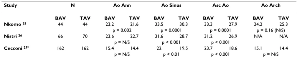
Table 2: Aortic dimensions (mm) in patients with haemodynamically normal AV.
Study N Ao Ann Ao Sinus Asc Ao Ao Arch
BAV TAV BAV TAV BAV TAV BAV TAV BAV TAV
Nkomo 25 44 44 23.2 21.6 33.5 30.3 33.3 27.9 24.2 25.3
p = 0.002 p = 0.0001 p = 0.0001 p = 0.16 (N/S)
Nistri 26 66 70 23.6 22.7 31.6 28.7 31.2 26.9 N/A N/A
p = N/S p < 0.001 p < 0.001
Cecconi 27* 162 162 15.4 14.4 22 19.5 23.7 18.6 15.1 14.4
p = N/S p < 0.01 p < 0.001 p = N/S
* Dimensions in mm/m2
Ao Ann = aortic annulus, Ao Sinus = aortic sinus, Asc Ao = ascending aorta, Ao Arch = aortic arch. BAV = Bicuspid aortic valve, TAV = Tricuspid aortic valve
consistently larger in patients with BAV. The largest differ-ence was seen in the dimensions of the proximal ascend-ing aorta. There was no significant difference between the BAV and control groups with respect to dimensions of the aortic arch. Other studies have confirmed these findings, of dilatation of the aortic root and ascending aorta in patients with BAV, and have also shown that there is no increased dilatation seen in the descending or abdominal aorta [26,27] (Table 2).
Aortic dilatation associated with aortic stenosis
Crawford and Roldan [3] carried out a study to determine the prevalence of dilated aortic root in patients with AS. They studied the echocardiograms of 118 patients with AS, with a valve area of <2.0 cm2. They were age-matched
to patients with aortic sclerosis, but no stenosis, and to normal controls. The aortic root diameter at the annulus, coronary sinus and STJ levels were measured using tran-sthoracic echocardiography. Dilated aortas were defined as 2 SDs above the mean values obtained in the normal group. This study concluded that aortic root dilatation is common in AS, but is not related to severity of the steno-sis. They did not, however, make any comparison between BAV and TAV. As stated earlier, one of the limitations of this study is that only patients with a valve area < 2.0 cm2
were included in the study.
Morgan-Hughes et al [28] carried out a prospective study measuring the aortic root and ascending aortic diameters,
using CT scan images, on patients (BAV, n = 10; TAV, n = 18) with severe AS prior to undergoing AVR. They con-cluded that patients with BAV and pure, severe AS have moderately dilated thoracic aorta compared to matched TAV (p < 0.005) (Table 3).
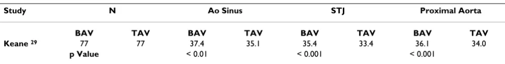
Keane et al [29] carried out a retrospective study to com-pare aortic size in BAV and controls with matched valvular lesions (AS, AR or mixed lesions). They measured the diameter of the left ventricular outflow tract (LVOT), sinus of Valsalva, STJ and proximal ascending aorta using tran-sthoracic echocardiograms. 118 consecutive patients with BAV were compared to controls with TAV. Paired analysis demonstrated significant aortic dilatation at all levels measured in BAV patients, with all degrees of AS and/or AR, compared to controls (Table 4). These differences were seen despite the significantly older age of the control group compared to the BAV group (mean 55.3 years (TAV) and 43.9 years (BAV)). This study also confirms the result mentioned above, that the degree of dilatation is not related to the severity of AS. It suggests that post-sten-otic dilatation is a feature of BAV but not of congenitally normal valves. It supports the theory that this is due, in part, to an underlying pathology within the aortic wall.
Aortic dilatation occurring post AVR
Patients that undergo AVR often have some degree of ascending aortic dilatation. At present, replacing the aortic root at the time of AVR is controversial as the risk of aortic
Table 3: Aortic dimensions (mm) in patients with AS
Study N Ao Ann Ao Sinus STJ
Crawford 3 No AS 108 25 36 30
Mild AS 47 25 36 36*
Moderate AS 29 24 36 36*
Severe AS 42 24 36 35*
BAV TAV BAV TAV BAV TAV BAV TAV
Morgan-Hughes 28 10 18 N/A N/A 41.1 33.8 39.1 31.1
p Value < 0.005 < 0.005
*p < 0.001 compared to normal/sclerotic AV.
Ao Ann = aortic annulus, Ao Sinus = aortic sinus, STJ = sinotubular junction. BAV = Bicuspid aortic valve, TAV = Tricuspid aortic valve
N/A = not applicable.
Table 4: Aortic dimensions (mm) of matched patients with AS/AR/Mixed valve disease.
Study N Ao Sinus STJ Proximal Aorta
BAV TAV BAV TAV BAV TAV BAV TAV
Keane 29 77 77 37.4 35.1 35.4 33.4 36.1 34.0
p Value < 0.01 < 0.001 < 0.001
dilatation following AVR is uncertain. Some studies have been carried out to determine the natural history of ascending aortic dilatation following AVR.
Yasuda et al [4] carried out a retrospective analysis of patients (BAV n = 13, TAV n = 14), using echocardiogra-phy, before and after AVR and 18 BAV patients without AVR. Diameters were measured at the sinus of Valsalva, STJ and the proximal ascending aorta. The annual dilata-tion rate was calculated by dividing changes of diameter during the follow-up period by the BSA and the observa-tional interval. Aortic dilatation in BAV patients was seen to be significantly faster than that of TAV at the proximal aorta only (0.18 compared to -0.08 mm/m2/year). There
was no significant difference in the dilatation rates of BAV patients with and without AVR (0.03 and 0.02 mm/m2/
year respectively). This study showed that AVR could not prevent progressive aortic dilatation in BAV. However, TAV patients did not show further aortic dilatation after AVR. It therefore seems that haemodynamic factors are not responsible for the ongoing dilatation in BAV.
Genetic/structural abnormality
Associations have been found between post-stenotic dila-tion and genetic and structural abnormalities within the AV and ascending aorta, as set out below. However, there is a lack of randomised trial data to establish whether these associations are in fact causal.
Fibrillin-1
Fibrillin-1 is a major protein component of extracellular matrix fibrils, called microfibrils. The FBN1 gene, found on chromosome 15, encodes for this large glycoprotein. Vascular tissues with deficient fibrillin-1 microfibrils release metalloproteinases. These enzymes weaken the vessel wall by degrading the elastic matrix components and leading to matrix disruption and consequent dilata-tion of the vessel.
Marfan's syndrome results from mutations in the FBN1 gene. The most common cardiovascular complication of this condition is progressive aortic root enlargement, ini-tially occurring at the sinus of Valsalva, and ascending aor-tic aneurysms. The aneurysms develop as a consequence of disruption of the medial and adventitial elastin and collagen in association with focci of cystic medial necrosis of the medial smooth muscle. There is also seen to be increased expression of MMPs (especially 2 and 9) within the aneurysmal aorta and, to a lesser extent, the AV [30].
Fedak et al [31] carried out a study to assess the vascular matrix remodelling in patients with BAV and its implica-tion for aortic dilataimplica-tion. Samples of aorta and pulmonary artery were obtained from patients undergoing surgery with BAV (n = 21) and TAV (n = 16). The amount of
fibril-lin-1, elastin and collagen was determined using quantita-tive immunohistochemical analysis, using fluorescence microscopy, for fibrillin-1, and hydroxyproline determi-nation. Fibrillin-1 content was significantly reduced, in both the aorta and pulmonary artery, of BAV patients compared to that seen in TAV suggesting a systemic defi-ciency of this. It was independent of valve function and patient age. However, the amount of matrix components, elastin and collagen, were unchanged. This decrease in the amount of fibrillin-1 in the vasculature of patients with BAV may trigger MMP production leading to matrix dis-ruption and vascular dilatation.
Recent studies have demonstrated that there is a difference in the expression of matrix proteins between the convexity and concavity of the dilated aorta in patients with BAV [32]. These results are consistent with the wall-stress asymmetry that has also been reported by Robicsek and colleagues [33]. They studied three explanted haemody-namically normal congenitally BAV, where they used a simulator to produce a computerised digital model. They showed excessive folding and creasing persistent through-out the cardiac cycle, extended area of leaflet contact, sig-nificant morphologic stenosis and asymmetrical flow patterns and turbulence. Furthermore, Richards et al [34] studied the influence of structural geometry on the sever-ity of BAV and stenosis using transthoracic, transoesopha-geal echocardiography and computer simulations. They demonstrated that for the same anatomic orifice area, functional severity is greater in BAV than in degenerative TAV patients with AS.
Matrix metalloproteinases (MMPs)
MMPs and is produced by fibroblasts or smooth muscle cells.
Degradation of ECM, especially elastin, within the aortic wall is a hallmark of abdominal aortic aneurysms (AAA). Studies have identified increased expression of MMP-1, MMP-2, MMP-9, TIMP-1, TIMP-2 and MMP/TIMP ratios as an important factor in the aetiology of AAA formation. However, the predominant MMP expressed in AAA is MMP-9, produced by macrophages [38].
Elastin and collagen degradation in thoracic aortic aneu-rysms is mediated by MMPs, particularly the gelatinases, MMP-2 and MMP-9.
Le Maire et al [39] studied MMP expression in ascending aortic aneurysms associated with BAV and TAV. Samples of ascending aorta were obtained from 29 patients (BAV n = 14, TAV n = 15). Histological and immunohistochemi-cal analysis was carried out on the specimens. They showed that ascending aortic aneurysms exhibited increased MMP expression when compared to controls (non-aneurysmal aortic tissue). The pattern of MMP expression, however, differed between aneurysms associ-ated with BAV and those with TAV. Another study, carried out by Boyum et al [40], showed that there was an increase in the level of MMP-2 and MMP-9 in thoracic aor-tic aneurysms associated with BAV compared to TAV. They did not, however, find any significant difference in the expression of TIMP-1 or TIMP-2.
Most studies have looked at the increased expression of MMPs within aneurysmal tissue. Ikonomidis et al [41] studied the effects of deletion of the TIMP-1 gene on the progression of murine thoracic aortic aneurysms (TAA). They used adult wild-type and TIMP-1 knockout mice. They showed that deletion of the TIMP-1 gene resulted in an increase in size and continued progression of TAA for-mation compared with wild-type mice. They concluded that this was, at least in part, due to the alteration of the balance between gelatinase activity and its endogenous inhibition. These results suggest that therapeutic strategies that can shift the MMP/TIMP stoichiometric balance away from net proteolysis may be used to inhibit the incidence and progression of TAA.
In diseased or degenerated congenitally deformed valves, increased local MMP activity could alter their elastic and collagen component leading to structural and functional failure.
Histological and immunohistochemical analysis of the AV in patients with nonrheumatic TAV and AS revealed an inflammatory infiltrate within the AV leaflets and an increase in expression of MMP-1, MMP-2 and MMP-3 in
AV of patients with severe AS. It was also found that MMP-9 was only present in the leaflets of patients with AS [42,43].
Koullias et al [44] carried out a study to semiquanitatively analyse the expression of MMPs (1, 2 and 9) and TIMPs (1 and 2) in AV tissue. The study group consisted of 26 patients, undergoing surgery for AS, AR, ascending aortic aneurysm or type A dissection (BAV n = 10, TAV n = 16, controls n = 4). They showed that MMP-9 expression was significantly higher in BAV compared to normal and dis-eased TAV. This incrdis-eased proteolytic presence in BAV may lead to the observed decrease in elastin and collagen content and their resultant functional failure. Others have reported similar results [45].
In summary, these findings suggest that an increase in MMP expression in the aorta of BAV patients may explain the predilection to aneurysm formation. Therefore, by modifying the MMP activity, it maybe possible to reduce or prevent the progression of thoracic aortic aneurysms (TAA). There may also be a role for altering the expression of MMPs to help reduce the progression of BAV disease.
Histological abnormalities
Congenital BAV is associated with cystic medial necrosis, that is, necrosis of the medial smooth muscle cells and accumulation of proteoglycan, of the aorta. De Sa et al [46] examined the histological changes in the ascending aorta and pulmonary trunk in patients with BAV. They studied 31 patients (BAV, n = 20 and TAV, n = 11) under-going AVR. Samples of ascending aorta and pulmonary trunk were collected at the time of surgery. The degenera-tive changes (medionecrosis, fibrosis, cystic medial necro-sis (mucoid material accumulation), changes in smooth muscle cell orientation and elastic fragmentation) in the ascending aorta and pulmonary trunk of patients with BAV disease were significantly more severe than in TAV patients. This severity was mainly related to degree of cystic medial necrosis, smooth muscle cell changes and elastic fragmentation. These findings may explain aortic root and ascending aortic dilatation in patients with BAV disease and pulmonary autograft dilation in certain patients following the Ross procedure.
Ascending aortic dilatation post AVR
defined as an increase in diameter of >0.3 cm from the preoperative measurement. This was observed in only 15% of the study population. No patients with baseline aortic dilatation (3.5–5.3 cm) dilated > 5.5 cm during the follow-up period (n = 107, mean 33.6 months). They found no clinical or valvular characteristics that predicted progressive aortic dilatation. Their conclusion was there-fore against routine replacement of the ascending aorta at the time of AVR.
Another clinical study, by Matsuyama et al [48], looked at the incidence of aortic complications in 35 patients fol-lowing AVR, with a preoperative dilated ascending aorta = 4.0 cm, assessed by computed tomography or operative findings. The baseline aortic diameter in the study popu-lation ranged from 4.0–5.5 cm. The mean follow -up period was 8.1 ± 3.5 years (range: 2.3–13 years). Aortic events occurred in 5 patients (1 aortic dissection, 2 aortic rupture and 2 reoperations). These complications occurred in patients with baseline aortic diameters 4.7– 5.0 cm. The authors concluded that the clinical course of patients with dilated ascending aorta is unpredictable and may occur even in patients with a baseline aortic diameter < 5.0 cm. They concluded that preventative aortic surgery at the time of AVR should be considered, to prevent aortic rupture and dissection, in patients with ascending aorta of 4.0–5.0 cm.
As mentioned earlier, Yasuda et al [4] showed that AVR in patients with BAV did not prevent progressive dilatation of the ascending aorta.
Surgical implications
At present there is controversy over the best management for mild to moderate aortic dilatation associated with AV disease, especially BAV. This is partly due to the lack of concrete evidence for progressive dilatation following AVR and partly due to the increased risk associated with aortic root replacement (ARR).
A retrospective study carried out by Shapira et al [49] over a 10 year period (1987–1997) showed that advances in non-invasive diagnosis and improved perioperative man-agement has lead to a decrease in morbidity and mortality of patients undergoing surgery on the proximal aorta with the operative mortality of thoracic aortic aneurysm repair reduced from between 9–17% to 2.6%.
Should the ascending aorta be replaced in patients with BAV?
AVR is the treatment of choice for patients with sympto-matic AV disease. Current recommendations for the surgi-cal replacement of ascending aortic aneurysms are diameter >5.5 cm or = 5.0 cm in patients with Marfan's syndrome [50]. Borger et al [51] studied 201 patients with BAV (mean age 56 ± 15 years) who underwent AVR. Patients were excluded if they had concomitant ascending aorta replacement. All BAV with ascending aorta > 5.0 cm had ascending aortic replacement and were therefore excluded. During the follow-up period of 10.3 years, 22 patients had long-term complications related to the ascending aorta including replacement of ascending aorta for aneurysm (n = 18), dissection (n = 1) and sudden car-diac death (n = 3). 44 patients had reoperations, mainly for AV prosthesis failure. The 15-year freedom from ascending aorta-related complications was 86%, 81% and 43% in patients with an aortic diameter of <4.0 cm, 4.0– 4.4 cm and 4.5–4.9 cm respectively. The authors therefore concluded that patients undergoing AVR for BAV disease should have their ascending aorta replaced if the preoper-ative diameter is >4.5 cm.
Ubranski et al [52] carried out a case-matched study that showed that replacement of the ascending aorta and AV can be performed with similar operative risk, valve-related mortality and late cardiac mortality as isolated AVR. They analysed 100 patients with AV disease and aneurysm (diameter >4.5 cm) of the aorta who underwent AVR and ascending aorta replacement (± complete root) and a
Table 5: Ascending aorta dilatation rate following AVR
Study N F/U (mo) Expansion Rate (cm/year)
Andrus 47 185 30.0 -0.03*
107 33.6 -0.01*
BAV TAV BAV TAV
21 164 30.0 +0.14 ** -0.05
Matsuyama 48 35 97.2 +0.058*
Yasuda 4 BAV TAV BAV TAV
13 14 116.4 +0.018*** -0.008
cm/(m2/yr) cm/(m2/yr)
*p = N/S, **p = 0.06, ***p = 0.03
matched group of patients undergoing just an AVR. There was no significant difference in the early mortality. 5-year survival seen in the ARR group was 60.7% compared to 86.3% in the AVR group (p = 0.13). At a mean follow-up period of 37 ± 17 months the freedom from cardiac deaths was almost identical in both groups. Similarly, Sundt and colleagues [53] in a retrospective analysis com-paring ARR with separate valve and ascending aorta replacement reported no significant difference in early mortality, but showed better survival for the complete root replacement at 5.6 years follow-up (p = 0.04).
Another surgical approach suggested for the management of BAV, associated with a dilated ascending aorta < 5.5 cm, is to carry out AVR with wrapping of the ascending aorta. This method has a low morbidity and mortality rate and was seen to decrease the risk of further dilatation, aneu-rysm formation and dissection [54,55]. More recently this procedure has been carried out using an external support made to fit the patient's aorta. This was done using digital information from magnetic resonance images to make a replica of the patient's aorta and then computer-aided design to produce the tailored graft [56].
Suitability of the Ross procedure in BAV
The Ross and Ross-Konno procedure allows the replace-ment of a stenotic or regurgitant AV in children and young adults with congenital AV disease. Dilatation of the pul-monary autograft root is a common complication follow-ing the Ross procedure. The pulmonary and aortic roots share a common embryological origin and it has been proposed that the dilatation of the pulmonary autograft may occur as a result of an intrinsic abnormality within the wall, as seen in the aorta of patients with congenital AV disease [57]. This theory is supported by the findings of de Sa et al [46], that the degenerative changes in the ascending aorta and pulmonary trunk of patients with BAV were more severe than those with TAV. However, this result has been contradicted by Schmid et al [58], who failed to show an association between morphological abnormalities in the dilated aorta and pulmonary artery, in patients with either BAV or TAV. They did, however, confirm the finding of more severe degenerative changes in the aorta associated with BAV. Risk factors for late dila-tation of the pulmonary autograft include younger patients, preoperative aortic aneurysm, BAV and those having ARR without support of the annulus and STJ [57,59]. Inclusion techniques have also been described whereby the pulmonary aoutograft is encased in a Dacron tube to prevent dilatation [60].
Hraska et al [61] analysed the mid-term results of 66 chil-dren who had undergone the Ross procedure. The mortal-ity rate approached zero in both simple and complex left heart lesions, including infants and neonates. Their main
concern was dilatation of the neo-aortic root leading to progression of AR, especially in patients with BAV. Bogers and colleagues analysed 123 patients, 81 with BAV who underwent the Ross procedure at a median follow-up of 5.3 years. Freedom from allograft and autograft interven-tion was similar in both groups [62].
Other therapeutic modalities
B-blockers
B-blockers decrease the sheer stress of the vessel wall distal to a stenotic lesion. B-blockers are used as long-term ther-apy in Marfan's syndrome to reduce the rate of pressure change in the aortic root. This is achieved by using their negative inotropic and chronotropic effects to reduce the impulse of the left ventricular ejection and decrease the heart rate. This has been shown to decrease the rate of aor-tic dilatation and reduce the development of aoraor-tic com-plications [63]. However, no studies have been carried out in BAV patients.
In animal models of AR β-blockers have been shown to reduce the ventricular dilatation and improve remode-ling.
Angiotensin-converting enzyme (ACE) inhibitors and statins
Aortic stenosis and atherosclerosis share several common risk factors: male sex, older age, hypertension, diabetes, smoking and elevated levels of low-density lipoprotein (LDL) cholesterol and lipoprotein(a) [1,7]. These obser-vations have led to the proposal of pharmacological strat-egies, already used in atherosclerosis, e.g. ACE inhibitors and hydroxymethylglutaryl-coenzyme A reductase inhibi-tors (statins), which may slow the progression of AS.
ACE has been shown to play an important role in devel-opment of atherosclerosis, presumably via its proinflam-matory effects. ACE has been found to be present in aortic sclerotic and stenotic valves, but is not found in normal aortic valves, where it may participate in lesion develop-ment, as is evidenced by the presence of its enzymatic product, angiotensin II. The observed association between ACE and LDL in both lesions and plasma suggests that LDL may deliver ACE to the lesions [64]. It has previously been shown that ACE inhibitors slow calcium accumula-tion in aortic valves but a recent study found that ACE inhibitors did not slow the haemodynamic progression rate of AS [65].
ste-nosis. This rate of haemodynamic progression was unre-lated to cholesterol levels and it is therefore thought that the effects of statins at the valvular level maybe due to their pleiotropic or anti-inflammatory properties rather than by their cholesterol-lowering effects [66]. However, as stated previously, the SALTIRE study group [8] found that treatment with atorvastatin did not halt the progres-sion of calcific aortic stenosis or induce its regresprogres-sion. The anti-inflammatory effects, independent of their lipid-low-ering effects, of statins have also been implicated as the mechanism by which they have been shown to suppress the development of experimental abdominal aortic aneu-rysms (AAA) in normal and hypercholesterolaemic mice. AAAs are associated with atherosclerosis, chronic inflam-mation and matrix metalloproteinase (MMP)-mediated connective tissue destruction. Statins were shown to pre-serve the medial elastin and smooth muscle cells and to alter the aortic wall expression of MMPs and their inhibi-tors [66].
Calcium antagonists
These have been used in Marfan's syndrome to reduce the rate of dilatation of the aortic root [67]. No studies have yet been carried out in BAV patients but they may have a role in decreasing the rate of post-stenotic dilatation.
N-terminal B-type natriuretic peptide
B-type natriuretic peptide (BNP) and N-terminal BNP (NtBNP) are neurohormones synthesized and secreted mainly by the ventricular myocardium. An increase in synthesis of NtBNP is associated with wall stress. Natriu-retic peptides have been reported to be independent pre-dictors of outcome in congestive heart failure, primary pulmonary hypertension, acute myocardial infarction and pulmonary embolism. Plasma levels of natriuretic pep-tides are known to be related to disease severity and symp-tomatic status in AS [68,69] and now preoperative NtBNP has been shown to predict postoperative outcome with regard to survival, symptomatic status and left ventricular function in severe AS [70]. As yet, this has not been meas-ured in aortic disease.
Synthetic MMP inhibitors (MMPIs)
The MMPs are involved in many physiological functions and, therefore, general inhibition may not be feasible and specific inhibition may be required. The activity of MMPs is kept under tight control at the level of transcription, activation of latent proenzymes and inhibition of proteo-lytic activity. Modulation of MMP regulation can occur at various biochemical sites. Therapeutic manipulation of the extracellular matrix has been used in other disease processes, including arthritis and malignancy. Possible mechanisms of MMP inhibition include: increasing the levels of the naturally occurring inhibitors (TIMPs), by either exogenous administration of recombinant TIMPs
or by increasing their local production; administration of synthetic inhibitors; and decreasing the production of MMPs.
Synthetic inhibitors of MMPs have been investigated in other disease processes, including the use of minocycline in rheumatoid arthritis, and several are currently under investigation for the use in cardiovascular disease [35].
Conclusion
Post-stenotic aortic dilatation is most commonly seen in patients with a BAV. The degree of this dilatation, how-ever, may not be related to the degree of AS and does not appear to be influenced by the occurrence of AVR. It is likely that the dilatation of the ascending aorta is due, mainly, to intrinsic pathology within the aortic wall rather than the haemodynamic effects of a dysfunctional AV.
There is controversy as to whether the ascending aorta should be replaced at the time of initial AV surgery, if the diameter of the aorta is < 5.5 cm. Most of the evidence suggests that the aorta will continue to dilate at an unknown rate. With improvements of surgical technique and perioperative management resulting in decreased morbidity and mortality following ARR, replacement of the ascending aorta should probably be considered for diameters of 4.5 – 5.5 cm. Other therapeutic strategies being investigated that may reduce the rate of dilation are β-blockers, statins and the new synthetic MMP inhibitors.
Competing interests
The author(s) declare that they have no competing inter-ests.
References
1. Stewart BF, Siscovick D, Lind BK, Gardin JM, Gottdiener JS, Smith VE, Kitzman DW, Otto CM: Clinical factors associated with calcific aortic valve disease: Cardiovascular Health Study. J Am Coll Cardiol 1997, 29(3):630-634.
2. Lindroos M, Kupari M, Heikkila J, Tilvis R: Prevalence of aortic valve abnormalities in the elderly: an echocardiographic study of a random population sample. J Am Coll Cardiol 1993,
21(5):1220-1225.
3. Crawford MH, Roldan CA: Prevalence of aortic root dilatation and small aortic roots in valvular aortic stenosis. Am J Cardiol
2001, 87(11):1311-1313.
4. Yasuda H, Nakatani S, Stugaard M, Tsujita-Kuroda Y, Bando K, Koba-yashi J, Yamagishi M, Kitakaze M, Kitamura S, Miyatake K: Failure to Prevent Progressive Dilation of Ascending Aorta by Aortic Valve Replacement in Patients With Bicuspid Aortic Valve: Comparison With Tricuspid Aortic Valve. Circulation 2003,
108:291-294.
5. Fedak PWM, Verma S, David TE, Leask RL, Weisel RD, Butany J: Clin-ical and pathophysiologClin-ical implications of a bicuspid aortic valve. Circulation 2002, 106:900-904.
6. Iung B, Baron G, Butchart EG, Delahaye F, Gohlke-Bärwolf C, Levang OW, Tornos P, Vanoverschelde J-L, Vermeer F, Boersma E, Ravaud P, Vahanian A: A prospective survey of patients with valvular heart disease in Europe: The Euro Heart Survey on Valvular Heart Disease. Eur Heart J 2003, 24:1231-1243.
atheroscle-rosis risk factors – a causal relationship? A clinical morphologic study. Clin Cardiol 1991, 14(12):995-999.
8. Cowell SJ, Newby DE, Prescott RJ, Bloomfield , Reid J, Northridge DB, Boon NA: A randomised trial of lipid-lowering therapy in calcific aortic stenosis. New Eng J Med 2005, 352(23):2389-2397. 9. Roberts WC: The congenitally bicuspid aortic valve: a study of
85 autopsy studies. Am J Cardiol 1970, 26:72-83.
10. Campbell M: Calcific aortic stenosis and congenital bicuspid aortic valve. Br Heart J 1968, 30:606-616.
11. Ward C: Clinical significance of the bicuspid aortic valve.
Heart 2000, 83:81-85.
12. Roberts WC, Ko JM: Frequency by Decades of Unicuspid, Bicuspid, and Tricuspid Aortic Valves in Adults Having Iso-lated Aortic Valve Replacement for Aortic Stenosis, With or Without Associated Aortic Regurgitation. Circulation 2005,
111(7):920-925.
13. Sans-Coma V, Fernandez B, Duran AC, Thiene G, Arque JM, Munoz-Chapuli R, Cardo M: Fusion of valve cushions as a key factor in the formation of congenital bicuspid aortic valves in Syrian hamsters. Anat Rec 1996, 244(4):490-498.
14. Fernandez MC, Duran AC, Real R, Lopez D, Fernandez B, de Andres AV, Arque JM, Gellego A, Sans-Coma V: Coronary artery anoma-lies and aortic valve morphology in the Syrian Hamster. Lab Anim 2000, 34(2):145-154.
15. Becker AE, Becker MJ, Edwards JE: Anomalies associated with coarctation of the aorta – Particular reference to infancy.
Circulation 1970, 41:1067-1075.
16. Stewart AB, Ahmed R, Travill CM, Newman CG: Coarctation of the aorta, life and health 20–44 years after surgical repair. Br Heart J 1993, 69:65-70.
17. Huntington K, Hunter AGW, Chan K-L: A prospective study to assess the frequency of familial clustering of congenital bicuspid aortic valve. J Am Coll Cardiol 1997, 30:1809-1812. 18. Hahn RT, Roman MJ, Mogtader AH, Devereux RB: Association of
aortic dilation with regurgitant, stenotic and functionally normal bicuspid aortic valves. J Am Coll Cadiol 1992, 19:283-8. 19. Kappetein AP, Gittenberger-de Groot AC, Zwinderman AH, Rohmer
J, Poelmann RE, Huysmans HA: The neural crest as a possible pathogenic factor in coarctation of the aorta and bicuspid aortic valve. J Thorac Cardiovasc Surg 1991, 102(6):830-836. 20. Schievink WI, Mokri B: Familial aorto-cervicocephalic arterial
dissections and congenitally bicuspid aortic valve. Stroke 1995,
26:1935-1940.
21. Lee TC, Zhao YD, Courtman DW, Stewart DJ: Abnormal aortic valve development in mice lacking endothelial nitric oxide synthase. Circulation 2000, 101:2345-2348.
22. Eagleton MJ, Peterson DA, Sullivan VV, Roelofs KJ, Ford JA, Stanley JC, Upchurch GR Jr: Nitric oxide inhibition increases aortic wall matrix metalloproteinase-9 expression. J Surg Res 2002,
104(1):15-21.
23. Gurvitz M, Chang R-K, Drant S, Allada V: Frequency of aortic root dilation in children with a bicuspid aortic valve. Am J Cardiol
2004, 94(10):1337-1340.
24. Okamoto RJ, Xu H, Kouchoukos NT, Moon MR, Sundt TM III: The influence of mechanical properties on the wall stress and dis-tensibility of the dilated ascending aorta. J Thorac Cardiovasc Surg 2003, 126(3):842-850.
25. Nkomo VT, Enriquez-Sarano M, Ammash NM, Melton LJ III, Bailey KR, Desjardins V, Horn RA, Tajik AJ: Bicuspid aortic valve associ-ated with aortic dilatation: A community-based study. Arteri-oscler Thromb Vasc Biol 2003, 23:351-356.
26. Nistri S, Sorbo MD, Marin M, Palisi M, Scognamiglio R, Thiene G:
Aortic root dilatation in young men with normally function-ing bicuspid aortic valves. Heart 1999, 82(1):19-22.
27. Cecconi M, Manfrin M, Moraca A, Zanoli R, Colonna PL, Bettuzzi MG, S Moretti, D Gabrielli, Perna GP: Aortic dimensions in patients with bicuspid aortic valve without significant valve dysfunc-tion. Am J Cardiol 2005, 95(2):292-294.
28. Morgan-Hughes GJ, Roobottom CA, Owens PE, Marshall AJ: Dilata-tion of the aorta in pure, severe, bicuspid aortic valve steno-sis. Am Heart J 2004, 147(4):736-740.
29. Keane MG, Wiegers SE, Plappert T, Pochettino A, Bavaria JE, John Sutton MG St: Bicuspid Aortic Valves Are Associated With Aortic Dilatation Out of Proportion to Coexistent Valvular Lesions. Circulation 2000, 102:35-39.
30. Segura AM, Luna RE, Horiba K, Stetler-Stevenson WG, McAllister HA, Willerson JT, Ferrans VJ: Immunohistochemistry of matrix metalloproteinases and their inhibitors in thoracic aortic aneurysms and aortic valves of patients with Marfan's syn-drome. Circulation Suppl 1998, 98:II-331-II-338.
31. Fedak PWM, de Sa MPL, Verma S, Nili N, Kazemian P, Butany J, Strauss BH, Weisel RD, David TE: Vascular matrix remodeling in patients with bicuspid aortic valve malformations: implica-tions for aortic dilatation. J Thorac Cardiovasc Surg 2003,
126(3):797-805.
32. Cotrufo M, Della Corte A, De Santo LS, Quarto C, De Feo M, Romano G, Amarelli C, Scardone M, Di Meglio F, Guerra G, Scarano M, Vitale S, Castaldo C, Montagnani S: Different patterns of extra-cellular matrix protein expression in the convexity and the concavity of the dilated aorta with bicuspid aortic valve: Pre-liminary results. J Thorac Cardiovasc Surg 2005, 130(2):504-511. 33. Robicsek F, Thubrikar MJ, Cook JW, Fowler B: The congenitally
bicuspid aortic valve: how does it function? Why does it fail?
Ann Thorac Surg 2004, 77:177-185.
34. Richards KE, Deserranno D, Donal E, Greenberg NL, Thomas JD, Garcia MJ: Influence of structural geometry on the severity of bicuspid aortic stenosis. Am J Physiol Heart Circ Physiol 2004,
287:H1410-H1416.
35. Dollery CM, McEwan JR, Henney AM: Matrix Metalloproteinases and cardiovascular disease. Circ Res 1995, 77:863-868. 36. Visse R, Nagase H: Matrix metalloproteinases and tissue
inhib-itors of metalloproteinases: structure, function, and bio-chemistry. Circ Res 2003, 92:827-839.
37. Nagase H, Woessner JF Jr: Matrix metalloproteinases. J Biol Chem
1999, 274:21491-21494.
38. Tamarina NA, McMillan WD, Shively VP, Pearce WH: Expression of matrix metalloproteinases and their inhibitors in aneurysms and normal aorta. Surg 1997, 122(2):264-271.
39. LeMaire SA, Wang X, Wilks JA, Carter SA, Wen S, Won T, Leon-ardelli D, Anand G, Conklin LD, Wang XL, Thompson RW, Coselli JS:
Matrix metalloproteinases in ascending aortic aneurysms: Bicuspid versus trileaflet aortic valves. J Surg Res 2005,
123(1):40-48.
40. Boyum J, Fellinger EK, Schmoker JD, Trombley L, McPartland K, Ittle-man FP, Howard AB: Matrix metalloproteinase activity in tho-racic aortic aneurysms associated with bicuspid and tricuspid aortic valves. J Thorac Cardiovasc Surg 2004,
127(3):686-691.
41. Ikonomidis JS, Gibson WC, Butler JE, McClister DM, Sweterlitsch SE, Thompson RP, Mukherjee R, Spinale FG: Effects of Deletion of the Tissue Inhibitor of Matrix Metalloproteinases-1 Gene on the Progression of Murine Thoracic Aortic Aneurysms. Circulation Suppl 2004, 110:II-268-273.
42. Kaden JJ, Dempfle C-E, Grobholz R, Fischer CS, Vocke DC, Kilic R, Sarikoc A, Pinol R, Hagl S, Lang S, Brueckmann M, Borggrefe M:
Inflammatory regulation of extracellular matrix remodeling in calcific aortic valve stenosis. Cardiovasc Pathol 2005,
14(2):80-87.
43. Edep ME, Shirani J, Wolf P, Brown DL: Matrix metalloproteinase expression in nonrheumatic aortic stenosis. Cardiovasc Pathol
2000, 9(5):281-286.
44. Koullias GJ, Korkolis DP, Ravichandran P, Psyrri A, Hatzaras I, Elefte-riades JA: Tissue microarray detection of matrix metallopro-teinases, in diseased tricuspid and bicuspid aortic valves with or without pathology of the ascending aorta. Eur J Cardiothorac Surg 2004, 26(6):1098-1103.
45. Soini Y, Satta J, Maatta M, Autio-Harmainen H: Expression of MMP2, MMP9, MT1-MMP, TIMP1 and TIMP2 mRNA in val-vular lesions of the heart. J Pathol 2001, 194(2):225-231. 46. de Sa M, Moshkovitz Y, Butany J, David TE: Histologic
abnormali-ties of the ascending aorta and pulmonary trunk in patients with bicuspid aortic valve disease: clinical relevance to the Ross procedure. J Thorac Cardiovasc Surg 1999, 118(4):588-596. 47. Andrus BW, O'Rourke DJ, Dacey LJ, Palac RT: Stability of
Ascend-ing Aortic Dilatation FollowAscend-ing Aortic Valve Replacement.
Circulation Suppl 2003, 108:II-295-II-229.
Publish with BioMed Central and every scientist can read your work free of charge "BioMed Central will be the most significant development for disseminating the results of biomedical researc h in our lifetime."
Sir Paul Nurse, Cancer Research UK
Your research papers will be:
available free of charge to the entire biomedical community
peer reviewed and published immediately upon acceptance
cited in PubMed and archived on PubMed Central
yours — you keep the copyright
Submit your manuscript here:
http://www.biomedcentral.com/info/publishing_adv.asp
BioMedcentral
49. Shapira OM, Aldea GS, Cutter SM, Fitzgerald CA, Lazar HL, Shemin RJ: Improved clinical outcomes after operation of the proxi-mal aorta: a 10-year experience. Ann Thorac Surg 1999,
67:1030-1037.
50. Elefteriades JA: Natural history of thoracic aortic aneurysms: indications for surgery, and surgical versus nonsurgical risks.
Ann Thorac Surg 2002, 74:S1877-S1880.
51. Borger MA, Preston M, Ivanov J, Fedak PWM, Davierwala P, Arm-strong S, David TE: Should the ascending aorta be replaced more frequently in patients with bicuspid aortic valve dis-ease? J Thorac Cardiovasc Surg 2004, 128(5):677-683.
52. Urbanski PP, Wagner M, Zacher M, Hacker RW: Aortic root replacement versus aortic valve replacement: a case-match study. Ann Thorac Surg 2001, 72(1):28-32.
53. Sundt TM 3rd, Mora BN, Moon MR, Bailey MS, Pasque MK, Gay WA Jr: Options for repair of a bicuspid aortic valve and ascending aortic aneurysm. Ann Thorac Surg 2000, 69:1333-1337.
54. Robicsek F, Thubrikar MJ: Conservative operation in the man-agement of annular dilatation and ascending aortic aneu-rysm. Ann Thorac Surg 1994, 57:1672-1674.
55. Kuralay E, Demirkilic U, özal E, Savas öz B, Cingöz F, Günay C, Ceylan S, Arslan M, Tatar H: Surgical approach to ascending aorta in bicuspid aortic valve. J Card Surg 2003, 18:173-180.
56. Golesworthy T, Lampérth M, Mohiaddin R, Pepper J, Thornton W, Treasure T: The tailor of Gloucester: a jacket for the Marfan's aorta. Lancet 2004, 364:1582-.
57. David TE, Omran A, Ivanov J, Armstrong S, de Sa MP, Sonnenberg B, Webb G: Dilatation of the pulmonary autograft after the Ross procedure. J Thorac Cardiovasc Surg 2000, 119(2):210-220. 58. Schmid F-X, Bielenberg K, Holmer S, Lehle K, Djavidani B, Prasser C,
Wiesenack C, Birnbaum D: Structural and biomolecular changes in aorta and pulmonary trunk of patients with aortic aneurysm and valve disease: implications for the Ross proce-dure. Eur J Cardiothorac Surg 2004, 25:748-753.
59. Luciani GB, Casali G, Favero A, Prioli MA, Barozzi L, Santini F, Maz-zucco A: Fate of the aortic root late after Ross operation. Cir-culation Suppl 2003, 108:II-61-II-67.
60. Slater M, Shen I, Welke K, Komanapalli C, Ungerleider R: Modifica-tion of the Ross procedure to prevent autograft dilataModifica-tion.
Semin Thorac Cardiovasc Surg Pediatr Card Surg Annu 2005, :181-184. 61. Hraska V, Krajci M, Haun C, Ntalakoura K, Razek V, Lacour-Gayet F,
Weil J, Reichenspurner H: Ross and Ross-Konno procedure in children and adolescents: mid-term results. Eur J Cardiothorac Surg 2004, 25(5):742-747.
62. Bogers AJ, Kappetein A-P, Roos-Hesselink JW, Takkenberg JJM: Is a bicuspid aortic valve a risk factor for adverse outcome after an autograft procedure? Ann Thorac Surg 2004, 77:1998-2003. 63. Shores J, Berger KR, Murphy EA, Pyeritz RE: Progression of aortic
dilatation and the benefit of long-term β-adrenergic block-ade in Marfan's syndrome. New Eng J Med 1994, 330:1335-1341. 64. O'Brien KD, Shavelle DM, Caulfield MT, McDonald TO, Olin-Lewis K, Otto CM, Probstfield JL: Association of angiotensin-converting enzyme with low-density lipoprotein in aortic valvular lesions and in human plasma. Circulation 2002, 106:2224-2230. 65. Rosenhek R, Rader F, Loho N, Gabriel H, Heger M, Klaar U,
Schemper M, Binder T, Maurer G, Baumgartner H: Statins but not angiotensin-converting enzyme inhibitors delay progression of aortic stenosis. Circulation 2004, 110:1291-1295.
66. Steinmetz EF, Buckley C, Shames ML, Ennis TL, Vanvickle-Chavez SJ, Mao D, Goeddel LA, Hawkins CJ, Thompson RW: Treatment with simvastatin suppresses the development of experimental abdominal aortic aneurysms in normal and hypercholester-olaemic mice. Ann Surg 2005, 241(1):92-101.
67. Rossi-Foulkes R, Roman MJ, Rosen SE, Kramer-Fox R, Ehlers KH, O'Loughlin JE, Davis JG, Devereux RB: Phenotypic features and impact of βeta blocker or calcium antagonist therapy on aor-tic lumen size in the Marfan syndrome. Am J Cardiol 1999,
83(9):1364-1368.
68. Weber M, Arnold R, Rau M, Brandt R, Berkovitsch A, Mitrovic V, Hamm C: Relation of N-terminal pro-B-type natriuretic pep-tide to severity of valvular aortic stenosis. Am J Cardiol 2004,
94:740-745.
69. Weber M, Arnold R, Rau M, Elsaesser A, Brandt R, Mitrovic V, Hamm C: Relation of N-terminal pro B-type natriuretic peptide to progression of aortic valve disease. Eur Heart J 2005,
26:1023-1030.