
R E V I E W
Open Access
An ongoing search for potential targets
and therapies for lethal sepsis
Guo-qiang Bao
1,2,3†, Li He
4†, David Lee
1, John D
’
Angelo
1and Hai-chao Wang
1,2*Abstract
Sepsis, which refers to a systemic inflammatory response syndrome resulting from a microbial infection, represents
the leading cause of death in intensive care units. The pathogenesis of sepsis remains poorly understood although
it is attributable to dysregulated immune responses orchestrated by innate immune cells that are sequentially
released early (e.g., tumor necrosis factor(TNF), interleukin-1(IL-1), and interferon-
γ
(IFN-
γ
)) and late (e.g., high mobility
group box 1(HMGB1)) pro-inflammatory mediators. As a ubiquitous nuclear protein, HMGB1 can be passively released
from pathologically damaged cells, thereby converging infection and injury on commonly dysregulated inflammatory
responses. We review evidence that supports extracellular HMGB1 as a late mediator of inflammatory diseases and
discuss the potential of several Chinese herbal components as HMGB1-targeting therapies. We propose that it is
important to develop strategies for specifically attenuating injury-elicited inflammatory responses without compromising
the infection-mediated innate immunity for the clinical management of sepsis and other inflammatory diseases.
Keywords:
Innate immune cells, Pathogen-associated molecular pattern molecules, High mobility group box 1, Herbal
components, Sepsis, Autophagy, Endocytosis, Double-stranded RNA-activated protein kinase R
Introduction
Cohabitating with various microbes, mammals have
de-veloped multiple strategies for combatting microbial
infections. As the first layer of defense, the epithelial
barriers effectively limit the access and growth of the
majority of pathogens. If they are breached, innate
im-mune cells immediately launch biological responses,
termed
“
inflammation,
”
to confine and remove these
pathogens [1]. These inflammatory responses are usually
appropriately propagated and often result in the successful
elimination of the invading pathogens. If unsuccessful, the
invading pathogens can leak into the bloodstream,
trigger-ing a widespread and systemic inflammatory response,
termed
“
sepsis.
”
Sepsis, which refers to a systemic
inflam-matory response syndrome resulting from a microbial
in-fection, represents the leading cause of death in intensive
care units. As a continuum of increasing clinical severity,
“
severe sepsis
”
is often associated with one or more acute
organ dysfunctions [2]. Despite recent advances in
anti-biotic therapy and intensive care, the overall mortality rate
of severe sepsis remains high [3].
The inflammatory responses are first initiated by
in-nate immune cells, such as macrophages and monocytes.
These innate immune cells are equipped with pattern
recognition receptors, such as the Toll-like receptors
(TLRs), TLR2, TLR3, TLR4, and TLR9 [4
–
8], for various
pathogen-associated molecular patterns (PAMPs), such
as bacterial peptidoglycan, double-stranded RNA,
endo-toxin, and CpG-DNA [9, 10]. The engagement of various
PAMPs with respective receptors triggers the sequential
release of early (TNF, IL-1 and IFN-
γ
) and late (HMGB1)
proinflammatory mediators [11
–
13]. Although early
pro-inflammatory cytokines contribute to the pathogenesis of
sepsis [14], their early kinetics of release makes them
diffi-cult to target in clinical settings.
Discovery of HMGB1 as a late mediator of lethal sepsis
Approximately 20 years ago, we aimed to search for
other late mediators that might contribute to the
patho-genesis of lethal sepsis. To identify such mediators, we
stimulated macrophages with early cytokines (e.g., TNF)
and screened the cell-conditioned medium for proteins
* Correspondence:[email protected]†Equal contributors
1
Department of Emergency Medicine, North Shore University Hospital, Manhasset, NY 11030, USA
2
The Feinstein Institute for Medical Research, 350 Community Drive, Manhasset, NY 11030, USA
Full list of author information is available at the end of the article
that were released late. The SDS-PAGE gel
electrophor-esis analysis revealed the release of 30-kDa protein with
an N-terminal amino acid sequence identical to HMGB1
[13], a member of the high mobility group-1
non-histone chromosomal protein family.
Active HMGB1 Secretion
HMGB1 is constitutively expressed to maintain a large
“
pool
”
of pre-formed protein in the majority of cells
[15, 16]. Bearing two nuclear-localization sequences
(NLS), HMGB1 is transported into the nucleus by the
nuclear import complexes, thereby maintaining a large
nuclear
“
pool
”
of pre-formed protein (Fig. 1) [17].
Within the nucleus, HMGB1 binds chromosomal DNA
and fulfills its nuclear functions, such as maintaining the
nucleosomal structure and regulating gene expression
[18]. The disruption of local HMGB1 expression renders
animals susceptible to infectious [19] or injurious insults
[20, 21], indicating an overall beneficial role of
intracellu-lar HMGB1 [22]. In response to exogenous PAMPs
(ds-RNA, CpG-DNA and endotoxin) [13, 23] or endogenous
cytokines (interferon (IFN)-
γ
, IFN-
β
and cold-inducible
RNA-binding protein (CIRP)) [24
–
26], macrophages/
monocytes actively release HMGB1. If dysregulated, the
excessive HMGB1 release adversely contributes to the
pathogenesis of infection- and injury-elicited
inflamma-tory diseases.
Lacking a leader signal sequence, HMGB1 cannot be
actively secreted via the classical ER-Golgi secretory
pathway [13]. Instead, the activated macrophages/
monocytes acetylate and phosphorylate HMGB1 at the
nuclear localization or export sequences (NLS or NES)
[22, 27
–
29] lead to the sequestration of HMGB1 within
cytoplasmic vesicles, which are destined for secreting
into the extracellular environment [16, 24, 30]. For
in-stance, in response to exogenous PAMPs (e.g.,
endo-toxin) or endogenous cytokines (e.g., IFNs), innate
immune cells acetylate lysine residues 28, 29, 42, 43,
179, 181, and 183 within the NLS sites lead to the
cyto-plasmic HMGB1 translocation in a JAK/STAT1-dependent
fashion (Fig. 2) [16, 24, 27, 30]. Indeed, the
pharmaco-logical inhibition or genetic interference with JAK/STAT1
signaling uniformly inhibits HMGB1 secretion induced
by IFN-
β
, IFN-
γ
or LPS. Notably, LPS might not
dir-ectly activate STAT1; however, it may trigger indirect
STAT1 activation through the intermediate production
of IFNs [31] that are capable of inducing HMGB1
re-lease [24, 25, 27].
After cytoplasmic translocation, HMGB1 is secreted
extracellularly through several pathways, including the
caspase-1/caspase-11-mediated inflammasome activation
and pyroptosis (Fig. 2). The pharmacological inhibition
with a broad-spectrum caspase inhibitor (Z-VAD-FMK)
or the genetic deletion of caspase-1/caspase-11
uni-formly reduces HMGB1 secretion from activated
macro-phages [32, 33]. Similarly, the genetic disruption of the
double-stranded RNA-activated protein kinase R (PKR)
or the pharmacological inhibition of PKR
phosphoryl-ation similarly reduces NLRP3 or NLRP1
agonists-induced inflammasome activation [34, 35], pyroptosis
[34, 35], and HMGB1 release [34]. In light of the likely
roles of PKR in the regulation of caspase-1-dependent
programmed cell death (pyroptosis) [35] and receptor
interacting protein (RIP)1/RIP3-dependent programmed
necrosis (necroptosis) (Fig. 2) [36], it is important to
ex-plore novel PKR inhibitors that may inhibit HMGB1
re-lease by preventing distinct cell death pathways.
Passive HMGB1 Release
In addition to active secretion, HMGB1 can be passively
released from damaged cells [37] after
ischemia/reperfu-sion [38, 39], trauma [40, 41], or toxemia [42
–
44],
thereby serving as damage-associated molecular pattern
molecule (DAMP). Necrosis can also be induced by
vari-ous viruses (e.g., West Nile, salmon anemia, dengue, and
influenza viruses) [45, 46] and cytokines (e.g., TNF, IFNs)
[36, 47]. Extracellular HMGB1 can also trigger
caspase-1-dependent programmed cell death, pyroptosis, which
is characterized by rapid plasma membrane rupture, and
the release of proinflammatory intracellular contents
(in-cluding HMGB1) [48], suggesting a pathogenic role of
pyroptosis in HMGB1 release during infection or injury.
Thus, infection and injury converge in a common
process, i.e., inflammation [49], which is orchestrated by
HMGB1 and other proinflammatory mediators (e.g.,
mitochondrial DNA and CIRP) released by activated
im-mune cells and damaged tissues [26, 50].
Once actively secreted or passively released,
extracellu-lar HMGB1 binds to various microbial products (e.g.,
CpG-DNA or LPS), thereby facilitating their recognition
by respective receptors to augment inflammatory
re-sponses [51]. Harboring three cysteine residues (C23,
C45 and C106) that are redox-sensitive, HMGB1 can be
modified into three isoforms termed
“
HMGB1
”
(all thiol
form),
“
disulfide HMGB1
”
(partially oxidized), and
oxi-dized HMGB1 (Fig. 1) [52, 53]. The
“
all-thiol
”
HMGB1
binds to other chemokines (e.g., CXCL12) and stimulates
leukocyte recruitment via the CXCR4 receptor [54] or
other signaling molecules [55
–
57] to the infection or
in-jury sites [58, 59]. In sharp contrast, disulfide HMGB1
can activate immune cells to produce
cytokines/chemo-kines via TLR4 or other receptors, such as RAGE [51],
TLR2, TLR4 [60
–
62], TLR9 [23, 51], cluster of
differenti-ation 24 (CD24)/Siglec-10 [63], Mac-1 [57],
thrombomo-dulin [64], or single transmembrane domain proteins
(e.g., syndecans) [65]. Once fully oxidized, HMGB1 is
devoid of either chemokine or cytokine activities (Fig. 1)
[52, 53]. Altogether, these studies suggest that extracellular
HMGB1 is a proinflammatory signal to recruit, alert, and
activate innate immune cells, thereby sustaining a
poten-tially injurious inflammatory response during sepsis.
Pathogenic role of HMGB1 in sepsis and injury
Experimental sepsis can be induced by several
tech-niques, including the infusion of exogenous bacterial
toxins (endotoxemia) and the disruption of host
epithe-lial barrier, to produce microbial translocation, e.g., cecal
ligation and puncture (CLP). In murine models of
endo-toxemia and CLP-sepsis, HMGB1 is first detected in the
circulation 8 h after the disease onset and is
subse-quently increased to plateau levels from 16 to 32 h
(Fig. 3a) [13, 66]. This late appearance of circulating
HMGB1 parallels with the onset of animal lethality from
endotoxemia or sepsis and distinguishes itself from TNF
and other early proinflammatory cytokines [67]. The
pathogenic role of HMGB1 in endotoxemia was inferred
from studies using HMGB1-neutralizing antibodies, which
conferred a dose-dependent protection against
endotoxin-induced tissue injury and lethality [13, 68]. In a more
clinically relevant CLP-induced sepsis, the delayed
ad-ministration of HMGB1-specific neutralizing antibodies
beginning 24 h
after
CLP dose-dependently rescued
ro-dents from lethal sepsis [32, 66, 69]. Moreover, the
tar-geted inhibition of HMGB1 expression in innate immune
cells (e.g., macrophages and dendritic cells) reduces
sys-temic HMGB1 accumulation and similarly rescues mice
from sepsis [70], supporting HMGB1 as a critical late
me-diator of experimental sepsis.
Notably, agents capable of inhibiting HMGB1 release
[71
–
73] or action [13, 66] confer protection against
sep-sis, particularly if administered in a delayed fashion to
strategically preserve the PAMPs-mediated early
inflam-matory response. At late-stage infection, the
PAMPs-mediated inflammatory response may be accompanied
by unintended cell injury and DAMPs release that amplify
the cytokine storm to precipitate organ dysfunction
(Fig. 3a) [29]. This likelihood is supported by recent
find-ings that HMGB1 is persistently elevated during late-stage
sepsis despite the cessation of initial infection [74] and
that it contributes to the long-term pathological
conse-quence of sepsis. Although microbial infection-induced
sepsis is indistinguishable from sterile injury-elicited
systemic inflammatory response syndrome [75, 76], it
may be more advantageous to develop strategies for
spe-cifically attenuating DAMPs-mediated inflammatory
re-sponses without compromising the PAMPs-mediated
innate immunity.
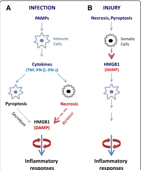
As a ubiquitous nuclear protein, HMGB1 can be
pas-sively released from necrotic cells [37] and can function
as a DAMP to elicit inflammatory responses (Fig. 3b).
Regardless of the origin, the actively secreted or
pas-sively released HMGB1 can similarly alert, recruit, and
activate immune cells [49, 77], triggering infection- and
injury-elicited systemic inflammatory responses that are
often indistinguishable in experimental or clinical
set-tings (Fig. 3) [76]. Indeed, HMGB1-neutralizing
anti-bodies are protective in animal models of ischemia/
reperfusion [38, 78, 79], trauma [80, 81], chemical
tox-emia [42, 82, 83], atherosclerosis [84], gastric ulcer [85]
and hyperoxia [86].
Discovery of Chinese herbs as HMGB1 inhibitors
The establishment of HMGB1 as a mediator of various
inflammatory diseases has prompted the search for
in-hibitors that can attenuate HMGB1 secretion or action
in various experimental settings. As summarized in
sev-eral recent reviews [22, 29, 87], a growing list of herbal
extracts (e.g., Danggui, Mung bean, and
Prunella
vul-garis
) [88, 89] and components have been demonstrated
to be effective in inhibiting endotoxin-induced HMGB1
secretion. In the present review, we compare the distinct
mechanisms by which several herbal therapies effectively
inhibit active HMGB1 secretion and action.
Glycyrrhizin (GZA) binds to HMGB1 to inhibit its secretion or
action
Radix Glycyrrhizae
(
Gancao
in Chinese, meaning
“
sweet
root
”
in Greek or
“
licorice
”
in English) has been
trad-itionally used for treating peptic ulcer, hepatitis, and
pul-monary bronchitis for many centuries. Its major
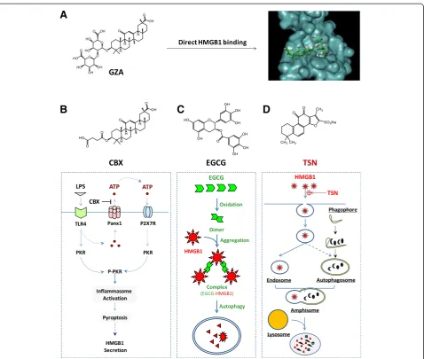
anti-inflammatory component, GZA (Fig. 4a), is protective in
animal models of hepatitis [90], hepatic
ischemia/reperfu-sion (I/R) injury [91, 92], endotoxin- and
acetaminophen-induced liver injury [93, 94]. Using biochemical techniques,
Sakamoto et al. (2001) first demonstrated that GZA directly
interacted with HMGB1 and impaired its DNA-binding
properties [95]. Subsequently, Mollica et al. (2007) used
A
B
nuclear magnetic resonance (NMR) and fluorescence
tech-niques to confirm that GZA directly docked into the
DNA-binding concaves of HMGB1 boxes (Fig. 4a) [96, 97].
Con-sistent with these findings, the GZA-mediated protection
has been associated with the inhibition of HMGB1 release
or its cytokine/chemokine activities [87].
Carbenoxolone (CBX) prevents PKR activation
Carbenoxolone (CBX) is a chemical derivative of
GZA, in which the glucuronic acid is replaced by
suc-cinic acid (Fig. 4b). As a medication previously
pre-scribed for esophageal ulceration and inflammation
[98], CBX has been demonstrated to dose-dependently
inhibit a variety of biological activities, including gap
junctions (50
–
100
μ
M) and panx1 channels (EC
50= 1
–
4
μΜ
) [99, 100]. We recently discovered that CBX
effectively inhibited LPS-induced HMGB1 secretion,
with estimated IC
50at 5
μ
M and IC
100at 10
μ
M [101].
It appears that CBX effectively inhibited
endotoxin-induced HMGB1 release by preventing PKR
up-regulation and phosphorylation (Fig. 4b). In light of
the findings that CBX (10
μ
M) could effectively inhibit
panx-1-mediated ATP release in response to hypoxia
[102], sheer stress [103] and low oxygen tension [104],
we propose that crude LPS (
containing trace amounts
of bacterial proteins and nucleic acids
) may prime
macrophages by up-regulating PKR expression and
simultaneously eliciting panx-1-mediated ATP release.
The extracellular ATP subsequently binds and activates
the purinergic P
2X
7receptor (P
2X
7R) [105], which
trig-gers PKR/inflammasome activation and HMGB1
secre-tion [87, 106].
A
B
C
D
Epigallocatechin-3-gallate (EGCG) stimulates autophagic
HMGB1 degradation
Green tea contains a class of biologically active
poly-phenolic catechins, including the most abundant
epigallocatechin-3-gallate (EGCG) (Fig. 4c). At low
concentrations, EGCG dose-dependently abrogates
LPS-induced HMGB1 secretion, with an estimated
IC
50< 1.0
μ
M [72]. Notably, the significant inhibition
of HMGB1 secretion is achieved when EGCG is added
2 h to 6 h post LPS stimulation [72], suggesting a
likeli-hood of delayed regimen for EGCG treatment. EGCG
appears to prevent LPS-induced HMGB1 secretion
stra-tegically by destroying HMGB1 in the cytoplasm via a
cellular degradation process, autophagy (
“
self-eating
”
)
(Fig. 4c).
EGCG can be trafficked into cytoplasmic vesicles
(pre-sumably autophagosomes) within 6 h and destined to
autophagolysosomes within 16 h [107]. Additionally,
EGCG can conjugate to cytoplasmic HMGB1, leading to
the formation of EGCG-HMGB1 complexes (dimmer,
trimmer, tetramer, and oligomer) (Fig. 4c) [107]. Because
these large EGCG-HMGB1 complexes cannot physically
pass through the narrow pore of the proteasome barrel of
the ubiquitin-proteasome pathway, they trigger the
au-tophagic degradation process. At concentrations effective
for inhibiting HMGB1 secretion, EGCG dramatically
en-hances the formation of autophagosomes [107]. Recently,
EGCG has proven to be effective in stimulating autophagy
in breast cancer cells [107], hepatocytes [108], retinal
pigment epithelial cells [109], and vascular endothelial
cells [110]. Given the likelihood that HMGB1 interacts
with autophagy regulators (e.g., beclin-1) in the
cyto-plasm [111, 112], it will be important to investigate
whether HMGB1 occupies a critical role in
EGCG-mediated autophagy. This assessment is relevant because
recent studies have indicated that bacterial endotoxin
in-duces significantly less autophagy in HMGB1-deficient
macrophages [19].
Tanshinone IIA sodium sulfonate (TSN) stimulates endocytic
HMGB1 uptake
Radix Salviae Miltiorrhizae
(
Danshen
in Chinese) is a
medicinal herb that contains several red pigments,
in-cluding tanshinone I, II, IV, and cryptotanshinone that
consist of various anti-inflammatory properties. As a
major component (representing 5 % to 6 % of the total
dry weight) of Danshen root, tanshinone IIA
dose-dependently attenuates LPS-induced HMGB1 secretion,
with an estimated IC
50< 25
μ
M. However, its poor water
solubility may adversely affect the bioavailability and
therapeutic efficacy of tanshinone IIA [73]. One
water-soluble derivative, tanshinone IIA sodium sulfonate (TSN)
(Fig. 4d), also dose-dependently inhibits LPS-induced
HMGB1 secretion with a lower IC
50< 10
μ
M. At doses
that completely prevent HMGB1 secretion, TSN does
not affect endotoxin-induced release of most other
cy-tokines and chemokines (such as IL-6, IL-12p40/p70,
KC, MCP-1, MIP-1
α
, MIP-2, and TNF), indicating a
se-lectivity for TSN in inhibiting HMGB1 secretion.
Although TSN itself is unable to stimulate autophagic
HMGB1 degradation [72], it induces the internalization
of exogenous HMGB1 into macrophage cytoplasmic
ves-icles likely through clathrin- and caveolin-dependent
endocytosis (Fig. 4d) [113]. The inhibition of
clathrin-dependent (e.g., chlorpromazine) and caveolin-clathrin-dependent
(e.g., nystatin and indomethacin) endocytosis uniformly
attenuates the TSN-mediated HMGB1 uptake.
Surpris-ingly, the depletion of several HMGB1 receptors (e.g.,
TLR2, TLR4, or RAGE) does not impair TSN-mediated
enhancement of HMGB1 uptake, suggesting that other
HMGB1-binding cell surface proteins (such as, Mac-1,
thrombomodulin, or syndecan) may be required for the
TSN-mediated HMGB1 internalization.
Intriguingly, emerging evidence has suggested that
cytoplasmic HMGB1 is a key activator of autophagy
[19, 111, 112], supporting a likely link between
TSN-mediated HMGB1 endocytosis and autophagy. When
occurring simultaneously, endosomes can fuse with
autop-hagosomes to form amphisomes [114, 115], which merge
with lysosomes to form autolysosomes to degrade the
amphisome contents [116]. Thus, endocytosis and
autoph-agy can converge on a common lysosome-dependent
path-way, leading to eventual HMGB1 degradation. TSN can
likely facilitate endocytosis of exogenous HMGB1, leading
to the subsequent HMGB1 degradation via a
lysosome-dependent pathway (Fig. 4d). It also explains why even
when administered several hours after the endotoxin
stimu-lation, TSN can still effectively block HMGB1 secretion.
Therapeutic efficacy of HMGB1-inhibiting herbs
interventions of crystalloid fluids, vasopressors, and
blood transfusions. Unfortunately, this simple intervention
was ineffective in reducing septic mortality [120, 121],
prompting the search for other HMGB1-targeting agents
for treating sepsis in humans.
Given that various herbal components are capable of
pre-venting endotoxin-induced HMGB1 secretion, we explored
their efficacy in animal models of CLP-induced sepsis.
Considering the late and prolonged kinetics of HMGB1
ac-cumulation in experimental sepsis [66], the first dose of
HMGB1 inhibitors was administered in a delayed fashion
24 h after the onset of sepsis. Repetitive intraperitoneal
ad-ministrations of EGCG [72], TSN [73], or CBX [101] at
24 h, 48 h, and 72 h post CLP significantly increased
ani-mal survival rates. When administered orally, EGCG
res-cued mice from lethal sepsis, significantly increasing
animal survival rates from 16 % to 44 % [107]. Intriguingly,
we found that EGCG facilitated bacterial elimination in
se-lective organs (e.g., liver and lung) in an animal model of
sepsis [122]. Importantly, these herbal components have
demonstrated to be beneficial in other models of
inflamma-tion, such as ischemia trauma, crush injury, radiainflamma-tion, and
chemical toxemia [87]. It is not yet known whether these
protective effects are associated with the inhibition of
HMGB1 release or chemokine/cytokine activities.
Recently, an herbal remedy consisting of five herbs
(
Radix Angelicae Sinensis
(
Danggui
in Chinese),
Radix
Salviae Miltiorrhizae
(
Danshen
in Chinese),
Flos Carthami
(
Honghua
in Chinese),
Rhizoma Ligustici Chuanxiong
(
Chuanxiong
in Chinese), and
Radix Paeoniae Rubra
(
Chishao
in Chinese)) has been developed in China for
treating septic patients. This combinational therapy,
termed
Xuebijing
(name in Chinese) has proven to be
pro-tective in animal models of sepsis [123] or in patients with
sepsis [124, 125]. Considering the distinct but likely
com-plementary mechanisms, HMGB1 inhibition and other
combinational therapy might also be associated with
im-proved therapeutic efficacy. For instance, the induction of
autophagy by EGCG may provide a negative feedback
regulation of inflammasome activation by eliminating
damaged mitochondria [126], removing active
inflamma-somes [126, 127], and destroying cytoplasmic HMGB1
[107]. It is thus important to test whether improved
pro-tection could be achieved by combinational therapy using
HMGB1 inhibitors that divergently modulate autophagy
(e.g., EGCG) and inflammasome (e.g., CBX). These
import-ant studies may pave the road to future clinical studies that
explore the therapeutic potential of additional herbal
cock-tails for treating sepsis and other inflammatory diseases.
Conclusions and outlook
For complex systemic inflammatory syndromes, it is
dif-ficult to translate successful animal studies into clinical
applications, in part, because of the pitfalls in the selection
of non-feasible therapeutic targets or non-realistic clinical
outcome measures, such as survival rates [1]. For instance,
therapeutic strategies targeting PAMPs (e.g., endotoxin)
[128] or PAMP signaling (e.g., eritoran) [129] fail to
im-prove survival in clinical trials of human sepsis, raising
questions regarding the feasibility of PAMPs-blocking
agents in the treatment of infectious diseases.
How-ever, the investigation of pathogenic cytokines in
ani-mal models of diseases has led to the development of
successful cytokine-targeting therapeutic strategies
(e.g., chimeric anti-TNF monoclonal antibody,
inflixi-mab, and a soluble TNF receptors-Fc fusion protein,
sTNF-R-Fc, etanercept) for autoimmune diseases, such
as rheumatoid arthritis [130]. Thus, there is ongoing
research for other clinically feasible therapeutic targets
(such as IL-3) and medications for human sepsis [131].
HMGB1, which is secreted from immunologically
acti-vated innate immune cells and is released from
patho-logically damaged cells, functions as a critically important
mediator in lethal infection and injury. In animal models
of sepsis, HMGB1-neutralizing antibodies or inhibitors
can rescue mice from the lethality, particularly if
adminis-tered in a delayed manner to preserve the potentially
beneficial early PAMPs-mediated inflammatory responses
[132]. Developing novel strategies for specifically
modulat-ing DAMP-elicited injurious inflammatory response
with-out impairing the PAMP-mediated beneficial innate
immunity against infection may be possible. Future
clin-ical studies are anticipated to test the efficacy of
HMGB1-neutralizing antibodies in the clinical
manage-ment of human inflammatory diseases.
However, humanized monoclonal antibodies (mAb) are
manufactured in low-yield and time-consuming
mamma-lian cells and are thus more expensive than small
mol-ecule chemical agents [29]. It is thus essential to develop
cost-effective, small molecule agents for the clinical
man-agement of human sepsis. One of the most selective
HMGB1 inhibitor, TSN, has already been used in China as
medication for patients with cardiovascular disorders. The
capacity to facilitate endocytic HMGB1 uptake by
profes-sional phagocytes may provide the basis for the treatment
of both infection- and injury-elicited inflammatory
dis-eases [29]. It is not yet known whether better protection
could be achieved by a combinational therapy with several
anti-HMGB1 agents. It is thus important to explore the
therapeutic potential of these HMGB1-inhibiting agents
in future studies.
Abbreviations
CBX:Carbenoxolone; CIRP: Cold-inducible RNA-binding protein; CLP: Cecal ligation and puncture; DAMP: Damage-associated molecule pattern molecules; EGCG: Epigallocatechin-3-gallate; GZA: Glycyrrhizin; HMGB1: High mobility group box 1; IFN: Interferon; IL: Interleukin; IC50: The half maximal
RAGE: Receptor for advanced glycation end products; RIP: Receptor interacting protein; STAT1: Signal transducer and activator of transcription 1; TLR: Toll-like receptor; TNF: Tumor necrosis factor; TSN: Tanshinone IIA sodium sulfonate.
Competing interests
The authors declare that they have no competing interests.
Authors’contributions
GB and HL generated the rough draft; DL and JD provided important comments about the clinical management of human sepsis. HW finalized the manuscript; All authors read and approved the final manuscript.
Acknowledgments
We are grateful to the peer reviewers for their critical and constructive comments. Work in authors’laboratory was supported by grants from the National Center of Complementary and Alternative Medicine (NCCAM, R01AT005076) and the National Institute of General Medical Sciences (NIGMS, R01GM063075).
Author details 1
Department of Emergency Medicine, North Shore University Hospital, Manhasset, NY 11030, USA.2The Feinstein Institute for Medical Research, 350
Community Drive, Manhasset, NY 11030, USA.3Department of General
Surgery, Tangdu Hospital, The 4th Military Medical University, Xi’an, Shaanxi 710032, China.4Department of Ophthalmology, University of Alabama at Birmingham, Birmingham, AL 35294, USA.
Received: 19 March 2015 Accepted: 20 July 2015
References
1. Wang H, Zhu S, Zhou R, Li W, Sama AE. Therapeutic potential of HMGB1-targeting agents in sepsis. Expert Rev Mol Med. 2008;10:e32. 2. Dellinger RP, Levy MM, Carlet JM, Bion J, Parker MM, Jaeschke R, et al.
Surviving Sepsis Campaign: international guidelines for management of severe sepsis and septic shock: 2008. Crit Care Med. 2008;36:296–327. 3. Angus DC, Linde-Zwirble WT, Lidicker J, Clermont G, Carcillo J, Pinsky MR.
Epidemiology of severe sepsis in the United States: analysis of incidence, outcome, and associated costs of care. Crit Care Med. 2001;29:1303–10. 4. Ha T, Lu C, Liu L, Hua F, Hu Y, Kelley J, et al. TLR2 ligands attenuate cardiac
dysfunction in polymicrobial sepsis via a phosphoinositide 3-kinase-dependent mechanism. Am J Physiol Heart Circ Physiol. 2010;298:H984–91. 5. Gao M, Ha T, Zhang X, Liu L, Wang X, Kelley J, et al. Toll-like receptor 3 plays
a central role in cardiac dysfunction during polymicrobial sepsis. Crit Care Med. 2012;40:2390–9.
6. Brightbill HD, Libraty DH, Krutzik SR, Yang RB, Belisle JT, Bleharski JR, et al. Host defense mechanisms triggered by microbial lipoproteins through toll-like receptors. Science. 1999;285:732–6.
7. Poltorak A, He X, Smirnova I, Liu MY, Huffel CV, Du X, et al. Defective LPS signaling in C3H/HeJ and C57BL/10ScCr mice: mutations in Tlr4 gene. Science. 1998;282:2085–8.
8. Hemmi H, Takeuchi O, Kawai T, Kaisho T, Sato S, Sanjo H, et al. A Toll-like receptor recognizes bacterial DNA. Nature. 2000;408:740–5.
9. Akira S, Takeda K. Toll-like receptor signalling. Nat Rev Immunol. 2004;4:499–511.
10. Baggiolini M, Loetscher P. Chemokines in inflammation and immunity. Immunol Today. 2000;21:418–20.
11. Chan ED, Riches DW. IFN-gamma + LPS induction of iNOS is modulated by ERK, JNK/SAPK, and p38 mapk in a mouse macrophage cell line. Am J Physiol Cell Physiol. 2001;280:C441–50.
12. Liu QY, Yao YM. Inflammatory response and immune regulation of high mobility group box-1 protein in treatment of sepsis. World J Emerg Med. 2010;1:93–8.
13. Wang H, Bloom O, Zhang M, Vishnubhakat JM, Ombrellino M, Che J, et al. HMG-1 as a late mediator of endotoxin lethality in mice. Science. 1999;285:248–51.
14. Tracey KJ, Fong Y, Hesse DG, Manogue KR, Lee AT, Kuo GC, et al. Anti-cachectin/ TNF monoclonal antibodies prevent septic shock during lethal bacteraemia. Nature. 1987;330:662–4.
15. Chen G, Li J, Qiang X, Czura CJ, Ochani M, Ochani K, et al. Suppression of HMGB1 release by stearoyl lysophosphatidylcholine:an additional mechanism for its therapeutic effects in experimental sepsis. J Lipid Res. 2005;46:623–7.
16. Bonaldi T, Talamo F, Scaffidi P, Ferrera D, Porto A, Bachi A, et al. Monocytic cells hyperacetylate chromatin protein HMGB1 to redirect it towards secretion. EMBO J. 2003;22:5551–60.
17. Kang R, Chen R, Zhang Q, Hou W, Wu S, Cao L, et al. HMGB1 in health and disease. Mol Aspects Med. 2014;40:1–116.
18. Bustin M. At the crossroads of necrosis and apoptosis: signaling to multiple cellular targets by HMGB1. Sci STKE. 2002;2002:E39.
19. Yanai H, Matsuda A, An J, Koshiba R, Nishio J, Negishi H, et al. Conditional ablation of HMGB1 in mice reveals its protective function against endotoxemia and bacterial infection. Proc Natl Acad Sci U S A. 2013;110:20699–704.
20. Kang R, Zhang Q, Hou W, Yan Z, Chen R, Bonaroti J, et al. Intracellular Hmgb1 inhibits inflammatory nucleosome release and limits acute pancreatitis in mice. Gastroenterology. 2014;146:1097–107.
21. Huang H, Nace GW, McDonald KA, Tai S, Klune JR, Rosborough BR, et al. Hepatocyte-specific high-mobility group box 1 deletion worsens the injury in liver ischemia/reperfusion: a role for intracellular high-mobility group box 1 in cellular protection. Hepatology. 2014;59:1984–97.
22. Lu B, Wang C, Wang M, Li W, Chen F, Tracey KJ, et al. Molecular mechanism and therapeutic modulation of high mobility group box 1 release and action: an updated review. Expert Rev Clin Immunol. 2014;10:713–27. 23. Ivanov S, Dragoi AM, Wang X, Dallacosta C, Louten J, Musco G, et al. A
novel role for HMGB1 in TLR9-mediated inflammatory responses to CpG-DNA. Blood. 2007;110:1970–81.
24. Rendon-Mitchell B, Ochani M, Li J, Han J, Wang H, Yang H, et al. IFN-gamma Induces High Mobility Group Box 1 Protein Release Partly Through a TNF-Dependent Mechanism. J Immunol. 2003;170:3890–7.
25. Kim JH, Kim SJ, Lee IS, Lee MS, Uematsu S, Akira S, et al. Bacterial endotoxin induces the release of high mobility group box 1 via the IFN-beta signaling pathway. J Immunol. 2009;182:2458–66.
26. Qiang X, Yang WL, Wu R, Zhou M, Jacob A, Dong W, et al. Cold-inducible RNA-binding protein CIRP triggers inflammatory responses in hemorrhagic shock and sepsis. Nat Med. 2013;19:1489–95.
27. Lu B, Antoine DJ, Kwan K, Lundback P, Wahamaa H, Schierbeck H, et al. JAK/STAT1 signaling promotes HMGB1 hyperacetylation and nuclear translocation. Proc Natl Acad Sci U S A. 2014;111:3068–73.
28. Youn JH, Shin JS. Nucleocytoplasmic shuttling of HMGB1 is regulated by phosphorylation that redirects it toward secretion. J Immunol. 2006;177:7889–97. 29. Wang H, Ward MF, Sama AE. Targeting HMGB1 in the treatment of sepsis.
Expert Opin Ther Targets. 2014;18:257–68.
30. Gardella S, Andrei C, Ferrera D, Lotti LV, Torrisi MR, Bianchi ME, et al. The nuclear protein HMGB1 is secreted by monocytes via a non-classical, vesicle-mediated secretory pathway. EMBO Rep. 2002;3:955–1001. 31. Ohmori Y, Hamilton TA. Requirement for STAT1 in LPS-induced gene
expression in macrophages. J Leukoc Biol. 2001;69:598–604.
32. Qin S, Wang H, Yuan R, Li H, Ochani M, Ochani K, et al. Role of HMGB1 in apoptosis-mediated sepsis lethality. J Exp Med. 2006;203:1637–42. 33. Lamkanfi M, Sarkar A, Vande WL, Vitari AC, Amer AO, Wewers MD, et al.
Inflammasome-dependent release of the alarmin HMGB1 in endotoxemia. J Immunol. 2010;185:4385–92.
34. Lu B, Nakamura T, Inouye K, Li J, Tang Y, Lundback P, et al. Novel role of PKR in inflammasome activation and HMGB1 release. Nature. 2012;488:670–4. 35. Hett EC, Slater LH, Mark KG, Kawate T, Monks BG, Stutz A, et al. Chemical
genetics reveals a kinase-independent role for protein kinase R in pyroptosis. Nat Chem Biol. 2013;9:398–405.
36. Thapa RJ, Nogusa S, Chen P, Maki JL, Lerro A, Andrake M, et al. Interferon-induced RIP1/RIP3-mediated necrosis requires PKR and is licensed by FADD and caspases. Proc Natl Acad Sci U S A. 2013;110:E3109–18.
37. Scaffidi P, Misteli T, Bianchi ME. Release of chromatin protein HMGB1 by necrotic cells triggers inflammation. Nature. 2002;418:191–5.
38. Tsung A, Sahai R, Tanaka H, Nakao A, Fink MP, Lotze MT, et al. The nuclear factor HMGB1 mediates hepatic injury after murine liver ischemia-reperfusion. J Exp Med. 2005;201:1135–43.
40. Cohen MJ, Brohi K, Calfee CS, Rahn P, Chesebro BB, Christiaans SC, et al. Early release of high mobility group box nuclear protein 1 after severe trauma in humans: role of injury severity and tissue hypoperfusion. Crit Care. 2009;13:R174. doi:10.1186/cc8152.
41. Peltz ED, Moore EE, Eckels PC, Damle SS, Tsuruta Y, Johnson JL, et al. HMGB1 is markedly elevated within 6 h of mechanical trauma in humans. Shock. 2009;32:17–22.
42. Zhou RR, Liu HB, Peng JP, Huang Y, Li N, Xiao MF, et al. High mobility group box chromosomal protein 1 in acute-on-chronic liver failure patients and mice with ConA-induced acute liver injury. Exp Mol Pathol. 2012;93:213–9.
43. Antoine DJ, Dear JW, Lewis PS, Platt V, Coyle J, Masson M, et al. Mechanistic biomarkers provide early and sensitive detection of acetaminophen-induced acute liver injury at first presentation to hospital. Hepatology. 2013;58:777–87. 44. Seo YS, Kwon JH, Yaqoob U, Yang L, de Assuncao TM, Simonetto DA, et al. HMGB1 recruits hepatic stellate cells and liver endothelial cells to sites of ethanol induced parenchymal cell injury. Am J Physiol Gastrointest Liver Physiol. 2013;305:G838–48.
45. Chen LC, Yeh TM, Wu HN, Lin YY, Shyu HW. Dengue virus infection induces passive release of high mobility group box 1 protein by epithelial cells. J Infect. 2008;56:143–50.
46. Whilding LM, Archibald KM, Kulbe H, Balkwill FR, Oberg D, McNeish IA. Vaccinia Virus Induces Programmed Necrosis in Ovarian Cancer Cells. Mol Ther. 2013;21:2074.
47. Cho YS, Challa S, Moquin D, Genga R, Ray TD, Guildford M, et al. Phosphorylation-driven assembly of the RIP1-RIP3 complex regulates programmed necrosis and virus-induced inflammation. Cell. 2009;137:1112–23. 48. Xu J, Jiang Y, Wang J, et al. Macrophage endocytosis of high-mobility group
box 1 triggers pyroptosis. Cell Death Differ. 2014;21:1229–39. 49. Andersson U, Tracey KJ. HMGB1 is a therapeutic target for sterile
inflammation and infection. Annu Rev Immunol. 2011;29:139–62. 50. Zhang Q, Raoof M, Chen Y, Sumi Y, Sursal T, Junger W, et al. Circulating
mitochondrial DAMPs cause inflammatory responses to injury. Nature. 2010;464:104–7.
51. Tian J, Avalos AM, Mao SY, Chen B, Senthil K, Wu H, et al. Toll-like receptor 9-dependent activation by DNA-containing immune complexes is mediated by HMGB1 and RAGE. Nat Immunol. 2007;8:487–96.
52. Venereau E, Casalgrandi M, Schiraldi M, Antoine DJ, Cattaneo A, De Marchis F, et al. Mutually exclusive redox forms of HMGB1 promote cell recruitment or proinflammatory cytokine release. J Exp Med. 2012;209:1519–28. 53. Yang H, Lundback P, Ottosson L, Erlandsson-Harris H, Venereau E, Bianchi
ME, et al. Redox modification of cysteine residues regulates the cytokine activity of high mobility group box-1 HMGB1. Mol Med. 2012;18:250–9. doi:10.2119/molmed.2011.00389.
54. Schiraldi M, Raucci A, Munoz LM, Livoti E, Celona B, Venereau E, et al. HMGB1 promotes recruitment of inflammatory cells to damaged tissues by forming a complex with CXCL12 and signaling via CXCR4. J Exp Med. 2012;209:551–63.
55. Yang D, Chen Q, Yang H, Tracey KJ, Bustin M, Oppenheim JJ. High mobility group box-1 protein induces the migration and activation of human dendritic cells and acts as an alarmin. J Leukoc Biol. 2007;81:59–66. 56. Dumitriu IE, Bianchi ME, Bacci M, Manfredi AA, Rovere-Querini P. The
secretion of HMGB1 is required for the migration of maturing dendritic cells. J Leukoc Biol. 2007;81:84–91.
57. Orlova VV, Choi EY, Xie C, Chavakis E, Bierhaus A, Ihanus E, et al. A novel pathway of HMGB1-mediated inflammatory cell recruitment that requires Mac-1-integrin. EMBO J. 2007;26:1129–39.
58. Degryse B, Bonaldi T, Scaffidi P, Muller S, Resnati M, Sanvito F, et al. The high mobility group HMG boxes of the nuclear protein HMG1 induce chemotaxis and cytoskeleton reorganization in rat smooth muscle cells. J Cell Biol. 2001;152:1197–206.
59. Degryse B, de Virgilio M. The nuclear protein HMGB1, a new kind of chemokine? FEBS Lett. 2003;553:11–7.
60. Yu M, Wang H, Ding A, Golenbock DT, Latz E, Czura CJ, et al. HMGB1 signals through Toll-like receptor TLR4 and TLR2. Shock. 2006;26:174–9.
61. Ha T, Xia Y, Liu X, Lu C, Liu L, Kelley J, et al. Glucan phosphate attenuates myocardial HMGB1 translocation in severe sepsis through inhibiting NF-kappaB activation. Am J Physiol Heart Circ Physiol. 2011;301:H848–55. 62. Xiang M, Shi X, Li Y, Xu J, Yin L, Xiao G, et al. Hemorrhagic shock activation
of NLRP3 inflammasome in lung endothelial cells. J Immunol. 2011;187:4809–17.
63. Chen GY, Tang J, Zheng P, Liu Y. CD24 and Siglec-10 selectively repress tissue damage-induced immune responses. Science. 2009;323:1722–5. 64. Abeyama K, Stern DM, Ito Y, Kawahara K, Yoshimoto Y, Tanaka M, et al.
The N-terminal domain of thrombomodulin sequesters high-mobility group-B1 protein, a novel antiinflammatory mechanism. J Clin Invest. 2005;115:1267–74.
65. Salmivirta M, Rauvala H, Elenius K, Jalkanen M. Neurite growth-promoting protein amphoterin, p30 binds syndecan. Exp Cell Res. 1992;200:444–51. 66. Yang H, Ochani M, Li J, Qiang X, Tanovic M, Harris HE, et al. Reversing
established sepsis with antagonists of endogenous high-mobility group box 1. Proc Natl Acad Sci U S A. 2004;101:296–301.
67. Wang H, Yang H, Czura CJ, Sama AE, Tracey KJ. HMGB1 as a Late Mediator of Lethal Systemic Inflammation. Am J Respir Crit Care Med. 2001;164:1768–73.
68. Abraham E, Arcaroli J, Carmody A, Wang H, Tracey KJ. HMG-1 as a mediator of acute lung inflammation. J Immunol. 2000;165:2950–4.
69. Suda K, Kitagawa Y, Ozawa S, Saikawa Y, Ueda M, Ebina M, et al. Anti-high-mobility group box chromosomal protein 1 antibodies improve survival of rats with sepsis. World J Surg. 2006;30:1755–62.
70. Ye C, Choi JG, Abraham S, Wu H, Diaz D, Terreros D, et al. Human macrophage and dendritic cell-specific silencing of high-mobility group protein B1 ameliorates sepsis in a humanized mouse model. Proc Natl Acad Sci U S A. 2012;109:21052–7.
71. Wang H, Liao H, Ochani M, Justiniani M, Lin X, Yang L, et al. Cholinergic agonists inhibit HMGB1 release and improve survival in experimental sepsis. Nat Med. 2004;10:1216–21.
72. Li W, Ashok M, Li J, Yang H, Sama AE, Wang H. A Major Ingredient of Green Tea Rescues Mice from Lethal Sepsis Partly by Inhibiting HMGB1. PLoS One. 2007;2:e1153.
73. Li W, Li J, Ashok M, Wu R, Chen D, Yang L, et al. A cardiovascular drug rescues mice from lethal sepsis by selectively attenuating a late-acting proinflammatory mediator, high mobility group box 1. J Immunol. 2007;178:3856–64. 74. Valdes-Ferrer SI, Rosas-Ballina M, Olofsson PS, Lu B, Dancho ME, Li J, et al.
High-mobility group box 1 mediates persistent splenocyte priming in sepsis survivors: evidence from a murine model. Shock. 2013;40:492–5.
75. Sursal T, Stearns-Kurosawa DJ, Itagaki K, Oh SY, Sun S, Kurosawa S, et al. Plasma bacterial and mitochondrial DNA distinguish bacterial sepsis from sterile systemic inflammatory response syndrome and quantify inflammatory tissue injury in nonhuman primates. Shock. 2013;39:55–62. 76. Vincent JL, Opal SM, Marshall JC, Tracey KJ. Sepsis definitions: time for
change. Lancet. 2013;381:774–5.
77. Zhu S, Li W, Ward MF, Sama AE, Wang H. High mobility group box 1 protein as a potential drug target for infection- and injury-elicited inflammation. Inflamm Allergy Drug Targets. 2010;9:60–72. 78. Wu H, Ma J, Wang P, Corpuz TM, Panchapakesan U, Wyburn KR, et al.
HMGB1 contributes to kidney ischemia reperfusion injury. J Am Soc Nephrol. 2010;21:1878–90.
79. Qiu J, Nishimura M, Wang Y, Sims JR, Qiu S, Savitz SI, et al. Early release of HMGB-1 from neurons after the onset of brain ischemia. J Cereb Blood Flow Metab. 2008;28:927–38.
80. Okuma Y, Liu K, Wake H, Zhang J, Maruo T, Date I, et al. Anti-high mobility group box-1 antibody therapy for traumatic brain injury. Ann Neurol. 2012;72:373–84.
81. Shimazaki J, Matsumoto N, Ogura H, Muroya T, Kuwagata Y, Nakagawa J, et al. Systemic involvement of high-mobility group box 1 protein and therapeutic effect of anti-high-mobility group box 1 protein antibody in a rat model of crush injury. Shock. 2012;37:634–8.
82. Yang R, Zhang S, Cotoia A, Oksala N, Zhu S, Tenhunen J. High mobility group B1 impairs hepatocyte regeneration in acetaminophen
hepatotoxicity. BMC Gastroenterol. 2012;12:45. doi:10.1186/1471-230X-12-45. 83. Nadatani Y, Watanabe T, Tanigawa T, Machida H, Okazaki H, Yamagami H,
et al. High mobility group box 1 promotes small intestinal damage induced by nonsteroidal anti-inflammatory drugs through Toll-like receptor 4. Am J Pathol. 2012;181:98–110.
84. Hirata Y, Kurobe H, Higashida M, Fukuda D, Shimabukuro M, Tanaka K, et al. HMGB1 plays a critical role in vascular inflammation and lesion formation via toll-like receptor 9. Atherosclerosis. 2013;231:227–33.
86. Patel VS, Sitapara RA, Gore A, Phan B, Sharma L, Sampat V, et al. High Mobility Group Box-1 mediates hyperoxia-induced impairment of Pseudomonas aeruginosa clearance and inflammatory lung injury in mice. Am J Respir Cell Mol Biol. 2013;48:280–7.
87. Wu A, He L, Long W, Zhou Q, Zhu S, Wang P, et al. Novel mechanisms of herbal therapies for inhibiting HMGB1 secretion or action.
EvidBasedComplement Alternat Med. 2015;2015:456305.
88. Wang H, Li W, Li J, Rendon-Mitchell B, Ochani M, Ashok M, et al. The aqueous extract of a popular herbal nutrient supplement, Angelica sinensis, protects mice against lethal endotoxemia and sepsis. J Nutr. 2006;136:360–5.
89. Zhu S, Li W, Li J, Jundoria A, Sama AE, Wang H. It Is Not Just Folklore: The Aqueous Extract of Mung Bean Coat Is Protective against Sepsis. Evid Based Complement Alternat Med. 2012;2012:498467. doi:10.1155/2012/498467. 90. Okamoto T, Kanda T. Glycyrrhizin protects mice from concanavalin
A-induced hepatitis without affecting cytokine expression. Int J Mol Med. 1999;4:149–52.
91. Mabuchi A, Wake K, Marlini M, Watanabe H, Wheatley AM. Protection by glycyrrhizin against warm ischemia-reperfusion-induced cellular injury and derangement of the microcirculatory blood flow in the rat liver. Microcirculation. 2009;16:364–76.
92. Ogiku M, Kono H, Hara M, Tsuchiya M, Fujii H. Glycyrrhizin prevents liver injury by inhibition of high-mobility group box 1 production by Kupffer cells after ischemia-reperfusion in rats. J Pharmacol Exp Ther. 2011;339:93–8. 93. Kuroda N, Inoue K, Ikeda T, Hara Y, Wake K, Sato T. Apoptotic response
through a high mobility box 1 protein-dependent mechanism in LPS/GalN-induced mouse liver failure and glycyrrhizin-mediated inhibition. PLoS One. 2014;9:e92884.
94. Wang X, Sun R, Wei H, Tian Z. High-mobility group box 1 HMGB1-Toll-like receptor TLR4-interleukin IL-23-IL-17A axis in drug-induced damage-associated lethal hepatitis: Interaction of gammadelta T cells with macrophages. Hepatology. 2013;57:373–84.
95. Sakamoto R, Okano M, Takena H, Ohtsuki K. Inhibitory effect of glycyrrhizin on the phosphorylation and DNA-binding abilities of high mobility group proteins 1 and 2 in vitro. Biol Pharm Bull. 2001;24:906–11.
96. Mollica L, De Marchis F, Spitaleri A, Dallacosta C, Pennacchini D, Zamai M, et al. Glycyrrhizin binds to high-mobility group box 1 protein and inhibits its cytokine activities. Chem Biol. 2007;14:431–41.
97. Yamaguchi H, Kidachi Y, Kamiie K, Noshita T, Umetsu H. Structural insight into the ligand-receptor interaction between glycyrrhetinic acid GA and the high-mobility group protein B1 HMGB1-DNA complex. Bioinformation. 2012;8:1147–53.
98. Shearman DJ, Hetzel D. The medical management of peptic ulcer. Annu Rev Med. 1979;30:61–79.
99. Ma W, Hui H, Pelegrin P, Surprenant A. Pharmacological characterization of pannexin-1 currents expressed in mammalian cells. J Pharmacol Exp Ther. 2009;328:409–18.
100. Poornima V, Madhupriya M, Kootar S, Sujatha G, Kumar A, Bera AK. P2X7 receptor-pannexin 1 hemichannel association: effect of extracellular calcium on membrane permeabilization. J Mol Neurosci. 2012;46:585–94.
101. Li W, Li J, Sama AE, Wang H. Carbenoxolone Blocks Endotoxin-Induced Protein Kinase R PKR Activation and High Mobility Group Box 1 HMGB1 Release. Mol Med. 2013;19:203–11.
102. Thompson RJ, Zhou N, MacVicar BA. Ischemia opens neuronal gap junction hemichannels. Science. 2006;312:924–7.
103. Reigada D, Lu W, Zhang M, Mitchell CH. Elevated pressure triggers a physiological release of ATP from the retina: Possible role for pannexin hemichannels. Neuroscience. 2008;157:396–404.
104. Sridharan M, Adderley SP, Bowles EA, Egan TM, Stephenson AH, Ellsworth ML, et al. Pannexin 1 is the conduit for low oxygen tension-induced ATP release from human erythrocytes. Am J Physiol Heart Circ Physiol. 2010;299:H1146–52.
105. Surprenant A, Rassendren F, Kawashima E, North RA, Buell G. The cytolytic P2Z receptor for extracellular ATP identified as a P2X receptor P2X7. Science. 1996;272:735–8.
106. Perregaux DG, McNiff P, Laliberte R, Conklyn M, Gabel CA. ATP acts as an agonist to promote stimulus-induced secretion of IL-1 beta and IL-18 in human blood. J Immunol. 2000;165:4615–23.
107. Li W, Zhu S, Li J, Assa A, Jundoria A, Xu J, et al. EGCG stimulates autophagy and reduces cytoplasmic HMGB1 levels in endotoxin-stimulated macrophages. Biochem Pharmacol. 2011;81:1152–63.
108. Zhou J, Farah BL, Sinha RA, Wu Y, Singh BK, Bay BH, et al. Epigallocatechin-3-gallate EGCG, a green tea polyphenol, stimulates hepatic autophagy and lipid clearance. PLoS One. 2014;9:e87161.
109. Li CP, Yao J, Tao ZF, Li XM, Jiang Q, Yan B. Epigallocatechin-gallate EGCG regulates autophagy in human retinal pigment epithelial cells: a potential role for reducing UVB light-induced retinal damage. Biochem Biophys Res Commun. 2013;438:739–45.
110. Kim HS, Montana V, Jang HJ, Parpura V, Kim JA. Epigallocatechin gallate EGCG stimulates autophagy in vascular endothelial cells: a potential role for reducing lipid accumulation. J Biol Chem. 2013;288:22693–705.
111. Tang D, Kang R, Livesey KM, Cheh CW, Farkas A, Loughran P, et al. Endogenous HMGB1 regulates autophagy. J Cell Biol. 2010;190:881–92. 112. Kang R, Livesey KM, Zeh HJ, Loze MT, Tang D. HMGB1: A novel Beclin
1-binding protein active in autophagy. Autophagy. 2010;6:1209–11. 113. Zhang Y, Li W, Zhu S, Jundoria A, Li J, Yang H, et al. Tanshinone IIA sodium
sulfonate facilitates endocytic HMGB1 uptake. Biochem Pharmacol. 2012;84:1492–500.
114. Gordon PB, Seglen PO. Prelysosomal convergence of autophagic and endocytic pathways. Biochem Biophys Res Commun. 1988;151:40–7. 115. Klionsky DJ. Autophagy: from phenomenology to molecular understanding
in less than a decade. Nat Rev Mol Cell Biol. 2007;8:931–7. 116. Berg TO, Fengsrud M, Stromhaug PE, Berg T, Seglen PO. Isolation and
characterization of rat liver amphisomes. Evidence for fusion of autophagosomes with both early and late endosomes. J Biol Chem. 1998;273:21883–92.
117. Lefering R, Neugebauer EA. Steroid controversy in sepsis and septic shock: a meta-analysis. Crit Care Med. 1995;23:1294–303.
118. Annane D, Sebille V, Charpentier C, Bollaert PE, Francois B, Korach JM, et al. Effect of treatment with low doses of hydrocortisone and fludrocortisone on mortality in patients with septic shock. JAMA. 2002;288:862–71. 119. Sprung CL, Annane D, Keh D, Moreno R, Singer M, Freivogel K, et al.
Hydrocortisone therapy for patients with septic shock. N Engl J Med. 2008;358:111–4.
120. Rivers E, Nguyen B, Havstad S, Ressler J, Muzzin A, Knoblich B, et al. Early goal-directed therapy in the treatment of severe sepsis and septic shock. N Engl J Med. 2001;345:1368–77.
121. Russell JA, Walley KR, Singer J, Gordon AC, Hebert PC, Cooper DJ, et al. Vasopressin versus norepinephrine infusion in patients with septic shock. N Engl J Med. 2008;358:877–87.
122. Zhao L, Li W, Zhu S, Tsai S, Li J, Tracey KJ, et al. Green tea catechins quench the fluorescence of bacteria-conjugated Alexa fluor dyes. Inflamm Allergy Drug Targets. 2013;12:308–14.
123. He XD, Wang Y, Wu Q, Wang HX, Chen ZD, Zheng RS, et al. Xuebijing Protects Rats from Sepsis Challenged with Acinetobacter baumannii by Promoting Annexin A1 Expression and Inhibiting Proinflammatory Cytokines Secretion. Evid Based Complement Alternat Med. 2013;2013:804940. doi:10.1155/2013/804940.
124. Yin Q, Li C. Treatment effects of xuebijing injection in severe septic patients with disseminated intravascular coagulation. Evid Based Complement Alternat Med. 2014;2014:949254. doi:10.1155/2014/949254.
125. Qi F, Liang ZX, She DY, Yan GT, Chen LA. A clinical study on the effects and mechanism of xuebijing injection in severe pneumonia patients. J Tradit Chin Med. 2011;31:46–9.
126. Nakahira K, Haspel JA, Rathinam VA, Lee SJ, Dolinay T, Lam HC, et al. Autophagy proteins regulate innate immune responses by inhibiting the release of mitochondrial DNA mediated by the NALP3 inflammasome. Nat Immunol. 2011;12:222–30.
127. Shi CS, Shenderov K, Huang NN, Kabat J, Abu-Asab M, Fitzgerald KA, et al. Activation of autophagy by inflammatory signals limits IL-1beta production by targeting ubiquitinated inflammasomes for destruction. Nat Immunol. 2012;13:255–63.
128. Ziegler EJ, Fisher Jr CJ, Sprung CL, Straube RC, Sadoff JC, Foulke GE, et al. Treatment of gram-negative bacteremia and septic shock with HA-1A human monoclonal antibody against endotoxin. A randomized, double-blind, placebo-controlled trial. The HA-1A Sepsis Study Group. N Engl J Med. 1991;324:429–36.
129. Opal SM, Laterre PF, Francois B, LaRosa SP, Angus DC, Mira JP, et al. Effect of eritoran, an antagonist of MD2-TLR4, on mortality in patients with severe sepsis: the ACCESS randomized trial. JAMA. 2013;309:1154–62.
131. Weber GF, Chousterman BG, He S, Fenn AM, Nairz M, Anzai A, et al. Interleukin-3 amplifies acute inflammation and is a potential therapeutic target in sepsis. Science. 2015;347:1260–5.
132. Yang H, Wang H, Ju Z, Ragab AA, Lundbäck P, Long W, et al. MD-2 is required for disulfide HMGB1-dependent TLR4 signaling. J Exp Med. 2015;212:5–14.
Submit your next manuscript to BioMed Central
and take full advantage of:
• Convenient online submission
• Thorough peer review
• No space constraints or color figure charges
• Immediate publication on acceptance
• Inclusion in PubMed, CAS, Scopus and Google Scholar
• Research which is freely available for redistribution


![(3R,4S,5R) Methyl 3,5 bis[(tert butyldimethylsilyl)oxy] 4 methoxycyclohex 1 enecarboxylate](data:image/gif;base64,R0lGODlhAQABAIAAAP///wAAACH5BAEAAAAALAAAAAABAAEAAAICRAEAOw==)