Neset Cerit
1, A, Asuman Arslan Onuk
1, B, Hamit Yasar Ellidag
2, C, D,
Esin Eren
3, E, Nurullah Bulbuller
4, E, Necat Yilmaz
2, FArylestarase and Oxidative Stress
in Operating Room Personnel
1 Anesthesia Clinic of the Antalya Education and Research Hospital of the Ministry of Health, Antalya, Turkey 2 Central Laboratories of the Antalya Education and Research Hospital of the Ministry of Health, Antalya, Turkey 3 Antalya Public Health Center of the Ministry of Health, Antalya, Turkey
4 General Surgery Clinic of the Antalya Education and Research Hospital of the Ministry of Health, Antalya,
Turkey
A – research concept and design; B – collection and/or assembly of data; C – data analysis and interpretation;
D – writing the article; E – critical revision of the article; F – final approval of article; G – other
Abstract
Background. Long-term occupational exposure to trace concentrations of volatile anesthetics is known to have adverse effects on the health of exposed personnel.
Objectives. We investigated paraoxonase-1 (PON1) and arylesterase (ARE), as well as antioxidant status (TAS) and total oxidant status (TOS) levels in anesthesia personnel (AP) who were chronically exposed to inhalation anesthetics, and compared them with levels in a control group.
Material and Methods. We designed a comparative prospective study with 50 female subjects. The first cohort included 25 full-time female workers in operating rooms in two locations in the Antalya Education and Research Hospital in Antalya, Turkey. The control group was comprised of 25 female individuals working in the same hos-pitals without any work-related exposure to hazardous agents.
Results. Serum ARE activity and TAS levels were significantly reduced (p = 0.04 and p < 0.0001, respectively), whereas TOS and OSI levels were found to be significantly higher (p = 0.01 and p < 0.0001, respectively) in AP. However, there were no significant differences in PON1 activity, PON1/HDL-C, ARE/HDL-C, and PON1/ /ARE (p = 0.30, p = 0.5, p = 0.1 and p = 0.7, respectively) when the two groups were contrasted.
Conclusions. According to the results of this study, depending on the putative role of PON/ARE in oxidant stress- -related diseases, particularly atherosclerosis and cancer, AP might be considered a risk group for the development of atherosclerosis and many other diseases (Adv Clin Exp Med 2014, 23, 1, 49–55).
Key words: anesthesia personnel, paraoxonase, arylesterase, oxidative stress, operating room.
Adv Clin Exp Med 2014, 23, 1, 49–55 ISSN 1899–5276
ORIGINAL PAPERS
© Copyright by Wroclaw Medical University
Inhalation anesthetics are still important sources of chemical hazards in operating theaters. Many factors influence workplace concentrations of anesthetic gases such as anesthetic procedures, air conditioning, apparatus leakage, fresh gas flow, and functioning of the scavenging system. Long- -term occupational exposure to trace levels of vol-atile anesthetics is known to have adverse effects on the health of exposed staff [1, 2]. The poten-tial detrimental chronic effects of anesthetic gas-es on neurological, hematological, immunologi-cal, reproductive, hepatic and renal systems, plus the possibilities of increased cancer risk, have been
subjects of previous research [3, 4]. The patho-physiology of these adverse effects of anesthet-ic gases is unknown, but several anesthetanesthet-ic agents produce free radicals and change the serum anti-oxidant levels in patients [5, 6].
substrates of this enzyme. These authors observed, using purified PON1, the prevention against cop-per-induced oxidation of low-density lipoprotein (LDL), which is provided by HDL. The authors showed that HDL, as well as PON1, prevented lipo-peroxide generation during the process of LDL oxi-dation. This suggested that the enzyme itself might be involved in the protective function attributed to HDL. Studies have provided evidence that PON1 has a broad range of biological substrate specificity such as oxidized cholesteryl esters, oxidized phos-pholipids, homocysteine-thiolactone, and a num-ber of drugs and pro-drugs [7, 8–10].
Oxidative stress is characterized as an imbal-ance between the production of free radicals and the elimination of these radicals by protective mechanisms via an agent known as antioxidants. This imbalance inevitably damages significant bio-molecules and potentially affects the health of the whole organism [11]. Therefore, studies should focus on the best biomarkers of oxidative stress. TAS, TOS, and OSI biomarkers may be thought to be disease biomarkers and effective supporters in treatment follow-up.
The aim of this study is to research the activ-ity of serum PON1 and ARE in anesthesia person-nel (AP) in comparison to healthy controls, and to investigate possible changes concerning oxida-tive stress.
Material and Methods
Subjects
We designed a comparative prospective study with 50 female subjects. The first cohort included 25 full-time female workers in operating rooms at 2 locations in the Antalya Education and Research Hospital in Antalya, Turkey. The control group was comprised of 25 female individuals working in the same hospitals without any work-related ex-posure to hazardous agents (e.g. radiation and an-esthetic gases). Only the subjects who had spent 5 or more years in the operating rooms were in-cluded in this study. For anesthesia, we used iso-flurane and sevoiso-flurane, which are used in advised minimum alveolar concentration (MAC) values for patients. Our centers used semi-closed circuit or normal-flow anesthesia.
The study was conducted in accordance with the declaration of Helsinki, and all participants were provided with specific written information on the aims of the study before written consent was obtained. All subjects in both groups filled in a structured questionnaire specifying gender, date of birth, smoking status, work-related exposure to
hazardous agents, previous exposure to diagnostic X-ray as a patient, recent local or general opera-tion with anesthesia, consumpopera-tion of vitamin sup-plements and other antioxidants, and use of ther-apeutic drugs.
Subjects, who had a history of surgery under anesthesia in the previous year, or received diag-nostic or therapeutic X-ray exposure or chemo-therapeutic drugs, were excluded. Vegetarians and those who used vitamin supplements, antioxidants, or any therapeutic drugs were also excluded.
Our prevention program does not include rou-tine monitoring of concentrations of in-room vol-atile anesthetics, but depends on air-conditioning and anesthetic scavenging systems. Hospital op-erating rooms, equipped with ventilation and an-esthetic scavenging facilities, are used for general surgical purposes. The operating theater systems involved ventilation with supplementary fresh air given by a pressure ventilation system (up to 6 air changes/h), pressure and evacuate ventilation sys-tems equipped with ventilation units supplying fresh air to, and discharging contaminated air outside, the operating room (more than 10 air changes/h), and complete air-conditioning systems with lami-nar air flow (more than 15 air changes/h).
Blood samples were acquired after an over-night fasting state. Serum examples were then sep-arated from the cells by centrifugation; lipid pa-rameters were measured freshly. Remaining serum were stocked at –80°C and used to analyze PON1, ARE, TAS, and TOS levels.
Analytical Methods
Measurement of Paraoxonase
and Arylesterase Enzyme
Activities in Serum
PON1 and ARE activities were determined by employing commercially available kits (Relas-say®, Turkey). The fully automated PON1 activity
to its products, namely phenol and acetic acid. The generated phenol is colorimetrically determined through oxidative linking with 4-aminoantipyrine and potassium ferricyanide. Spontaneous hydroly-sis of PHACET was subtracted from the total rate of hydrolysis. The molar absorptivity of the col-ored complex is 4000 M-1 cm-1, and one unit of ARE activity is equivalent to 1 mmol of PNP hy-drolyzed per L/min 37°C [13].
Measurement of the Total
Antioxidant Status of Serum
The total antioxidant status of the serum was determined using an automated colorimetric mea-surement method for total antioxidant status de-veloped by Erel [14]. In this method, antioxidants in the serum (plasma) reduced dark blue-green colored 2, 2′-azino-bis (3-ethylbenzthiazoline-6- -sulphonic acid) (ABTS) radical to colorless de-creased ABTS form. The alteration of absorbance at 660 nm is related to the total antioxidant con-centration of the serum. The method identifies the antioxidative effect of the serum against the po-tent free-radical reactions initiated by the gener-ated hydroxyl radical. The results are shown as the micromolar trolox equivalent per liter.
Measurement of the Total
Oxidant Status of Serum
The total oxidant status of the serum (or plas-ma) was determined using an automated colo-rimetric measurement method for total oxidant status developed by Erel [15]. In this method, ox-idants present in the serum (or plasma) oxidize the ferrous ion (Fe+2)-chelator complex to fer-ric ion (Fe+3), which occurs a colored complex with a chromogen in an acidic medium. The color, which can be determined spectrophotometrically, is related to the total amount of oxidant compound available in the serum. The results are shown in point of micromolar hydrogen peroxide equivalent per liter (μmol H2O2 Equiv./L).
Oxidative Stress Index
The percentage ratio of total oxidant status lev-els to total antioxidant status levlev-els were accepted as oxidative stress index (OSI) [16]. OSI value was calculated according to the following formula: oxi-dative stress index (arbitrary unit) = total oxidant status (micromolar hydrogen peroxide equivalent per L)/total antioxidant status (micromolar trolox equivalent per L).
Routine Parameters
The levels of serum lipid parameters [total cholesterol (TC), triglycerides (TG), LDL-choles-terol (LDL-C) and HDL-cholesLDL-choles-terol (HDL-C)], were measured through using commercially avail-able assay kits (Abbott) with an autoanalyzer (Ar-chitect® c16000, Abbott Diagnostics).
Statistical Analysis
Statistical analyses were carried out using Med-Calc statistical software (MedMed-Calc, Mariakerke, Belgium). The results were shown as median (95% confidence interval). The importance of the differ-ences between groups was calculated by Wilcoxon test and the Mann-Whitney U-test. P values less than 0.05 were accepted as the significance level.
Results
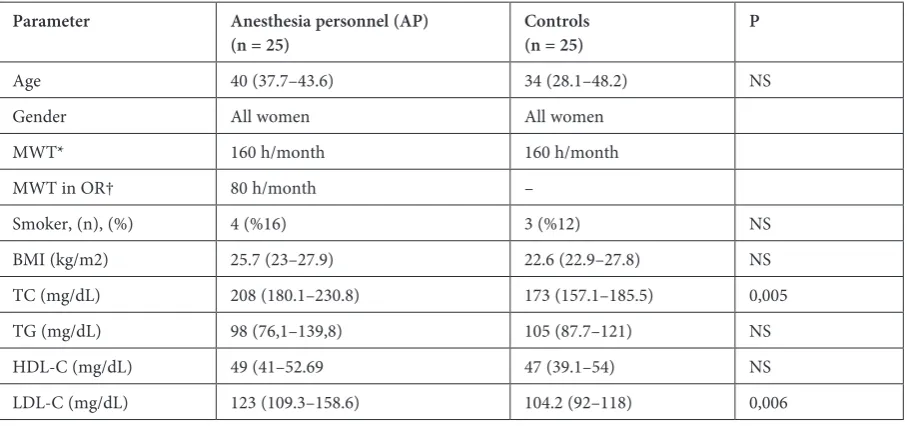
There were no significant differences in age or gender (all female) between AP and controls (p > 0.05). Smokers were more common in AP (16%) than in controls (12%), but this did not show a significant difference (p > 0.05). Body mass indi-ces (BMI) were similar in both groups (p = 0.74). When lipid parameters were compared, total cho-lesterol (TC) and LDL chocho-lesterol (LDL-C) levels were significantly increased between the AP and the control group [(p = 0.005) and (p = 0.006), re-spectively], whereas HDL cholesterol (HDL-C) and triglycerides (TG) levels were not statistically significant (p > 0.05). Demographic and laboratory findings obtained from AP and controls are sum-marized in Table 1.
Serum ARE activity and TAS levels were sig-nificantly reduced (p = 0.04, p < 0.0001, respec-tively), whereas TOS and OSI levels were found to be significantly higher (p < 0.01, p < 0.0001, re-spectively) in AP (Fig. 1). However, there were no significant differences in PON1 activity, PON1/ /HDL-C, ARE/HDL-C, and PON1/ARE (p = 0.30, p = 0.5, p = 0.1, and p = 0.7, respectively), when the 2 groups were compared (Table 2). This implies that PON1, ARE, and HDL-C values are indepen-dent of each other.
Discussion
this is the first study to interpret the association between impaired oxidative balance and HDL-re-lated PON1, ARE enzyme activity, and AP. The outcomes of our study showed that the mean TOS and OSI levels were significantly increased; where-as the mean ARE enzyme activity and TAS levels were significantly decreased, in female AP com-pared to the control female subjects.
Although cancerogenicity, mutagenicity, and many harmful effects are discussed as effects of long-time exposure to anesthetic gases, several re-view articles have suspected the results of stud-ies, finding positive correlations of the incidence of occupational disease with exposure to the vola-tile and gaseous substances. Studies have suggested that long-term exposure to trace levels of anesthet-ic gas is detrimental to operating-room personnel despite developed scavenging systems. Free radi-cals are one of the harmful effects related to vola-tile anesthesia [21–23].
Reactive oxygen species (ROS) and free radi-cals are the results of a cellular metabolism, which is a vital process in the stimulation of signaling
pathways in animal cells in response to changes in environmental conditions [24]. Important biomol-ecules such as proteins, lipids, and DNA are targets for free radicals, and modification of these mole-cules can raise the risk of mutagenesis [25]. Under sustained environmental stress, ROS are produced over a long time, and thus important damage may occur to the cell structure and functions. In recent years, investigators have raced to confirm that “in-creased oxidative stress due to the impaired oxi-dative/antioxidative balance” was the absolute impact in the pathogenesis of human disease, in-cluding cancer [26].
PON1 and ARE are enzymes that have lipo-philic esterase antioxidant characteristics. PON1 binds to HDL and contributes to the elimination of various chemical compounds such as paraox-on and lipid soluble free radicals from lipid per-oxidation. PON1, together with ARE, have been shown to function as a one enzyme [27–29]. Stud-ies have already defined that ARE enzyme activ-ity reflects the antioxidant property of PON1, al-though it is not directly responsible for it [30, 31]. Fig. 1. Serum ARE activity and TAS levels were reduced statistically and significantly, whereas TOS and OSI levels were found to be significantly higher in AP. However, there were no significant differences among PON1 and other parameters
AP_ARES Control_ARES
AP_TAS Control_TAS
Interestingly, Tang et al. [32] has shown that low ARE activity is a strong prognostic value for car-diovascular risk. In addition, diminished serum ARE activity provides increased prognostic value, and clinical reclassification of stable subjects are at risk of developing death, myocardial infarction, and stroke.
However, hypercholesterolemia is among the risk factors for atherosclerosis. LDL, the major cholesterol carrier in circulation, can undergo ox-idative modification in vascular cells, and cellular uptake of oxidized LDL leads to the generation of ROS [32–34]. LDL oxidation affects its lipid, and oxidative modifications in these molecules are in-volved in the mechanisms leading to endotheli-al dysfunction during atherosclerosis [35]. In this study, we found that serum cholesterol and LDL
levels of AP patients were significantly increased compared to the control group. These results sug-gest that AP might be considered a risk group for the development of cardiovascular risk.
Many clinical observations using different methods have reached the conclusion that oxida-tive stress had increased in AP [2, 17–20]. Serum (or plasma) concentrations of different oxidants and antioxidants can be measured separately in laboratories, but these measurements are labor-in-tensive, time-consuming, costly, and require spe-cialized personnel. As measuring different oxi-dants and antioxidant molecules separately is not easy, in order to acquire a simple detection of the oxidative stress in patients, it is enough to only as-sess the level of TAS and TOS and to calculate the OSI [14, 15].
Table 1. Demographic and laboratory finding of AP and controls were similar except TC and LDL of AP
Parameter Anesthesia personnel (AP)
(n = 25) Controls(n = 25) P
Age 40 (37.7–43.6) 34 (28.1–48.2) NS
Gender All women All women
MWT* 160 h/month 160 h/month
MWT in OR† 80 h/month –
Smoker, (n), (%) 4 (%16) 3 (%12) NS
BMI (kg/m2) 25.7 (23–27.9) 22.6 (22.9–27.8) NS TC (mg/dL) 208 (180.1–230.8) 173 (157.1–185.5) 0,005 TG (mg/dL) 98 (76,1–139,8) 105 (87.7–121) NS HDL-C (mg/dL) 49 (41–52.69 47 (39.1–54) NS LDL-C (mg/dL) 123 (109.3–158.6) 104.2 (92–118) 0,006
* Mean Working time, † Operating Room, NS – Non significant, TC – Total Cholesterol, TG – Triglycerid, LDL-C – Low Density Lipoprotein Cholesterol, HDL-C – High Density Lipoprotein Cholesterol. Median (%95 CI for median) for all parameters.
Table 2. Oxidative and antioxidative parameters of AP and controls
Parameter Anesthesia personel (AP)
(n = 25) Controls(n = 25) P
PON1 (U/L) 120.6 (109.7–146.5) 130.4 (108.5–154) 0.3 ARE (kU/L) 254.5 (207.4–274) 281.3 (240–293) 0.04 TAS (nmol Troloks/L) 2.03 (1.91–2.17) 2.29 (2.19–2.3) < 0.0001 TOS (μmol H2O2 Equiv./L) 12.9 (11.4–14.3) 6.1 (4.01–14.5) 0.01
OSI 6.1 (4.01–14.5) 0.3 (0.1–0.5) < 0.0001 PON1/HDL-C 2.4 (1.9–3.6) 2.7 (1.9–3.9) 0.5 ARE/HDL-C 5.2 (3.9–6.1) 5.5 (4.8–6.3) 0.1 PON/ARE 0.5 (0.4–0.6) 0.5 (0.4–0.7) 0.7
In conclusion, no biological monitoring of the exposure or direct exposure measurements were performed. In fact, the staff of operating theaters is exposed to many other factors, such as chemical agents other than volatile anesthetics, infected bi-ological material, potential injuries, radiation, ar-tificial lightning, fatigue, and psychosocial stress, which may constitute substantial health hazards. As many of these factors might potentially disturb
redox homeostasis, it is risky to assume that the observed changes resulted solely from anesthet-ic gas exposure, whanesthet-ich actually might only be dis-cussed as one possible, though important, reason for the observed phenomena. However, one major limitation of the study is the small number of sam-ples. Apparently, larger studies are needed to con-stitute the relationship of oxidative stress and oth-er factors in AP.
References
[1] Vessey MP: Epidemiological studies of the occupational hazards of anaesthesia—a review. Anaesthesia 1978, 33, 430–438.
[2] Türkan H, Aydin A, Sayal A: Effect of volatile anesthetics on oxidative stress due to occupational exposure. World J Surg 2005, 29, 540–542.
[3] Cohen EN, Gift HC, Brown BW, Greenfield W, Wu ML, Jones TW, Whitcher CE, Driscoll EJ, Brodsky JB:
Occupational disease in dentistry and chronic exposure to trace anesthetic gases. J Am Dent Assoc 1980, 101, 21–31.
[4] Venables H, Cherry N, Waldron HA, Buck L, Edling C,Wilson HK: Effects of trace levels of nitrous oxide on psychomotor performance. Scand J Work Environ Health 1983, 9, 391–396.
[5] Plummer JL, Beckwith AL, Bastin FN, Adams JF, Cousins MJ, Hall P: Free radical formation in vivo and hepa-totoxicity due to anesthesia with halothane. Anesthesiology 1982, 57, 160–166.
[6] Malekirad AA, Ranjbar A, Rahzani K, Kadkhodaee M, Rezaie A, Taghavi B, Abdollahi M: Oxidative stress in operating room personnel: occupational exposure to anesthetic gases. Hum Exp Toxicol 2005, 24, 597–601.
[7] Yılmaz N: Relationship between paraoxonase and homocystein: crossroads of oxidative disease. Arch Med Sci 2012, 8, 138–153.
[8] Mackness MI, Arrol S, Durrington PN: Paraoxonase prevents accumulation of lipoperoxides in low-density lipo-protein. FEBS Letters 1991, 286, 152–154.
[9] Mackness MI, Durrington PN, Mackness B: How high-density lipoprotein protects against the effects of lipid peroxidation. Cur Opin Lipidol 2000, 11, 383–388.
[10] Ahmed Z, Ravandi A, Maguire GF, Emili A, Draganov D, La Du BN, Kuksis A, Connelly PW: Apolipoprotein A-I promotes the formation of phosphatidylcholine core aldehydes that are hydrolyzed by paraoxonase (PON-1) during high density lipoprotein oxidation with a peroxynitrite donor. J Biol Chem 2001, 276, 24473–24481.
[11] Valko M, Rhodes CJ, Moncola J, Izakovic M, Mazura M: Free radicals, metals and antioxidants in oxidative stress-induced cancer. Mini-review. Chemico-Biological Interactions 2006, 160, 1–40.
[12] Eckerson HW, Wyte MC, La Du BN: The human serum paraoxonase/arylesterase polymorphism. Am J Hum Genet 1983, 35, 1126–1138.
[13] Haagen L, Brock A: A new automated method for phenotyping arylesterase (E.C.3.1.1.2.) based upon inhibition of enzymatic hydrolisis of 4-nitrophenyl acetate. Eur J Clin Chem Clin Biochem 1992, 30, 391–395.
[14] Erel O: A novel automated direct measurement method for total antioxidant capacity using a new generationmore stable ABTS radical cation. Clin Biochem 2004, 37, 277–285.
[15] Erel O: A new automated colorimetric method for measuring total oxidant status. Clin Biochem 2005, 38, 1103–1111.
[16] Harma M, Harma M, Erel O: Increased oxidative stress in patients with hydatidiform mole. Swiss MedWkly 2003, 133, 563–566.
[17] Chandrasekhar M, Rekhadevi PV, Sailaja N, Rahman MF, Reddy JP, Mahboob M, Grover P: Evaluation of genetic damage in operating room personnel exposed to anaesthetic gases. Mutagenesis 2006, 21, 249–254.
[18] Peric M, Vranes Z, Marusic M: Immunological disturbances in anaesthetic personnel chronically exposed to high occupational concentrations of nitrous oxide and halothane. Anaesthesia 1991, 46, 531–537.
[19] Wrońska-Nofer T, Nofer JR, Jajte J, Dziubałtowska E, Szymczak W, Krajewski W, Wąsowicz W, Rydzyński K:
Oxidative DNA damage and oxidative stress in subjects occupationally exposed to nitrous oxide (N(2)O). Mutat Res 2012, 731, 58–63.
[20] Baysal Z, Cengiz M, Ozgonul A, Cakir M, Celik H, Kocyigit A: Oxidative status and DNA damage in operating room personel. Clin Biochem 2009, 42, 189–193.
[21] Sinclair AJ, Barnett AH, Lunec J: Free radicals and antioxidant systems in health and disease. Br J Hosp Med 1990, 43, 334–344.
[22] Andreoli TE: Free radicals and oxidative stress. Am J Med 2000, 108, 650–651.
[23] Kudou M, Kudou T, Matsuki A: Changes of plasma superoxide dismutase like activity during general anesthesia and surgery in man. Masui 1990, 39, 1172–1177.
[24] Durackova Z: Some current insights into oxidative stress. Physiol Res 2010, 59, 459–469.
[26] Crawford A, Fassett RG, Geraghty DP, Kunde DA, Ball MJ, Robertson IK, Coombes JS: Relationships between single nucleotide polymorphisms of antioxidant enzymes and disease. Gene 2012, 501, 89–103.
[27] Aviram M, Rosenblat M, Bisgaier CL, Newton RS, Primo-Parmo SL, La Du BN: Paraoxonase inhibits high-density lipoprotein oxidation and preserves its functions. A possible peroxidative role for paraoxonase. J Clin Invest 1998, 101, 1581–1590.
[28] Li HL, Liu DP, Liang CC: Paraoxonase gene polymorphisms, oxidative stress, and diseases. J Mol Med 2003, 81, 766–779.
[29] Gan KN, Smolen A, Eckerson HW, La Du BN: Purification of human serum paraoxonase/arylesterase. Evidence for one esterase catalyzing both activities. Drug Metab Dispos 1991, 19, 100–106.
[30] Rosenblat M, Gaidukov L, Khersonsky O, Vaya J, Oren R, Tawfik DS, Aviram M: The catalytic histidine dyad of high density lipoprotein-associated serum paraoxonase-1 (PON1) is essential for PON1-mediated inhibition of low density lipoprotein oxidation and stimulation of macrophage cholesterol efflux. J Biol Chem 2006, 281, 7657–7665.
[31] Otocka-Kmiecik A, Orłowska-Majdak M: The role of genetic (PON1 polymorphism) and environmental factors, especially physical activity, in antioxidant function of paraoxonase. Post Hig Med Dosw 2009, 63, 668–677.
[32] Tang WH, Hartiala J, Fan Y, Wu Y, Stewart AF, Erdmann J, Kathiresan S; CARDIoGRAM Consortium, Roberts R, McPherson R, Allayee H, Hazen SL: Clinical and genetic association of serum paraoxonase and aryles-terase activities with cardiovascular risk. Arterioscler Thromb Vasc Biol 2012, 32, 2803–2812.
[33] Kruth HS: Lipoprotein cholesterol and atherosclerosis. Curr Mol Med 2001, 1, 633–653.
[34] Berlier JA, Navab M, Fogelman AM,Frank JS, Demer LL, Edwards PA, Watson AD, Lusis AJ: Atherosclerosis: basic mechanism, oxidation, inflammation, genetics. Circulation 1995, 91, 2488–2496.
[35] Eren E, Aydın O, Yilmaz N: High Density Lipoprotein and it’s DysfunctionThe Open Biochemistry Journal 2012, 6, 1–10.
Address for correspondence:
Hamit Yasar Ellidag
Antalya Education and Research Hospital Clinical Biochemistry Central Laboratory Ministry of Health
Varlik Mahallesi Kazim Karabekir Caddesi Soguksu 07050 Antalya
Turkey
Tel.: 00 905 054 952 155
E-mail: [email protected]
Conflict of interest: None declared