90
Comparative study of In Vitro and In Vivo antioxidant property of different Ixora species
Sankhadip Bose*1, Sushomasri Maji2, Pranabesh Chakraborty2
1. Gupta College of Technological Sciences, College of Pharmacy, Ashram More, G.T.
Road, Asansol, West Bengal, India.
2. Bengal School of Technology, Hooghly, West Bengal, India.
*Corresponding author: [email protected]
ABSTRACT:
Ixora species (Rubiaceae) contains many important phytoconstituents in the various
parts of it and they are also responsible for some important biological activities like antitumour activity, wound healing and antimicrobial activity. The antioxidant capacity of I. coccinea and I. purviflora leaves were assayed for their scavenging abilities against superoxide anion radicals, hydroxyl radical, nitric oxide radical, hydrogen peroxide, metal chelation and reducing power. All the extracts inhibited all above said free radicals in a dose-dependent manner. As antioxidant action has been reported to play a
crucial role in the hepatoprotection and excellent result of these plants in In Vitro model
of antioxidant activity, an attempt has been taken to elucidate the effect on CCl4
induced hepatic damage with reference to biochemical marker enzymes & histopathology. In the present study all extracts of both the plants have been found to
reduce both serum ALP and Bilirubin. Treatment with CCl4 increases the levels of total
lipid, total triglycerides and total cholesterol in liver. Presence of significantly high
concentration of total lipid and cholesterol in the serum of CCl4 treated animals and
maintains of these towards near normal values in different extracts administered rats demonstrates the hepatoprotective effect of both the Ixora species.
Key words: Antioxidant, hepatoprotection, Ixora coccinea, Ixora perviflora, aspartate amino transferase, L-alanine amino transferase.
INTRODUCTION:
The plants belonging to Rubiaceae family are generally a rich source of substances of phytochemical interest. Numbers of plants from this family are used in traditional
system of medicine. [1] From the large list of various plants of rubiaceae family we have
91
plants are generally used as the ornamental plants in gardens and parks. But the review of literature showed that there are so many important phytoconstituents present in the various parts of the above said plants and they are also responsible for some important biological activities like antitumour activity, wound healing and antimicrobial activity.
Some preliminary phytochemical investigations reported that flavonoids are present in the flowers and leaves of the same. Therefore, the objectives of the present study were
to investigate the In Vitro antioxidant activity of I. coccinea and I. perviflora leaves
through the free radical scavenging, superoxide anion radical scavenging, nitric oxide scavenging, metal chelation and reducing power assay.
Liver diseases, especially viral hepatitis occurs predominantly in the developing world
with an enormous impact on public health & economy. [2] Carbon tetrachloride (CCl4) is
widely used in animal models to induce acute liver injury. [3-5] It is generally believed
that the toxicity of CCl4 results from its reductive dehalogenation by the cytochrome
P450 enzyme system into the highly reactive free radical trichloromethyl redical. [6] Antioxidant action has been reported to play a crucial role in the hepatoprotection. [7] It has been suggested that natural antioxidants are more safe and healthy than
synthetic antioxidants which used in foods. [8] In the present study an attempt has
been taken to elucidate the effect of the different extracts of Ixora species on CCl4
induced hepatic damage with reference to biochemical marker enzymes &
histopathology. The result of this In Vivo study may support these two plants to be good
herbal antioxidant drugs.
MATERIALS AND METHODS: Plant materials
The fresh leaves of Ixora coccinea Linn and Ixora perviflora Vahl were collected from the
Medicinal Plants’ Garden of Gupta College Of Technological Sciences, in the morning between 9 to 10 a.m. during the months of November and December. The herbariums of Ixora coccinea Linn and Ixora perviflora Vahl were submitted to Botanical Survey of India, Shibpur Botanical Garden, Howrah and was authenticated by the Director of the same. Reference number of the Authentication letter is CNH/ 1-1/ (201)/ 2007/ Tech. II.
Extraction of plant materials
92
were concentrated in a rotary flash evaporator. The residue (semisolid) was dried in a desicator over sodium sulfite and kept in refrigerator for further study.
In other hand, fresh leaves of each plant were collected and shade dried at room temperature, pulverized and 100 gm (each batch) leaf powder of each plant was extracted with distilled water (500 ml) through cold maceration. The extract was concentrated in a rotary flash evaporator. The residue (semisolid) was dried in a desicator over sodium sulfite and kept in refrigerator for further study.
In Vitro Antioxidant Activity
Superoxide anion scavenging activity assay
The scavenging activity of various extracts of Ixora coccinea leaves towards superoxide
anion radicals was measured by colorimetric method. [9, 30] Superoxide anion was
generated in a non-enzymatic phenazine methosulfate-nicotinamide adenine
dinucleotide (PMS-NADH) system through the reaction of PMS, NADH and oxygen. It was assayed by the reduction of nitroblue tetrazolium (NBT). In these experiments the superoxide anion was generated in 3ml of Tris-HCl buffer (100mM, pH 7.4) containing 0.75ml of NBT (300µM) solution, 0.75ml of NADH (936 µM) solution and 0.3ml of different concentrations of each extract. The reaction was initiated by adding 0.75ml of PMS (120 µM) to the mixture. After 5 min of incubation at room temperature, the absorbance at 560nm was measured in spectrophotometer. The superoxide anion scavenging activity was calculated according to the following equation:
% inhibition = [(A0-A1)/A0] ×100
Where A0 was the absorbance of the control (blank, without extract) and A1 was the
absorbance in the presence of the extract.
Hydroxyl radical scavenging activity assay
The scavenging activity for hydroxyl radicals was measured. [10, 30] Reaction mixture
contained 60µl of 1.0 mM FeCl3, 90 µl of 1mM 1,10-phenanthroline, 2.4 ml of 0.2 M
phosphate buffer (pH 7.8), 150 µl of 0.17 M H2O2 and 1.5 ml of extract at various
concentrations. Adding H2O2 started the reaction. After incubation at room temperature
for 5 min, the absorbance of the mixture at 560nm was measured with a spectrophotometer. The hydroxyl radicals scavenging activity was calculated according to the following equation:
% inhibition = [(A0-A1)/A0] ×100
Where A0 was the absorbance of the control (blank, without extract) and A1 was the
93
Nitric oxide scavenging activity assay
Sodium nitroprusside in aqueous solution at physiological pH spontaneously generates nitric oxide, which interacts with oxygen to produce nitrite ions, which can be determined by the use of the Griess lllosvoy reaction. [11-12, 30] 2 ml of 10 mM sodium nitroprusside in 0.5 ml of phosphate buffer saline (pH 7.4) was mixed with 0.5 ml of extract at various concentrations and the mixture incubated at 25 ˚C for 150 min. From the incubated mixture 0.5 ml was taken out and added into 1.0 ml of sulfanilic acidreagent (33% in 20% glacial acetic acid) and incubated at room temperature for 5 min. Finally, 1.0 ml naphthylenediamine dihydrochloride (0.1% w/v) was mixed and incubated at room temperature for 30 min. The absorbance at 540nm was measured with a spectrophotometer. The nitric oxide radicals scavenging activity was calculated according to the following equation:
% inhibition = [(A0-A1)/A0] ×100
Where A0 was the absorbance of the control (blank, without extract) and A1 was the
absorbance in the presence of the extract.
Hydrogen peroxide scavenging activity assay
Hydrogen peroxide scavenging activity of the extract was estimated by replacement
titration. [13, 30] Aliquot of 1.0 ml of 0.1mM H2O2 and 1.0 ml of various concentrations
of extracts were mixed, followed by 2 drops of 3% ammonium molybdate, 10 ml of 2M
H2SO4 and 7.0 ml of 1.8 M KI. The mixed solution was titrated with 5.09 mM NaS2O3
until yellow colour disappeared. Percentage of scavenging of hydrogen peroxide was calculated as:
% inhibition = [(V0-V1)/V0] ×100
Where V0 was Volume of NaS2O3 solution used to titrate the control sample in the
presence of hydrogen peroxide (without extract), V1 was the volume of NaS2O3 solution
used in the presence of extract.
Fe2+ chelating activity assay
To 0.5 ml of extract, 1.6 ml of deionized water and 0.05 ml of FeCl2 (2mM) was added.
After 30 s, 0.1 ml ferrozine (5mM) was added. Ferrozine reacted with the divalent iron to form stable magenta complex species that were very soluble in water. After 10 min of
room temperature, the absorbance of the Fe2+ - Ferrozine complex was measured at
562nm. [14, 30] The chelating activity of the extract for Fe2+ was calculated as:
Chelating rate (%) = [(A0-A1)/A0] ×100
Where A0 was the absorbance of the control (blank, without extract) and A1 was the
94
Reducing power assay
The extract (0.75 ml) at various concentrations was mixed with 0.75 ml of phosphate
buffer (0.2M, pH 6.6) and 0.75 ml of potassium hexacyanoferrate (K3Fe(CN)6) (1%, w/v),
followed by incubating at 50° C in a water bath for 20 min. The reaction was stopped by
adding 0.75 ml of trichloroacetic acid (TCA) solution (10%) and then centrifuged at 800g for 10 min. 1.5 ml of the supernatant was mixed with 1.5 ml of distilled water and 0.1 ml of ferric chloride solution (0.1%, w/v) for 10 min. The absorbance at 700nm was measured as the reducing power. [15, 30] Higher absorbance of the reaction mixture indicated greater reducing power.
In Vivo antioxidant activity Experimental Animal
Wister strain albino rats of 150-200 g body weight were used in study. Animals were procured from Institutional Animal house of Gupta College Of Technological Sciences, Asansol. All animals were kept in polyacrylic cages and maintained under standard
housing conditions (room temperature 24-270C and humidity 60-65% with 12:12 light:
dark cycles. Food was provided in the form of dry pellets and water ad libitum). All the
experiments were conducted between 9.00 and 15.00 hours. The animals were allowed to get acclimatized to the laboratory conditions for 7 days before the commencement of the experiment. Food was withdrawn 18 hours prior to the commencement of the experiment. All experiments involving animals comply with the ethical standards of animal handling and approved by Institutional Animal Ethics Committee (Regd.No.– 955/a/06/ CPCSEA).
Experimental Procedure
Hepatopathy was induced in animals by administration of CCl4 interperitoneally (i.p) at
the dose of 1.25 ml/kg,i.p, in liquid paraffin for 14 days .The rats were equally divided into Six groups, each group contains six animals. [18,23]
Group-I was considered as control, Group-II was considered as CCl4 (1.25 ml/kg,i.p,/14
days) treated animals, Group-III was considered as CCl4 and aqueous extract
(250mg/kg,p.o/14 days) treated animals, where Group-IV was considered as CCl4 and
hydro-alcoholic extract (250mg/kg,p.o/14 days) treated animals, Group-V was treated
as CCl4 and ethyl acetate extract (250mg/kg,p.o/14 days) treated animals and at last
Group-VI was considered as CCl4 and silymarin (100 mg/kg, p.o/14 days) treated
animals.
95
enzymes and other parameters, Liver was dissected out and immediately preserved in 10% formaldehyde solution for histopathological study.
Serum biochemical estimations
Triglycerides, Cholesterol, Total Protein, Total Bilirubin, Serum glutamate- oxaloacetate transaminase (S.G.O.T) or Aspartate amino transferase (AST), Serum glutamate- pyruvate transminase (S.G.P.T) or L-alanine amino transferase (ALT), Alkaline phosphate (ALP) were estimated using standard commercial kits from SPAN India Ltd. Surat, India. [16-19,21-29]
Statistical Analysis
In in-vitro activity tests were carried out in triplicate. The amount of extract needed to
inhibit free radicals concentration by 50%, IC50, was graphically determined and means,
standard deviation & correlation were computed by using Microsoft Excel. In in-vivo activity the values are expressed in terms of Mean ± S.E.M. of (n=6).
Prism 3.0 software was used to perform all statistical analysis. It was carried out by one-way analysis of variance (ANOVA), followed by Tukey Kramer multiple comparison post test. P values < 0.05 were considered as significant.
RESULTS AND DISCUSSION:
In Vitro activity
Superoxide anion scavenging activity
Superoxide is biologically important since it can be decomposed to form stronger oxidative species such as singlet oxygen and hydroxyl radicals, is very harmful to the cellular components in a biological system. The Superoxide anion scavenging activity of
the extracts from Ixora coccinea and Ixora perviflora assayed by the PMS-NADH system
is shown. The Superoxide anion scavenging activities of all the extracts of Ixora coccinea were increased markedly with the concentrations. Now in between three extracts ethyl acetate and aqueous extracts showed better result than hydro-alcoholic
extract. The half inhibition concentration (IC50) of hydro-alcoholic, aqueous and ethyl
acetate extracts ware 95 µg/ml, 45 µg/ml and below 10 µg/ml respectively for I.
coccinea. Where for I. perviflora the half inhibition concentration (IC50) of
hydro-alcoholic, aqueous and ethyl acetate extracts ware 98 µg/ml, 60 µg/ml and 20 µg/ml
respectively. These results suggested that all the extracts of Ixora coccinea and Ixora
96
extract and ethyl acetate extract of I. coccinea showed better superoxide anion
scavenging activity than any other extracts of I. perviflora.
Hydroxyl radical scavenging activity
Hydroxyl radical is very reactive and can be generated in biological cells through the
Fenton reaction. The result showed the above three extracts of I.coccinea and Ixora
perviflora leaves exhibited concentration dependent scavenging activities against
hydroxyl radicals generated in a Fenton reaction system. The IC50 of hydro-alcoholic,
aqueous and ethyl acetate extracts ware 120µg/ml, 55 µg/ml and 20 µg/ml respectively for I. coccinea and for I. perviflora the IC50 of hydro-alcoholic, aqueous and ethyl acetate
extracts ware 150µg/ml, 78 µg/ml and 15 µg/ml respectively. The potential scavenging abilities of phenolic substances might be due to the active hydrogen donor ability of hydroxyl substitution. Similarly, high molecular weight and the proximity of many aromatic rings and hydroxyl groups are more important for the free radical scavenging by specific functional groups.
Nitric oxide scavenging activity
Nitric oxide (NO) is a potent pleiotropic mediator of physiological process such as smooth muscle relaxation, neuronal signalling, inhibition of platelet aggregation and regulation of cell mediated toxicity. It is a diffusible free radical which plays many roles as an effector molecule in diverse biological systems including neuronal messenger, vasodilation and antimicrobial and antitumor activities. All the three extracts of I.coccinea and I. perviflora moderately inhibited nitric oxide in dose dependent manner.
The IC50 of hydro-alcoholic, aqueous and ethyl acetate extracts ware 920 µg/ml, 620
µg/ml and more then 1000 µg/ml respectively for I. coccinea and for I. perviflora the
IC50 of hydro-alcoholic, aqueous and ethyl acetate extracts ware 930 µg/ml, 510 µg/ml
and more then 1000 µg/ml respectively. Now in between three extracts aqueous extract showed better result than hydro-alcoholic and ethyl acetate extracts.
Hydrogen peroxide scavenging activity
Hydrogen peroxide is a weak oxidizing agent and can inactivate a few enzymes directly, usually by oxidation of essential thiol (-SH) groups. Hydrogen peroxide can cross cell
membranes rapidly, once inside the cell, H2O2 can probably react with Fe2+, and
possibly Cu2+ ions to form hydroxyl radical and this may be the origin of many of its
toxic effects. It is therefore biologically advantageous for cells to control the amount of
hydrogen peroxide that is allowed to accumulate. All the three extracts of I.coccinea
hydro-97
alcoholic, aqueous and ethyl acetate extracts ware 800 µg/ml, 720 µg/ml and more
then 1000 µg/ml respectively for I. coccinea and for I. perviflora The IC50 of
hydro-alcoholic, aqueous and ethyl acetate extracts ware 680 µg/ml, 540 µg/ml and more then 1000 µg/ml respectively. Now in between three extracts aqueous extract showed better result than hydro-alcoholic and ethyl acetate extracts.
Fe2+ chelating activity
All the three extracts of I. coccinea and I. perviflora inhibited ferrous ion in dose
dependent manner. The IC50 of hydro-alcoholic, aqueous and ethyl acetate extracts ware
225 µg/ml, 160 µg/ml and 80 µg/ml respectively for I. coccinea and for I. perviflora the
IC50 of hydro-alcoholic, aqueous and ethyl acetate extracts ware 210 µg/ml, 200 µg/ml
and 90 µg/ml respectively. In between that ethyl acetate extract showed more effective scavenging of ferrous ion than hydro-alcoholic and aqueous extracts.
Reducing power activity
It depict the reductive effect of Ixora coccinea and Ixora perviflora similar to the
antioxidant activity, the reducing power of both I. coccinea and I. perviflora increased
with increasing dosage. High absorbance at 700nm indicates high reducing power.
In-vivo activity
Liver injury induced by CCl4 is the best characterized system of xenobiotic –induced
hepatotoxicity and is commonly used models for the screening of anti hepatotoxic and
/or hepatoprotective activities of drugs. The changes associated with CCl4 induced liver
damage are similar to that of acute viral hepatitis. It has been established that CCl4
acumulates in hepatic parenchymal cells and gets metabolically activated by CYP P-450
depended monoxygenases form trichloromethyl free radical (CCl3). These free radical
alkylates cellular proteins and other macromolecules with a simultaneous attact on poly unsaturated fatty acids in the presence of oxygen to produce lipid peroxides. Leading to liver damage, Lipid peroxidation will initiate pathological changes such as depression of protein synthesis, elevation of serum marker enzymes such as AST, ALT and ALP. SGPT is more specific to the liver and a better parameter for detecting liver damage.
All the extracts at the dose of 250 mg/kg decreased the level of both AST and ALT
significantly in CCl4 treated rats indicating maintenance of functional integrity of
98
In the present study all extracts of both the plants have been found to reduce both serum ALP and Bilirubin in the treated groups compared with the untreated ones. The site specific oxidative damage of some of the susceptible amino acids of proteins is regarded as the major cause of metabolic dysfunction during pathogenesis. Hypoalbuminaemia is most frequent in the presence of advanced chronic diseases. Hence decline in total protein content can be deemed as a useful index of the severity of cellular dysfunction in chronic liver diseases. The lowered level of total proteins
recorded in the serum as well as lever of CCl4 treated rats reveals the severity of
hepatopathy. Different extracts treated rats maintained near normally of total protein
level. Treatment with CCl4 increases the levels of total lipid, total triglycerides and total
cholesterol in liver. Presence of significantly high concentration of total lipid and
cholesterol in the serum of CCl4 treated animals and maintains of these towards near
normal values in different extracts administered rats demonstrates the hepatoprotective effect of both the Ixora species. In between all the extracts, the aqueous extract has shown better result than other ethyl acetate and hydro-alcoholic extracts (Table 1 and Table 2).
Table 1: Serum Biochemical Estimations of Ixora coccinea Extracts
Groups Treatment
T ri g ly c e ri d e s ( m g / d l. ) C h o le s te ro l ( m g / d l. ) T o t a l P r o t e in ( g / d l. ) T o t a l B il ir u b in ( m g / d l. ) S .G .O .T ( IU / L ) S .G .P .T ( IU / L ) A lk a li n e p h o s p h a te ( K A U n it s )
I Normal
Control 151.72± 2.57 131.2± 1.61 7.29± 0.12 0.50± 0.02 38.26± 2.13 37.4±
2.15 7.36± 2.02
II CCl4 262.06±
4.09
281.2±
3.3
4.94 ±
0.27 1.85± 0.25 111.30 ±3.55 88.4± 2.55 25.26± 2.20
III CCl4 +
Aqueous Extract 165.51± 2.60 225.0± 1.41 5.88± 1.08 1.10± 0.13 62.60± 2.44 40.8± 2.97
13.15 ±
3.57
IV CCl4 +
Hydro-alcoholic Extract 217.93± 1.41 243.7± 2.07 6.35± 1.03 1.25± 0.03 68.11± 2.66 51.0± 1.80 15.78± 3.19
V CCl4 + Ethyl
acetate Extract 190.21± 2.60 233.4± 1.72 6.11± 0.16 1.15± 0.03 63.10± 1.23 56.2± 1.92 14.77± 0.22
VI CCl4 +
99
Table 2: Serum Biochemical Estimations of Ixora perviflora Extracts
Groups Treatment
T ri g ly c e ri d e s ( m g / d l. ) C h o le s te ro l ( m g / d l. ) T o ta l P ro te in ( g / d l. ) T o ta l B il ir u b in ( m g / d l. ) S .G .O .T ( IU / L ) S .G .P .T ( IU / L ) A lk a li n e p h o s p h a te ( K A U n it s )
I Normal
Control 152.32± 1.15 130.5± 1.01 7.52± 0.26 0.60± 0.03 40.11± 1.33 35.3±
2.22 7.00± 1.03
II CCl4 282.16±
3.11
285.1±
2.3
4.00 ±
0.33 2.00± 0.05 121.2± 3.01 85.5± 1.05 30.02± 1.10
III CCl4 +
Aqueous Extract 160.01± 2.01 235.0± 1.04 6.02± 1.05 1.15± 0.10 71.30± 2.02 47.3± 2.07
21.01 ±
3.27
IV CCl4 +
Hydro-alcoholic Extract 220.00± 1.04 244.2± 2.21 5.85± 0.03 1.35± 1.03 78.11± 2.01 52.0± 1.20 25.54± 2.11
V CCl4 + Ethyl
acetate Extract 190.01± 1.00 232.2± 1.02 6.10± 0.15 1.25± 1.03 60.01± 0.06 51.1± 1.02 22.21± 0.54
VI CCl4 +
Silymarin 154.92± 2.43 133.8± 1.92 7.40± 0.16 0.51± 0.03 41.02± 2.60 39.41± 1.72 9.11± 0.16 Histopathological Study
Histopathological studies also provided supportive evidence for the biochemical analysis. The different extracts treated groups and Silymarin treated group showed the
normal parenchymal architecture without noticeable alteration compared to CCl4
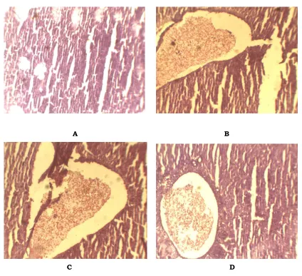
treated group. Centrilobular necrosis accompanied by fatty changes and ballooning
degeneration were observed in group treated with CCl4 (Fig. 1).
CONCLUSION:
Both I. coccinea and I. perviflora showed a promising result in In Vitro and In Vivo
100
A B
C D
Fig. 1: Histpathological Section of A) CCl4 treated Liver , B) I. coccinea Extract Treated Liver, C) I. perviflora Extract Treated Liver , D) Silymarin Treated Liver
ACKNOWLEDGEMENT:
Authors thank Mr. M. S. Mondal, Joint Director, Botanical Survey of India, Shibpur, Kolkata, for his help in identification of the plants. Authors are also thankful to Prof. T.K. Basu, Retired Head, Department of Botany, Netajee Mahavidyalaya, Arambag, Hoogly for his ideas about the geographical source of the selected plants and preparation of their herbarium.
REFERENCE:
1. Kirtikar, K. R., Basu, B. D. Indian Medicinal Plants. 2nd ed., Oriental Enterprises:
Dehradun, Uttaranchal, India, 2001.
2. Simonsen L., Kan A., Lloy J. Unsafe infection in the developing world and
101
3. Mizuoka H., Shikata N., Yang J., Takasu M., Inoue K, Tsubura A. Biphasic effect of
colchicines on acute liver injury induced by carbon tetrachloride or by dimethylnitrosamine in mice. J Hepatol. 1999; 1: 825-833.
4. Rao P. S., Mangipudy R. S., Mehendale M. H. Tissue injury and repair as paralal
and opposing responses to CCl4 hepatotoxicity: A novel dose-response. Toxicology.
1997; 118: 181-193.
5. Czaja M. J., Xu J., Alt E. Prevention of carbon tetrachloride induced rat liver injury
by soluble tumor necrosis factor receptor.Gastroenterology. 1995; 108: 849-854.
6. Recknagel R. O., Glende E. A., Dolak J. A. Waller R. L. Mechanisms of carbon
tetrachloride Toxicity. Pharmacol Ther. 1989; 43: 139-154.
7. Galati E. M., Mondello M. R., Lauriano E. R., Taviano M. F., Galluzzo, Miceli N. O.
Fruit juice protects liver from carbon tetrachloride-induced injury. Phytother Res. 2005; 19(9): 796-800.
8. Xiao-Yu S. In Vitro and In Vivo antioxidant activity of Pinus korainensis seed extract
containing phenolic compounds.Food Chemistry. 2009: 117: 681-686.
9. Liu F., Ooi V. E. C., Chang S. T., Free radical scavenging activity of mushroom
polysaccharide extracts. Life Sci. 1997; 60: 763-771.
10. Yu W., Zhao Y., Shu B. The radical scavenging activities of radix puerariae
isoflavonoids: A chemiluminescence study. Food Chem. 2004; 86: 525-529.
11. Garrat, D. C. The qualitative analysis of drugs. Vol. 3, Chapman and Hall: Japan,
1964.
12. Bhatt L. R., Baek S. H. Antioxidant capacity of crude extract and fractions from Woodfordia fruticosa flower. Oriental Pharmacy and Experimental Medicine. 2007; 7(2): 162-170.
13. Zhang, X. Y. Principles of Chemical Analysis. Chine Science Press: Beijing, 2000.
14. Dinis T. C. P., Madeira V. M. C., Almeidam L. M. Action of phenolic derivatives
(acetoaminophen, salycilate and 5-aminosalycilate) as inhibitors of membrane lipid peroxidation and peroxyl radicals scavengers. Ach. Biochem. And Biophy. 1994; 315: 161-169.
15. Oyaizu M. Studies on products of browning reactions: antioxidant activities of products of browning reaction prepared from glucose amine. Jap. J. Nutr. 1986; 44: 307-315.
16. Koneri R., Balaraman R., Fridous, Vinodh K. M., Hepatoprotective effects of
102
17. Krishnaraju A. V., Rao C. V., Reddy K. N., Trimurtutlu G. In Vitro and In Vivo
antioxidant activity of Aphanmixis poystachya bark, Ameri. Journ. of Infec.
Diseases. 2009;5(2): 60-67.
18. Verma A. R., Vijayakumar M., Rao C. V., Mathela C. S. In Vitro and In Vivo
antioxidant properties and DNA damage protective activity of green fruit of Ficus
glomerata,Food and Chemical Toxicology. 2010; 48: 704-709.
19. Zhang Q., Li N., Liu X., Zhao Z., Li Z., Xu Z. The structure of a sulfated galactan
from Porphyra haitanensis and its In Vivo antioxidant activity. Carbohydrate
Research. 2004; 339:105-111.
20. Xiao-Yu S., Zhen-Yu W., Jia-Ren L. In Vitro and In Vivo antioxidant activity of Pinus
korainensis seed extract containing phenolic compounds. Food Chemistry. 2009; 117: 681-686.
21. Qingming Y., Pan X., Kong W., Yang H., Su Y., Li Z., Zhang Y., Yang Y., Ding L., Liu
G. Antioxidant activities of malt extract from barley (Hordeum vulgare L.) towards
various oxidative stress in vitro and in vivo,Food Chemistry. 2010;118: 84-89.
22. Wang H., Xiang D. G., Gao C. Z., Lei C., Wen B. Y. In Vitro and In Vivo antioxidant
activity of aqueous extract from Choerospondias axillaries fruit. Food Chemistry.
2008; 106: 888-895.
23. Murthy K. N. C. , Vanitha A., Rajesha J., Swamy M. M., Sowmya P. R., Gokare A. R. In vivo antioxidant activity of carotenoids from Dunaliella salina-a green microalga. Life Sciences. 2005; 76: 1381–1390.
24. Meng F., Zhou B., Lin R., Jia L., Liu X., Deng P., Fan K., Wang G., Wang L., Zhang J. Extraction optimization and in vivo antioxidant activities of exopolysaccharide by Morchella esculenta SO-01, Bioresource Technology. 2010:1- 6.
25. Haenen G. R. M. M., Arts M. J. T. J., Bast A., Coleman M. D. Structure and activity in assessing antioxidant activity in vitro and in vivo A critical appraisal illustrated with the flavonoids. Environmental Toxicology and Pharmacology. 2006; 21: 191– 198.
26. Wang D., Wang L., Zhu F., Chen X. D., Zou L., Saito M., Li L. In vitro and in vivo
studies on the antioxidant activities of the aqueous extracts of Douchi (a traditional Chinese salt-fermented soybean food). Food Chemistry. 2008; 107: 1421–1428. 27. Galetta F., Franzoni F., Cervetti G., Regoli F., Fallahi P., Tocchini L., Carpi A.,
Antonelli A., Petrini M., Santoro G. In vitro and in vivo study on the antioxidant
activity of dexrazoxane. Biomedicine & Pharmacotherapy. 2009: 1-5.
28. Fritz K. L., Seppanen C. M., Kurzer M. S., Csallany A. S. The in vivo antioxidant
103
29. Zhang Q., Li N., Zhou G., Lu X., Xu Z., Li Z. In vivo antioxidant activity of
polysaccharide fraction from Porphyra haitanesis (Rhodephyta) in aging mice.
Pharmacological Research. 2003; 48: 151–155.