K
RYSTYNAŚ
REDZIŃSKA1, A
NNAG
ALICKA1, J
ANUSZP
OPKO2,H
ALINAP
OROWSKA1,
A
NDRZEJG
INDZIEŃSKI1Elastase and Gelatinase Activities in Synovial Fluid of Patients
with Reactive Arthritis, Osteoarthritis, and Rheumatoid Arthritis
Aktywność elastazy i żelatynazy w płynie stawowym pacjentów
z reaktywnym zapaleniem stawów, chorobą zwyrodnieniową
i reumatoidalnym zapaleniem stawów
1 Department of Medical Chemistry, Medical University of Bialystok, Białystok, Poland
2 Department of Pediatric Orthopaedics and Traumatology, Medical University of Bialystok, Białystok, Poland
Adv Clin Exp Med 2008, 17, 5, 559–564 ISSN 1230−025X
ORIGINAL PAPERS
© Copyright by Silesian Piasts University of Medicine in Wrocław
Abstract
Background.Rheumatoid arthritis (RA), reactive arthritis (ReA), and osteoarthritis (OA) are a group of joint dis− eases which differ in pathogenesis, intensity, and rate of progression. They are characterized by different stages of irreversible damage to the extracellular matrix of the cartilage caused by enzymatic degradation. The aim was to compare the activities of elastase and gelatinase in synovial fluid (SF) samples from patients with reactive arthri− tis with those of patients with OA and RA and determine correlations between elastase activity and cartilage matrix degradation.
Material and Methods. Elastase activity was determined using the low−molecular−weight synthetic substrate MeOSuc−Ala−Ala−Pro−Val−p−NA and gelatinolytic activity using zymography. The concentrations of collagen and proteoglycan degradation products were measured by spectrophotometric methods.
Results.Elastase activity was detectable in 92% of the patients with ReA and RA and in 75% of those with OA. The elastase activity in SF samples from ReA patients, mainly due to metalloenzymatic activity, amounted to about 66% of this activity in RA patients and was 2.3−times higher than in the OA patients. Activation of prometallopro− teases resulted in a 4.5−fold increase in elastinolytic activity in ReA compared with a 3.2−fold increase in OA and a 4−fold increase in RA fluids. In contrast to OA, significant correlations between elastase activity and both gly− cosaminoglycans and type II collagen concentrations were found in SF samples of patients with ReA and RA. Furthermore, the active forms of two gelatinase proenzymes (92 and 72 kDa, respectively) were detected in ReA and RA synovial fluids.
Conclusions.These results indicate that in ReA and RA, both elastase and gelatinases can contribute in similar degree to the degradation of major cartilage components (Adv Clin Exp Med 2008, 17, 5, 559–564).
Key words:elastase, gelatinase, synovial fluid, joint diseases.
Streszczenie
Wprowadzenie. Reumatoidalne zapalenie stawów (RA), reaktywne zapalenie stawów (ReA) i zwyrodnieniowa choroba stawów (OA) są grupą chorób stawów o różnej patogenezie, intensywności i szybkości rozwoju. Choro− bom stawów towarzyszy nieodwracalne uszkodzenie macierzy pozakomórkowej chrząstki spowodowane degrada− cją enzymatyczną.
Cel pracy. Porównawcze badania aktywności elastazy i żelatynazy w płynie stawowym pacjentów z ReA, OA i RA, a także określenie ewentualnej korelacji między aktywnością elastazy i stężeniem produktów degradacji chrząstki.
Materiał i metody. Aktywność elastazy określano, używając drobnocząsteczkowego syntetycznego substratu MeOSuc−Ala−Ala−Pro−Val−p−NA. Aktywność żelatynolityczną oznaczano metodą zymografii, a stężenie produk− tów degradacji chrząstki określano spektrofotometrycznie.
Chronic arthritic conditions such as osteo− arthritis (OA) and rheumatoid arthritis (RA) are characterized by irreversible damage to the carti− lage extracellular matrix (ECM) caused of the enzymatic degradation of the two major compo− nents of cartilage: collagen type II (CII) and aggre− can [1, 2]. Degradation of the cartilage matrix is a complex process which is due in part to the action of several matrix metalloproteinases (MMPs) such as collagenases, stromelysins, and aggrecanases [3], cysteine proteases such as cathepsins [4], and exoglycosidases [5, 6]. MMPs are mostly synthesized as proenzymes appearing in cartilage or joint fluid in inactive forms [7]. Larbre et al. indicated that elastase, which belongs to the serine proteinase family, is also one of the destructive enzymes involved in inflammatory dis− eases [8]. It is able to cleave the main components of the matrix of connective tissues, such as elastic fibers, collagen, and fibronectin, and can release proteoglycans from rabbit articular cartilage [9, 10]. As a result of this breakdown, molecular markers of depolymerized aggrecan and CII appear in the synovial fluid (SF) of affected joints. A measure of aggrecan degradation is the presence of sulfated glycosaminoglycans (GAGs) in SF [1]. The activity of free elastase is modified by pro− tease inhibitors such as α1 protease inhibitor
(α1PI) and α2macroglobulin (α2MG) in body flu−
ids; however, according to data from Schalkwijk et al., elastase decomposes cartilage even in the presence of these inhibitors [11].
In this study the levels of elastase and gelati− nase activity were evaluated in SF from patients with reactive arthritis (ReA), RA, and OA. ReA is an immune−mediated arthritis resulting from the presence of bacteria−derived antigens in the SF as a result of chronic bacterial infiltration of different body organs [12, 13]. The activities of elastase and gelatinase in SF from the three patient groups were studied and compared. Correlation between elas− tase activity and cartilage matrix degradation was investigated.
Material and Methods
Synovial Fluids
The study comprised patients with ReA, OA, and RA (n = 12 in each group). The group of patients with ReA consisted of 4 women and 8 men (mean age: 17.4 years, range: 15–21 years) with disease duration of 4–20 weeks. The patients were classified according to Sieper and Braun [14]. Seven patients had prior infection with Chlamydia trachomatis (respiratory infection) and 5 patients with Yersinia enterocolitica (alimentary tract dis− ease). The OA group consisted of 8 women and 4 men (mean age: 63.2, range: 55–76 years) with primary medium gravidity knee OA fulfilling the classical and radiological criteria of the American College of Rheumatology [15]. All the patients with knee OA had radiological evidence of nar− rowing of joint space and osteophyte formation in one or more knee compartments. The patients (8 women and 4 men, mean age: 45.3 years, range: 25–72 years) with RA (according to the 1987 year criteria of the American College of Rheumatology, formerly the American Rheumatism Association [16]) had a clinically inflamed knee joint with effu− sion, joint swelling, and pain.
SF samples were aspirated from the knee joints of the patients during routine outpatient therapeutic procedures. Immediately after aspira− tion the fluids were centrifuged at 1700 × g for 15 minutes at 4oC. The supernatants were collected and stored at –70oC until use. All the patients had given their written consent to participation in the study.
Elastase Activity in SF
For elastase activity determination, the syn− thetic substrate MeOSuc−Ala−Ala−Pro−Val−p−NA (Sigma) was used and the amount of released 4−ni− troaniline was measured at 410 nm using a spec− trophotometer [17]. One unit of enzyme was defined as 1 µmol of substrate hydrolyzed per minute at 25oC assuming for nitroaniline a molar extinction coefficient of ε = 8.8 × 10³/mol/cm. Elastase activity of SF samples was expressed as ności elastolitycznej w płynie stawowym chorych z RA w porównaniu z 3,2−krotnym w SF OA i 4−krotnym w SF RA. W płynie stawowym chorych z RA i ReA wykazano statystycznie istotną korelację między aktywnością ela− stazy i stężeniem zarówno GAG, jak i kolagenu typu II, której nie stwierdzono w płynie stawowym chorych z OA. W płynie stawowym pacjentów z ReA i RA stwierdzono występowanie aktywnych form dwóch proenzymów że− latynaz (92 i 72 kDa).
Wnioski. Uzyskane wyniki wskazują, że elastaza i żelatynazy, wykazujące aktywność w płynie stawowym cho− rych z ReA i RA, w podobnym stopniu wpływają na degradację głównych składników chrząstki (Adv Clin Exp Med 2008, 17, 5, 559–564).
the number of microunits per milliliter. Elastase from human leukocytes (80 U/mg, Sigma) was used to construct a standard curve. In another set of experiments, SF samples were pre−incubated with 1 mM aminophenyl mercuric acid (p−APMA) for 4 hours at 37oC to activate proMMPs. To inhib− it metal−dependent elastase activity, SF samples were pre−incubated with a 10 mM EDTA solution for 3 hours at 37oC and then the elastase activity was measured as described above.
Zymography
Gelatinolytic activity of the SF samples was determined according to the method of Unemori and Werb [18]. Synovial fluids were mixed with Laemmli sampling buffer containing 2.5% SDS (without reducing agent). Equal volumes of fluids (3 µl each) were electrophoresed on 10% polyacry− lamide gels polymerized with gelatin (1 mg/mL). After electrophoresis, the gels were incubated in a 2% Triton X−100 solution for 30 minutes at 37oC to remove SDS and then incubated for 18 hours at 37oC in incubation buffer solution (50 mM Tris/HCl buffer, pH 8.0, containing 5 mM CaCl2). After staining the gels with Coomassie brilliant blue R 250, gelatin−degrading activity was identi− fied as clear zones on a blue background.
Collagen Determination
Collagen concentration in SF samples was determined according to the method of Komsa− −Penkova et al. [19] using as a standard collagen II from mouse sternum cartilage (Sigma).
Sulfated GAG Assay
Sulfated GAGs were assayed by the 1,9−di− methylmethylene blue binding method of Farndale et al. [20] using chondroitin 4−sulfate (Sigma) as a standard.
Statistical Analysis
The results were subjected to statistical analy− sis by the STATISTICA 6.0 PL program. Elastase activity is expressed as the mean value in each group ± the standard deviation (SD) because the values met the criteria of a normal distribution. Student’s t−test was used to compare results between groups, with p< 0.05 considered statisti− cally significant. Correlations between elastase activity level and both collagen II and GAG con− centrations in the SF samples were assessed by Spearman’s coefficients (level of significance:
p< 0.05).
Results
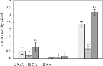
Elastase activity in the synovial fluid samples from ReA, RA, and OA joints was determined using a synthetic substrate and expressed in µU/mL of synovial fluid. Ninety−two percent of the patients with ReA or RA demonstrated detectable elastase activity compared with 75% of the patients with OA. The highest elastase activity (0.8 ± 0.35 µU/mL) was found in SF from RA patients (Fig. 1 column 1). In the SF samples from the patients with ReA, elastase activity amounted to about 66% of the activity of those with RA and was 2.3 times higher than in the SF samples of the patients with OA. To investigate the source of elastase activity in the SF samples, EDTA was added to abolish metal−dependent elastase activity. The mean inhibition of elastase activity by EDTA in ReA was found to be approximately 82%, com− pared with 65% in OA and 78% in RA SF samples (Fig. 1 column 2). A significant increase in elasti− nolytic activity was observed after enzyme activa− tion of prometalloproteases by p−APMA (Fig. 1 column 3). In ReA, elastase activity was increased approximately 4.5−fold compared with 3.2−fold in OA and 4−fold in RA fluids.
Elevation of metal−dependent elastase activity may contribute to collagen and proteoglycan degradation and the release of degradation prod− ucts to SF. The present study found that the con− centrations of collagen and proteoglycan degrada− tion products were comparable in ReA and RA, but the concentration was higher than in OA fluids
0 0.5 1 1.5 2 2.5 3 3.5 4
ReA OA RA
**
* *
** **
elastase activity uU/mL
*
Fig. 1.Elastase activity, using low−molecular−weight substrates, expressed in µU/mL of SF in ReA, OA, and RA samples (column 1) after incubation with 10 mM EDTA (column 2) and after activation with 1 mM p−APMA (column 3). Mean value and standard devia− tion for the different samples are shown. *P< 0.01, **P< 0.05 compared with ReA
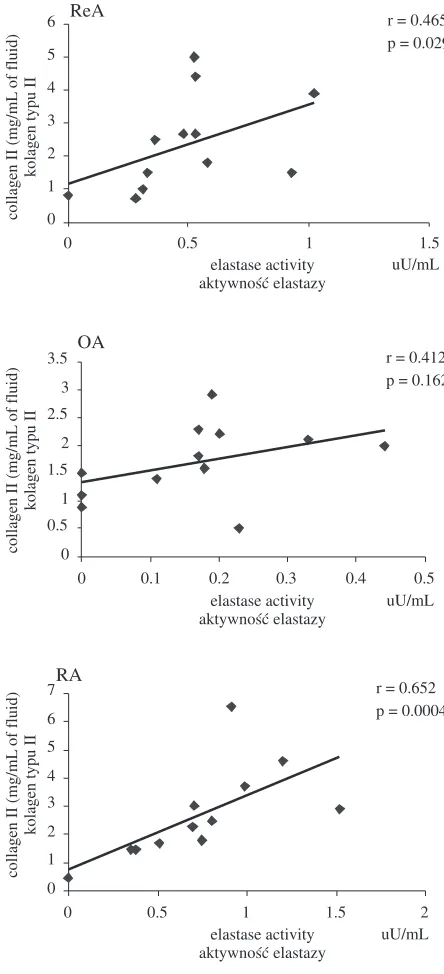
(data not shown). Correlations between the activi− ty of elastase and the concentration of degradation products of type II collagen and GAGs were com− pared in the SF samples of ReA, OA, and RA (Figs. 2 and 3). Significant correlations were found between elastase activity and GAG concentration in RA (r = 0.65, p= 0.006) and ReA (r = 0.676,
p= 0.02) as well as between enzyme activity and CII degradation product concentrations in RA (r = 0.652, p = 0.0004) and ReA (r = 0.465,
p= 0.029) SF samples. In contrast, the correlation between enzyme activity and CII concentration in the OA SF samples was not significant.
Gelatinolytic activity determined by zymogra− phy and two forms of proenzyme (92 and 72 kDa) were detected in the SF samples of the three inves− tigated groups (Fig. 4). However, in the ReA and RA, in contrast to the OA SF samples, with the exception of proenzymes, the low−molecular− −weight active forms of both gelatinases were also detectable.
0 0.2 0.4 0.6 0.8 1 1.2 1.4 1.6
0 0.5 1 1.5
elastase activity aktywność elastazy
uU/mL
ReA r = 0.676
p = 0.02
0 0.2 0.4 0.6 0.8 1 1.2 1.4
0 0.1 0.2 0.3 0.4 0.5
OA r = 0.357
p = 0.041
0 0.2 0.4 0.6 0.8 1 1.2 1.4
0 0.5 1 1.5 2
RA r = 0.650
p = 0.006 elastase activity
aktywność elastazy
uU/mL
elastase activity aktywność elastazy
uU/mL
GAGs (mg/mL
of fluid)
GAGs (mg/mL
of fluid)
GAGs (mg/mL
of fluid)
Fig. 2.Correlations between elastase activity (µU/mL) and GAGs concentration (mg/ml) in SF from patients with ReA, OA, and RA
Ryc. 2. Korelacja między aktywnością elastazy (µU/ml) i stężeniem GAGs (mg/ml) w SF pacjentów z ReA OA i RA
0 1 2 3 4 5 6
0 0.5 1 1.5
r = 0.465 p = 0.029 ReA
0 0.5 1 1.5 2 2.5 3 3.5
0 0.1 0.2 0.3 0.4 0.5
elastase activity aktywność elastazy
uU/mL OA
r = 0.412 p = 0.162
0 1 2 3 4 5 6 7
0 0.5 1 1.5 2
RA
r = 0.652 p = 0.0004
elastase activity aktywność elastazy
uU/mL elastase activity
aktywność elastazy
uU/mL
collagen II (mg/mL
of fluid)
kolagen typu II
collagen II (mg/mL
of fluid)
kolagen typu II
collagen II (mg/mL
of fluid)
kolagen typu II
Fig. 3.Correlations between elastase activity (µU/mL) and collagen type II concentration (mg/mL) in SF from patients with ReA, OA, and RA
Discussion
The destruction of joint cartilage is of central importance in human arthritic diseases. In patients with RA and OA there is clear evidence for increased expression and synthesis of enzymes, including MMPs, in synovial tissue and cartilage and increased concentrations of the same proteas− es in joint fluids [3, 21]. These enzymes are syn− thesized as proenzymes and the majority of the enzyme is found in its inactive form in cartilage and joint fluids [3, 7]. Several studies indicate that the main enzymes involved in the destruction of joint cartilage are leukocytic elastase and collage− nase, which are secreted by inflammatory cells [8–11].
The present study demonstrated that the elas− tase activity found in ReA and RA SF samples was mostly due to metallodependent enzymes because EDTA inhibited about 80% of this activity. In OA, about 65% of the elastase activity was metallode− pendent. Using a low−molecular−weight synthetic
substrate, a higher level of elastase activity was found in RA than in OA SF samples, in accordance with previous reports [22, 23]. Patients diagnosed with ReA represent a new population included in comparative studies of elastase and gelatinase activity along with OA and RA groups. Synovial fluid from patients with ReA, as from those with with RA, showed higher elastase activity than that from patients with OA. Furthermore, in both ReA and RA SF samples, the present study found the active forms of both gelatinases (72 kDa and 92 kDa), in contrast to OA SF, where only proen− zymes were detectable. It has been demonstrated that both the 72 kDa and 92 kDa gelatinases also showed elastase activity [24]. To establish the rela− tionship between elastase activity and collagen and proteoglycans degradation in SF from patients with ReA, OA, and RA, correlation coefficients of elastase activity to concentration of degradation products of CII and GAGs were calculated These correlations were significant in the RA and ReA SF samples. In contrast, in OA SF samples the cor− relation between enzyme activity and CII concen− tration was not significant. In synovial fluids of RA, where large numbers of polymorphonuclear phagocytes are present together with immune complexes and rheumatoid factor, these complex− es may stimulate lysosomal enzyme release [3, 8, 17, 22, 23].
In ReA, the intra−articular persistence of bac− terial antigens may explain the appearance of an inflammatory reaction in the synovium [12, 13]. Bacterial lipopolysaccharide contributes to the recruitment of leukocytes into the synovium and leads to the persistence of mononuclear phago− cytes within the inflamed synovium. Therefore, in ReA, as in RA, lysosomal enzymes can be released in large quantities by inflammatory cells. In OA, where inflammatory phenomena are less intense, enzymes such as elastase and collagenase are produced in limited quantities by chondrocytes and are neutralized by inhibitors when secreted [1, 21]. The present comparative study showed that in ReA as in RA patients, elastases and gelatinases can contribute in similar degrees to the degrada− tion of the major components of cartilage and play a role in the pathogenesis of joint damage.
ReA
OA
RA
92 kDa
72 kDa
ReA
OA
RA
92 kDa
72 kDa
Fig. 4.Zymography of synovial fluids from ReA, OA,
and RA. Equal amounts (about 3 µL of fluid) were electrophoresed on 10% polyacrylamide gels polymer− ized with gelatin (1 mg/mL). Gelatin−degrading enzymes were identified as clear zones with molecular masses of 72 kDa and 92 kDa. Gels are representative of experiments with three SF samples from patients with ReA, OA, and RA
Ryc. 4. Zymografia płynu stawowego pacjentów z ReA, OA i RA. Jednakowe ilości płynu (około 3 µl) poddawano elektroforezie w 10% żelu poliakryloami− dowym zawierającym żelatynę (1 mg/ml). Enzymy trawiące żelatynę identyfikowano na podstawie ja− snych pasm żelu w miejscach odpowiadających biał− kom wzorcowym o masie cząsteczkowej 72 kDa i 92 kDa. Elektroforogram przedstawia typowy obraz uzyskany z 3 płynów stawowych od poszczególnych grup pacjentów
References
[1] Lark MW, Bayne EK, Flanagan J, Harper CF, Hoerrner LA, Hutchinson NI, Singer II, Donatelli SA, Weidner JR, Williams HR, Mumford RA, Lohmander LS:Aggrecan degradation in human cartilage. Evidence for both matrix metalloproteinase and aggrecanase activity in normal, osteoarthritic, and rheumatoid joints. J Clin Invest 1997, 100, 93–106.
[3] Walakovits LA, Moore VL, Bhardwaj N, Gallic GS, Lark MW:Detection of stromelysin and collagenase in syn− ovial fluid from patients with rheumatoid arthritis and posttraumatic knee injury. Arthritis Rheum 1992, 35, 35–42.
[4] Lemaire R, Huet G, Zerimech F, Grard G, Fontaine C, Duquesnoy B, Flipo RM:Selective induction of the secretion of cathepsins B and L by cytokines in synovial fibroblast−like cells. Br J Rheumatol 1997, 36, 735–43.
[5] Popko J, Zalewska A, Brycka R, Macias T, Knas M, Zwierz:Activity of N−acetyl−β−hexosaminidase and its isoenzymes in joint fluid from a knee with an injured anterior cruciate ligament. Biol Sport 2002, 19, 43–49.
[6] Popko J, Zalewska A, Olszewski S, Górska A, Sierakowski S, Zwierz K, Urban M:Activity of N−acetyl−beta hexosaminidase in serum and joint fluid of the knees of patients with juvenile idiopathic arthritis. Clin Exp Rheumatol 2003, 21, 675.
[7] Dean DD, Martel−Pelletier J, Pelletier JP, Howell DS, Woessner JFJr:Evidence for metalloproteinase and metalloproteinase inhibitor imbalance in human osteoarthritic cartilage. J Clin Invest 1989, 84, 678–685.
[8] Larbre JP, Moore AR, Da Silva JAP, Iwamura H, Ionnou Y, Willoughby A: Direct degradation of articular cartilage by rheumatoid synovial fluid: contribution of proteolytic enzymes. J Rheumatol 1994, 21, 1796–1801.
[9] Gadher SJ, Eyre DR, Duance VC, Wotton SF, Heck LW, Schmid TM, Woolley DE:Susceptibility of cartilage collagens type II, IX, X, and XI to human synovial collagenase and neutrophil elastase. Eur J Biochem 1988, 175, 1–7.
[10] McDonald JA, Kelley DG: Degradation of fibronectin by human leukocyte elastase. Release of biologically active fragments. J Biol Chem 1980, 25, 8848–8858.
[11] Schalkwijk J, van den Berg WB, van de Putte LB, Joosten LA:Elastase secreted by activated polymorphonu− clear leucocytes causes chondrocyte damage and matrix degradation in intact articular cartilage: escape from inac− tivation by alpha−1−proteinase inhibitor. Br J Exp Pathol 1987, 68, 81–88.
[12] Sieper J, Braun J: Reactive arthritis. Curr Opin Rheumatol 1999, 11, 238–243.
[13] Toivanen P:From reactive arthritis to rheumatoid arthritis. J Autoimmun 2001,16, 369–371.
[14] Sieper J, Braun J:Problems and advances in the diagnosis of reactive arthritis. J Rheumatol 1999, 26, 1224–1234.
[15] Brandt KD, Fife RS, Braunstein EM, Katz B: Radiological grading of the severity of knee osteoarthritis: rela− tion of the Kellgren and Lawrence grade to a grade based on joint space narrowing, and correlation with arthro− scopic evidence of articular cartilage degradation. Arthritis Rheum 1991, 34, 1381–1386.
[16] Arnett FC, Edworthy SM, Bloch DA, Shane DJ, Fries JH, Cooper NS, Healey LA, Kaplan SR: The American Rheumatism Association 1987 revised criteria for the classification of rheumatoid arthritis. Arthritis Rheum 1988, 31, 315–324.
[17] Chevalier X, Groult N, Texier JM, Larget−Piet B, Hornebeck W:Elastase activity in cartilage extracts and syn− ovial fluids from subjects with osteoarthritis or rheumatoid arthritis: the prominent role of metallproteinases. Clin Exp Rheum 1996, 14, 235–241.
[18] Unemori EN, Werb Z:Reorganization of polymerized actin: a possible trigger for induction of procollagenase in fibroblasts cultured in and on collagen gels. J Cell Biol 1986,103, 1021–1031.
[19] Komsa−Penkova R, Spirova R, Bechev B: Modification of Lowry's method for collagen concentration mea− surement. J Biochem Biophys Methods 1996, 32, 33–43.
[20] Farndale RW, Sayers CA, Barrett AJ:A direct spectrophotometric microassay for sulfated glycosaminoglycans in cartilage cultures. Connect Tissue Res 1982, 9, 247–248.
[21] Yoshikara Y, Nakamura H, Obata K, Yamada H, Hayakawa T, Fujikawa K, Okada Y: Matrix metallopro− teinases and tissue inhibitors of metalloproteinases in synovial fluids from patients with rheumatoid arthritis or osteoarthritis. Ann Rheum Dis 2000, 59, 455–464.
[22] Al−Haik N, Lewis DA, Struthers G:Neutral protease, collagenase and elastase activities in synovial fluids from arthritic patients. Agents Actions 1984, 15, 436–442.
[23] Breedveld F, Lafeber G, Siegert C, Vlemming LJ, Cats A:Elastase and collagenase activities in synovial fluid of patients with arthritis. J Rheumatol 1987, 14, 1008–1012.
[24] Senior RM, Griffin GL, Fliszar CJ, Shapiro SD, Golldberg GI, Welgus HG:Human 92 and 72−kilodalton type IV collagenases are elastases. J Biol Chem 1991, 12, 7870–7875.
Address for correspondence:
Krystyna Średzińska
Department of Medical Chemistry Medical University
15−230 Białystok 8 Poland
Tel.: +48 85 748 56 73 E−mail: [email protected]
Conflict of interest: None declared