Transcription of telomeric DNA leads to high levels of
homologous recombination and t-loops
Anirban Kar, Smaranda Willcox and Jack D. Griffith
*Lineberger Comprehensive Cancer Center, University of North Carolina, Chapel Hill, NC 27599-7295, USA
Received May 06, 2016; Revised August 22, 2016; Accepted August 24, 2016
ABSTRACT
The formation of DNA loops at chromosome ends (t-loops) and the transcription of telomeres produc-ing G-rich RNA (TERRA) represent two central fea-tures of telomeres. To explore a possible link be-tween them we employed artificial human telomeres containing long arrays of TTAGGG repeats flanked by the T7 or T3 promoters. Transcription of these DNAs generates a high frequency of t-loops within in-dividual molecules and homologous recombination events between different DNAs at their telomeric se-quences. T-loop formation does not require a single strand overhang, arguing that both terminal strands insert into the preceding duplex. The loops are very stable and some RNase H resistant TERRA remains at the t-loop, likely adding to their stability. Tran-scription of DNAs containing TTAGTG or TGAGTG re-peats showed greatly reduced loop formation. While in the cell multiple pathways may lead to t-loop for-mation, the pathway revealed here does not depend on the shelterins but rather on the unique character of telomeric DNA when it is opened for transcription. Hence, telomeric sequences may have evolved to fa-cilitate their ability to loop back on themselves.
INTRODUCTION
The ends of eukaryotic chromosomes are capped by DNA– protein complexes termed telomeres, which in most species consist of a repeating DNA element bound by general chro-matin proteins and telomere-specific factors. Telomeres may be as short as a few hundred base pairs (bp) in yeasts and as long as 150 kb in plants. In humans, telomeres approach 15 kb at birth and diminish as we age. The telomere specific proteins in higher eukaryotes are present in one or more multi-protein complexes termed shelterins, as the DNA at the end of the telomere must be sheltered from erroneous recognition as a double strand break (1). This function is be-lieved to be accomplished by the combination of shelterin binding and an architectural solution in which the DNA
terminus folds back to generate a duplex loop (t-loop) that hides the DNA end (2,3).
T-loops have been isolated and visualized by electron mi-croscopy (EM) from species ranging from yeast to humans as reviewed in (2,4). The t-loop junction can occur anywhere along the length of the telomere, generating a spectrum of circle sizes; in peas, t-loops as large as 120 kb with an 80 kb circular portion were observed (5). Recently, using a fluores-cent PNA probe to telomeric sequences, STORM imaged fields of t-loops from mouse cells were observed (4) and the dimensions of the t-loop molecules were close to those ob-served by EM. The percentage of looped species relative to linear telomeric restriction fragments has ranged from 15 to 30% in the EM studies and up to 40% in the STORM analysis.
Our earlier suggestion that telomeres might loop back on themselves (2,3) was based on the fact that most eukaryotic telomeres end with a 3single stranded (ss) tail on the G-rich strand which has the same sequence as the preceding du-plex (ds) telomeric DNA (6). Based on our understanding of homologous recombination (HR) reactions, the 3ss tail would be expected to invade the preceding duplex segment to generate a lasso structure with a D-loop at the junction. This represents a simple intramolecular HR product. Using a model telomere template containing 1–2 kb of TTAGGG repeats and terminating with a 3ss tail on the G rich strand, we observed that purified TRF2 protein would generate t-loops in 15–30% of the DNA, but that TRF1 would not (3). Loop formation required the presence of a homologous 3 G rich ss tail (7), and the junctions were stained with a sin-gle strand binding protein (3) pointing to a D-loop at the junction; however, it remained possible that both strands could be inserted. The recent STORM studies showed that using an inducible system, elimination of functional TRF2 in cells resulted in a several fold reduction in the number of t-loop molecules, while elimination of other shelterins, in-cluding TRF1 and Pot1, did not (4). Because TRF2 is part of a multiprotein complex, the other shelterin components may work with TRF2 to either actively form t-loops or per-haps stabilize them once formed by a variety of mechanisms. Indeed, in the cell there may be multiple pathways leading to t-loop formation and stabilization and one potential path-way might involve transcription.
*To whom correspondence should be addressed. Tel: +1 919 966 8563; Fax: +1 919 966 3015; Email: [email protected]
C
The Author(s) 2016. Published by Oxford University Press on behalf of Nucleic Acids Research.
Telomeres from yeast to humans are transcribed by RNA polymerase II from sub-telomeric promoters. The C-rich telomeric strand is predominantly transcribed, generating a G-rich RNA termed TERRA as first described by the Lingner and Blasco groups (8,9). TERRA transcripts vary in length but can be as long as 9000 nt in humans; however, it is possible that much of the TERRA is relatively small (8– 10). TERRA is now accepted as a key structural element of telomeres, likely providing a structural scaffold upon which proteins involved in telomere maintenance bind. TRF1 and TRF2 (11), hnRNPA1 (12) and replication factors such as Orc1 (11) bind TERRA. Cell cycle studies show that TERRA peaks in G1, declines through S phase and then rises in G2 (10).
Biological studies have shown that TERRA levels are in-creased in human cancer cells (13) and in cells employing the ALT pathway (Alternative Lengthening of Telomeres) (14– 16). TERRA may be involved in telomere length signaling as depletion of TRF2 and shortening of telomeres lead to el-evated levels of TERRA (17,18). Indeed, Porroet al. (2014) showed that reducing TRF2 experimentally in cells having long telomeres results in an increase in TERRA, suggesting that TERRA helps remodel telomeres after loss of TRF2 (18).
TERRA transcribed from mammalian telomeres con-tains repeating runs of 3 guanine bases that can form G quartets. We showed that 576 nt (UUAGGG)n TERRA transcripts are arranged into chains of 24 nt particles linked by a 3 nt bridge (19). Of particular relevance here is the find-ing that transcription of telomeres can result in RNA–DNA hybrids termed R loops in which the G-rich strand is dis-placed and TERRA base pairs to the C-rich template DNA strand, as reviewed in (20). Telomeric R loops have been de-tected at the telomeres of yeast, human and human cancer cells including ALT cells (20). In ALT cells, a direct corre-lation has been observed between enhanced HR, elevated TERRA levels and repression of RNase H1 (14). R-loops may promote HR by opening the helix and providing an en-try site for another homologous DNA strand. Indeed, upon expression of RNase H1 in ALT cells, the level of TERRA diminished and the telomeres were no longer maintained via HR (14). Thus, transcription of a telomere could directly in-fluence its architecture by generating R loops that facilitate intramolecular looping as well as telomere–telomere asso-ciations.
To begin to probe these possibilities in anin vitrosystem we employed T7 RNA polymerase and two sets of DNA templates containing the T7 promoter. One contains a 576 bp tract of TTAGGG repeats at the end of a 3.5 kb plasmid and a second consists of very long ds telomeric DNA ap-proaching the size of human telomeres. Transcription leads to a high frequency of HR products consisting of highly sta-ble t-loops and molecules that have undergone intermolec-ular recombination at their telomeric sequences. This work suggests that t-loop formation is driven by the unusual na-ture of telomeric DNA when it is opened for transcription and that shelterin proteins may have evolved to protect the loops once formed by a variety of mechanisms.
Figure 1.DNA templates. The pRST5 plasmid (7) contains a 576 bp track of (TTAGGG)nrepeats in a pGEM background. (A) Cleavage with BsmBI
generates a linear DNA with the repeats at one end and the T7 RNA poly-merase promoter at the beginning of the repeats oriented as shown. The 4 nt 5overhang allows ligation of ss tails. (B) Cleavage with BbsI results in the opposite orientation and the T3 RNA polymerase promoter oriented as shown. (C) Cleavage with ScaI places the repeats in a central location. (D) Ligation of the linear DNA molecules shown in A generates a minia-ture chromosome with the telomeric sequences at both ends.
MATERIALS AND METHODS
Plasmid DNAs
Construction of pRST5 (Figure 1) containing 576 bp of TTAGGG repeats and T7/T3 promoters was previously de-scribed (7,19). In some experiments the linear DNA con-tained 54 nt or 94 nt 3 TTAGGG overhangs (oligonu-cleotides synthesized by MWG Operon Inc.).
Proteins
For some experiments transcription was carried out using the T7 and T3 Maxiscript kits from Ambion Inc. In addi-tion, T7 RNA polymerase was purified in this laboratory from a plasmid containing a hexahistidine-tagged form of T7 RNA polymerase expressed inE. coli(gift of laboratory of Thomas Cech) using their purification protocol. TRF1 and TRF2 proteins were purified and characterized in this laboratory as N-terminally hexahistidine tagged forms, pre-viously described (21). TheEscherichia coliexpression vec-tors generated (deposited in Addgene.org, Cambridge, MA, plasmid #53209 TRF1 and plasmid #50488 for TRF2) were based on vectors generously provided by the laboratory of E. Gilson (University of Nice, Nice, France). Both proteins were purified by multiple HisTrap HP, HiTrap Heparin HP and HiTrap QFF columns using an AKTA purifier FPLC (GE Healthcare Lifesciences) and were free of any detectible nucleases.
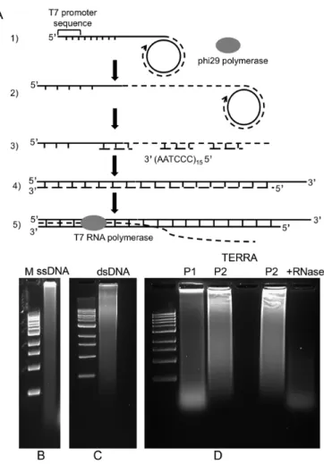
Generation of long telomeric dsDNA
5CACTAA3 (quadruplex mutant) was circularized using CircLigase (Epicentre, Illumina) under conditions de-scribed by the vendor. A 115 nt oligonucleotide consisting of the T7 promoter (5TAATACGACTCACTATAGG3) followed by 16 5TTAGGG3 repeats (wild type), 5TGAGTG3 repeats (telomeric sequence mutant) or 5TTAGTG3repeats (quadruplex mutant) (Eurofins) was annealed to the circle to generate a dsDNA circle with a displaced tail containing the T7 promoter. Incubation for 16 h at 30◦C with 29 DNA polymerase (New England Biolabs) in a buffer containing 10 mM dNTPs yielded G-rich telomeric ssDNA up to at least 15 000 nt long as measured on agarose gels. The newly synthesized DNA was deproteinized with 20g Proteinase K (Invitrogen), 0.02M EDTA and 0.4% SDS in a 50l volume for 1 h at 55◦C and purified by ethanol precipitation for further processing. A complementary 90 nt oligonucleotide consisting of 15 5CCCTAA3 (wild type), 5CACTCA3 (telomeric sequence mutant) or 5CACTAA3 (quadruplex mutant) repeats (Eurofins) was then annealed following heating to 100◦C to release the small circle and the sample was then incubated with DNA polymerase I (New England Biolabs) for 2 h following the manufacturer’s protocol. This newly synthesized dsDNA was deproteinized with 20 g Proteinase K (Invitrogen), 0.02M EDTA and 0.4% SDS in a 50l volume for 1 h at 55◦C and purified by DNA Clean & ConcentratorTM-25 kit (Zymo research). The size of the
resulting telomeric dsDNA was analyzed by 0.8% agarose gel electrophoresis and measured by EM.
Transcription of the telomere templates
Using the commercial T7 or T3 Maxiscript kits (Ambion Inc.) transcription was carried out at 37◦C for 1 to 60 min in the buffers supplied by the vendor. Transcription by T7 RNA polymerase purified in this laboratory was carried out using a buffer containing: 40 mM Tris-Cl, 10 mM MgCl2, 4
mM spermidine, 10 mM DTT, 10 mM NaCl for 5 to 60 min. Typical transcription reactions contained 100 ng of DNA in a 30l volume. Following incubation of the DNA at 37◦C, the DNA was treated with 70g/ml trioxsalen (Sigma) and long wave UV (∼360 nm, 1 cm from sample) to crosslink the DNA, followed by treatment with 660g/ml RNase A (Sigma) or in some experiments 10 units of RNase H (New England Biolabs) or 100 units of RNase T1 (Thermo Sci-entific) followed by addition of Proteinase K (500g/ml), SDS (to 0.5%) and EDTA (5 mM) and incubation for 60 min at 37 or 55◦C. The DNA was then passed over 2 ml columns of 2% agarose beads (Agarose Bead Technolo-gies) previously equilibrated in 10 mM Tris pH 7.5, 0.1 mM EDTA. The DNA in the excluded volume was examined by EM. In some transcription experiments, the pRST5 DNA was treated with 10U S1 nuclease (Thermo Scientific) for 30 min at room temperature, followed by enzyme inactiva-tion and DNA cleaning with DNA clean and concentrator kits (Zymo Research) prior to transcription. Transcription efficiencies of different long template DNAs were checked after transcription by treating them with TURBO DNAse (Ambion) as per manufacturer’s protocol and running the samples on a 1% agarose gel. The electrophoresis was run at 140 V in a 10 cm tray and the gel stained with SYBR Gold
(Thermo Scientific) and examined in a UV transilluminator (BioRad.).
Iron particle staining of TERRA
Transcription reactions with pRST5 DNA were carried out for 30 min at 37◦C using ribonucleotides at 10 mM concen-tration including a mixture of biotin-16-UTP (Roche Di-agnostics GmbH) and unmodified UTP at a molar ratio of 1:4. The samples were then treated with RNase A and H for 1 h at 37◦C followed by deproteinization with Proteinase K and SDS at 37◦C. The samples were filtered through agarose beads and the peak fractions pooled. Paramagnetic iron particles (MACS streptavidin microbeads, Miltenyi Inc.) (3l) were added to a 50l aliquot of the purified t-loop molecules and allowed to incubate overnight at 4◦C. The samples were then filtered through agarose beads to remove the unbound beads and prepared for EM.
Binding and transcription with TRF1 and TRF2
Preincubation of pRST5 with TRF1 or TRF2 was done in a buffer of 20 mM Hepes, pH 8.0, 150 mM KCL, 1 mM DTT and 0.1 mM EDTA. For TRF1, 480 ng of protein were added to 100 ng of DNA, and for TRF2, 72, 144 or 288 ng of protein were incubated with 100 ng of DNA for 20 min at room temperature. Salts were then increased to achieve 40 mM Tris pH 7.5, 10 mM MgCl2, 4 mM spermidine, 10 mM
DTT, 10 mM NaCl. Ribotriphosphates followed by RNA polymerase T7 were added.
Gel electrophoresis
Transcription reactions were carried out with 100 ng DNA substrate in a volume of 20l, processed and deproteinized as carried out for EM and loaded on a 0.8% agarose gel in TAE buffer. Electrophoresis was run at 140 V in a 10 cm tray and the gel stained with SYBR Gold (Thermo Scientific) and examined in a UV transilluminator (BioRad.).
Electron microscopy
DNA samples to be examined by spreading in a mono-layer of denatured cytochrome C at an air-water interface were prepared as described (22) including rotary shadow casting in a high vacuum with platinum–palladium. DNA and DNA–RNA complexes to be examined directly were adsorbed to thin carbon foils in the presence of spermi-dine, washed with a water-ethanol series, air dried and ro-tary shadow cast with tungsten at 10−7 torr (23). Images
were captured using a Gatan Orius real time CCD cam-era (Pleasanton, CA, USA) attached to an FEI Tecnai T12 TEM/STEM instrument (Hillsboro, OR, USA) operated at 40 kV.
RESULTS
Transcription of telomeric DNA produces high levels of t-loops and HR products
Figure 2. Agarose gel electrophoretic analysis of transcription mediated events.Agarose gel electrophoresis of linear pRST5 (BsmBI cut, no 3 overhang) DNA in transcription buffer alone (U). If the DNA was tran-scribed for 30 min (Materials and Methods) and then deproteinized but not treated with RNase A, the DNA with RNA bound was present as a smear whether or not it was crosslinked with psoralen and UV (1,2). Following transcription, deproteinization and treatment with RNase A (3) the DNA was present as a ladder of bands with the monomer band shifted upward.
flanked on one side by a T7 promoter and on the other by a T3 promoter. Cleavage by BsmBI places the TTAGGG repeats at one end of the DNA leaving a 4 nt 5 over-hang on the C rich strand allowing ligation of oligonu-cleotides to generate 3TTAGGG ss overhangs on the G-rich strand. Transcription by T7 RNA polymerase gener-ates G-rich TERRA (19). Cleavage with BbsI places the re-peats at the other end with the T3 promoter oriented into the repeats and transcription by T3 RNA polymerase gen-erates C-rich transcripts. Ligation of the BsmBI cut DNA will generate dimeric molecules with the T7 promoter and telomeric tracts at both ends. In all T7 RNA polymerase transcriptions below (unless otherwise noted) the pRST5 template was cleaved with BsmBI.
Linear pRST5 containing a 54 nt (TTAGGG)9 3
over-hang was transcribed with T7 RNA polymerase at 37◦C for 30 min (using the enzyme purified in this laboratory or the Ambion Megascript kit with similar results) followed by photo crosslinking with psoralen and UV. The samples were then deproteinized with Proteinase K and SDS. Agarose gel electrophoresis (Figure 2, lanes 1,2) showed that irrespec-tive of crosslinking, the linear DNA appeared as a smear of slower migrating material due presumably to RNA re-maining bound to the DNA. When the samples were treated with RNase A (30 min, 37◦C) following transcription, and then deproteinized, a distinct ladder of bands was observed (Figure2, lane 3). The most rapidly migrating band was at a position slightly retarded from the non-transcribed DNA
and this was followed by sharp multimer bands, as well as material near or in the well of the gel.
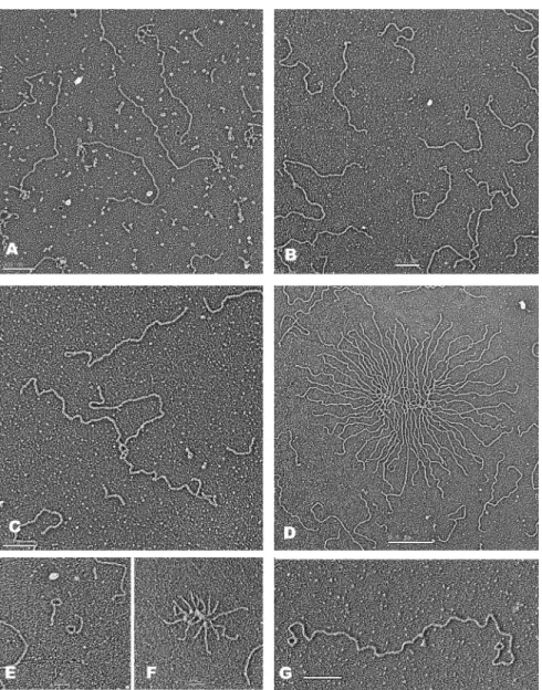
To directly image DNA in these experiments, following deproteinization the samples were filtered through agarose beads to separate the nucleic acids for EM examination (Materials and Methods). The DNA was spread on a film of denatured cytochrome C protein at an air–liquid interface (‘surface spreading’, Materials and Methods). Examination of samples that had not undergone RNase A treatment (as in Figure2, lane 2), revealed fields in which, in one experi-ment, 92% (n=212) of the linear DNA contained an RNA ‘bush’ at one end (Figure3A). When the RNase A treatment was included (as in Figure2, lane 3), the RNA was no longer visible and up to one half of the linear DNAs showed a small loop at one end (Figure3B) (Table1A). In addition, multi-mers involving 2, 3 or more DNAs joined at one end (Figure 3C) were present, as well as clusters in which a large number of DNAs were fused at one end leaving the other end spread out from a dense central core (Figure3D). We termed these ‘DNA bouquets’. From 25 to 50% of the DNA was present in multimers and bouquets, which were not seen in the ab-sence of transcription. The EM results thus correlated per-fectly with the results from agarose gel electrophoresis and the slight retardation of the monomer band is presumably due to the end being looped.
To demonstrate that the small loops and sites where the multimers were fused were at the telomeric sequences, sam-ples were prepared as above (Figure3B–D) and then treated with NaeI. This generates a 935 bp fragment containing 359 bp of plasmid sequences followed by the TTAGGG repeats. EM revealed∼1 kb fragments containing the loops (Fig-ure3E) and bouquets with the DNA arms reduced to∼1 kb (Figure3F). Mapping the location of the loops along the pRST5 DNA showed that the loops were localized ex-clusively to the telomeric sequences with a distribution of loop sizes between 200 and 550 bp and a peak near 400 bp (Supplementary Figure S1). Using a miniature chromo-some containing the telomeric tracts at both ends (Figure 1D), transcription generated frequent molecules with small loops at both ends (Figure3G).
When pRST5 DNA with the 54 nt 3overhang was incu-bated for 30 min in the transcription buffer without RNA polymerase and then prepared for EM, only 4% (n=338) of the linear DNAs had a loop at one end. If RNA poly-merase was included but the incubations lacked rNTPs, or contained ATP and UTP but not GTP, the percentage of DNAs with a loop at one end was 1% (n=100) and 1% (n
=101), respectively, and multimers or bouquets were not observed. Incubation of pRST5 DNA (100 ng) containing the 54 nt overhang for 30 min at 37◦C with 100 ng of pu-rified TERRA under transcription conditions but without RNA polymerase did not generate t-loops (2% looped, n= 200) (Table1A).
exam-Figure 3. EM visualization of transcription induced loops and HR products. (A) If the DNA templates were transcribed but not treated with RNAse A prior to preparation for EM, over 70% of the DNA had a bush of RNA attached at one end (see text for details). (B–G) With RNase A treatment, fields of linear molecules were present many of which contained a (B) tiny loop at one end and in the same fields examples of (C) several DNAs joined at one end were present, along with DNA bouquets containing (D) many molecules fused at one end. (E) When the samples above (B–G) were cleaved with NaeI that cuts 935 bp in from the telomeric end∼1 kb fragments with loops (E) as well as bouquets with∼1 kb arms (F) were observed. (G) Transcription of the minichromosome template shown in Figure1D and processing as in B yielded DNAs with loops at both ends. Samples were prepared by surface spreading with cytochrome C (Materials and Methods) followed by rotary metal shadow casting. Bars indicating magnification are shown in each panel.
ples were seen following transcription in which one or both ends were fused back to the middle of the DNA. However, when this DNA was mixed with an equal amount of linear DNA with the telomeric repeats at the end, and the mix-ture transcribed, bimolecular HR products were observed in which the end of the DNA with the terminal repeats in-vaded the middle of the DNA with the central repeats (Sup-plementary Figure S2A). This argues that the multimers and bouquets involve HR strand invasions at the telomeric sequences. While most of the incubations were carried out at 37◦C for 15 or 30 min, a significant number of looped DNAs were seen as early as 1 and 2 min (14% and 23%, respectively), with an increase over the remaining 60 min. Agarose gel electrophoresis verified that the dimer, trimer and multimer bands were only seen following transcription,
and further, that these bands began to appear as early as 2 min after initiation of the reactions (Figure4, lanes 1–3).
Transcription-mediated looping does not require a 3ss over-hang
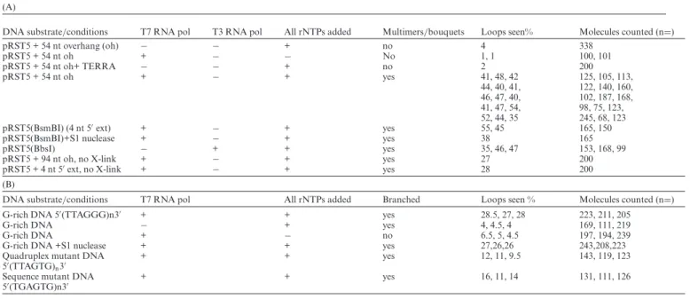
Table 1. Summary of EM scoring of over 6500 single molecules from the transcription experiments. Multiple entries in the rows represent replicate ex-periments under the same conditions but done on different days. Corresponding number of molecules scored for each is listed in order in the left column. X-link refers to psoralen and UV photo crosslinking. See text for specific details
(A)
DNA substrate/conditions T7 RNA pol T3 RNA pol All rNTPs added Multimers/bouquets Loops seen% Molecules counted (n=)
pRST5 + 54 nt overhang (oh) − − + no 4 338
pRST5 + 54 nt oh + − − No 1, 1 100, 101
pRST5 + 54 nt oh+ TERRA − − + no 2 200
pRST5 + 54 nt oh + − + yes 41, 48, 42 125, 105, 113,
44, 40, 41, 122, 140, 160, 46, 47, 40, 102, 187, 168, 41, 47, 54, 98, 75, 123,
52, 44, 35 245, 68, 123
pRST5(BsmBI) (4 nt 5ext) + − + yes 55, 45 165, 150
pRST5(BsmBI)+S1 nuclease + − + yes 38 165
pRST5(BbsI) − + + yes 35, 46, 47 153, 168, 99
pRST5 + 94 nt oh, no X-link + − + yes 27 200
pRST5 + 4 nt 5ext, no X-link + − + yes 28 200
(B)
DNA substrate/conditions T7 RNA pol All rNTPs added Branched Loops seen % Molecules counted (n=)
G-rich DNA 5(TTAGGG)n3 + + yes 28.5, 27, 28 223, 211, 205
G-rich DNA − + yes 4, 4.5, 4 169, 111, 219
G-rich DNA + − no 6.5, 5, 4.5 197, 194, 239
G-rich DNA +S1 nuclease + + yes 27,26,26 243,208,223
Quadruplex mutant DNA 5(TTAGTG)n3
+ + yes 12, 11, 9.5 143, 119, 123
Sequence mutant DNA 5(TGAGTG)n3
+ + yes 16, 11, 14 131, 111, 126
Figure 4. Agarose gel electrophoretic analysis of DNA transcribed in the presence of TRF1 or TRF2. (A) Agarose gel electrophoresis of lin-ear pRST5 (BsmBI cut no 3overhang) DNA in transcription buffer alone (U). Lanes 1–6: the same DNA transcribed with T7 polymerase for for 2 min (1,4), 5 min (2,5) and 20 min (3,6), then treated with RNase A, and deproteinized. In lanes 4–6 the DNA was preincubated for 20 min with TRF1 protein (text, Materials and Methods) prior to transcription. (B) pRST5 DNA (BsmBI cut, 54 nt sss 3overhang) was preincubated with 0, 2, 4 or 8 TRF2 monomers per terminal ss/ds junction, respectively, (lanes 1–4) prior to transcription for 30 min with T7 RNA polymerase, RNase treatment and deproteinization.
the DNA with a 54 nt 3tail (data not shown). Fully blunt ended DNAs were also efficient templates for loop forma-tion as transcripforma-tion of S1 treated DNA yielded 38% (n= 165) of the linear DNA having terminal loops (n = 165) (Table1A).
Cleavage of pRST5 with BbsI produces a linear DNA with a 4 nt 5extension and with the telomeric repeats at the other end as contrasted to BsmBI cleavage (Figure1B). This DNA contains a T3 RNA polymerase promoter at the be-ginning of the repeats oriented to produce a C-rich telomere transcript. Upon transcription with T3 RNA polymerase
(Materials and Methods) in three separate experiments the percentage of monomer DNAs with a loop at one end was 35%, 46% and 47% (n=420) (Table1A). Multimers were present at a similar frequency as in the experiments above. These results show that transcription mediated t-loop for-mation can occur using two different RNA polymerases, and provides another example in which a 3ss overhang was not required.
Transcription mediated t-loops are highly stable
loops when the RNA remained present, pRST5 with a 54 nt 3ss overhang was transcribed with T7 RNA polymerase for 30 min followed by deproteinization and agarose chro-matography, but not treated with RNase A or H. The sam-ple was then left at 4◦C for 72 h followed by photo crosslink-ing and RNase A treatment. A high percentage of t-loops was observed (57% of the monomer DNA) which was as high as seen in experiments when crosslinking was done at the end of the transcription reactions. In other studies (data not shown) t-loops were equally stable with 4 nt 5 exten-sions as with the 54 nt 3extension.
It was of interest to examine the thermal stability of the t-loop molecules. Linear pRST5 (4 nt 5extension) was tran-scribed for 30 min and the DNA processed for EM but with-out photo crosslinking. Aliquots were then heated at 21◦C, 37◦C, 42◦C, 55◦C and 65◦C for 20 min followed by prepara-tion at room temperature for EM by direct adsorpprepara-tion. As the temperature increased, the percentage of monomers in t-loops declined: 46% (n=102) at 21◦C, 24% (n=75) at 37◦C, 25% (n=141) 42◦C, 16% (n=128) at 55◦C and 14% (n= 129) at 65◦C, revealing a significant thermal stability. From these experiments we conclude that transcription-mediated t-loops are highly stable, further arguing that both strands are inserted back into the preceding telomeric duplex. This high stability will open the door for future experiments us-ing non-crosslinked t-loop preparations to probe their res-olution by junction resolving enzymes.
TRF1 and TRF2 do not impede looping in these reactions
It was of interest to carry out initial investigations of how preincubation of pRST5 with TRF1 or TRF2 might affect its subsequent transcription. Clearly a detailed study for the future will require a eukaryotic RNA polymerase II tran-scription system and a chromatinized DNA template. The TRF2 protein used was previously characterized in anin vitrostrand assimilation assay (21) and the amount used (2, 4 and 8 protein monomers per telomeric ss/ds junction) was based on previous studies (26) and on an EM determina-tion of the maximal amount of TRF2 that could be added without generating large DNA aggregates. The DNA con-taining a 54 nt 3overhang was preincubated at room tem-perature for 20 min with TRF2 in binding buffer and then transcribed with T7 RNA polymerase following addition of salts optimal for transcription (Materials and Methods). Parallel experiments were carried out using TRF1. Incuba-tion condiIncuba-tions were taken from our study of TRF1 binding to telomeric DNA (27). In agreement with that study, a sto-ichiometry of 2 monomers per TTAGGG repeat was found by EM to fully coat >75% of the telomeric tracts on the pRST5 DNA. Preincubation of pRST5 (4 nt 5overhang) for 20 min at room temperature under optimal TRF1 bind-ing conditions was followed by transcription (Materials and Methods), purification of the products and agarose gel elec-trophoresis. As shown (Figure4A and B), no significant dif-ference in the pattern of monomer and multimer bands was observed between the samples preincubated with or with-out either TRF1 or TRF2. EM examination did not reveal any significant effects of preincubation with either protein.
A beaded particle is frequently present at the t-loop junction and may contain TERRA
The high stability of the t-loop molecules suggested the pos-sibility that some TERRA may remain tightly bound even following purification. To examine this, pRST5 DNA was transcribed with T7 RNA polymerase in reactions contain-ing biotin-16-UTP. The samples were treated with RNase A and H for 1 h, deproteinized, filtered through agarose beads and then mixed with∼50 nm paramagnetic iron particles coated with streptavidin (Materials and Methods) and fil-tered again to remove the unbound iron particles. Prepara-tion for EM involved directly adsorbing the samples to thin carbon foils followed by washing, drying and rotary shadow casting with tungsten (Materials and Methods). Examina-tion of fields of molecules showed that 40% (n=84) of the DNA contained iron particles at one end, a percentage close to the fraction in t-loops. When the particles did not obscure the loop (Figure5A), they were located at the t-loop junc-tion. When two or more DNAs were joined by intermolec-ular HR, iron particles were bound exclusively at the DNA junctions (Figure5B). Thus, the presence of TERRA may account in part for the high stability of the t-loop molecules. The higher resolution of tungsten shadow casting made it possible to obtain a better picture of the t-loops and junc-tions, and showed that the loops frequently contain a small, usually oblong bead at the loop junction (Figure5C–G, J; and Supplementary Figure S2B–F). Measurement of the di-mensions of the beads revealed an average length of 13.3 nm and width of 10.8 nm (n=17). In experiments using T7 RNA polymerase, from 30 to 75% of the loop junctions showed these beads while the remaining junctions did not. In reactions employing T3 RNA polymerase, similar beads were observed in one experiment at 46% of the junctions (n
=154). Treatment with both RNase A and T1 (which will digest TERRA if it is not bound to DNA) did not reduce the frequency or size of the beads; however, treatment with RNase A and RNase H reduced the fraction of loop junc-tions with a bead by 20% and 26% in two different exper-iments. Nonetheless, a significant portion retained a bead. The samples had undergone extensive deproteinization with Proteinase K and SDS and thus it is unlikely that these par-ticles represent remaining RNA polymerase. The loop junc-tions often contained a short DNA stem typical of regressed forks (chicken foot structures) (Figure 5I, J, Supplemen-tary Figure S2E and F). These were more commonly found in the samples that had not undergone photo crosslinking where they were present in 17% (n=197) of the loop junc-tions.
Generation of human-sized telomeric and mutant telomeric DNAs
Figure 5. Architecture and composition of the telomeric tracts following transcription.(AandB) Linear pRST5 with no overhang or (C–J) a 54 nt 3 overhang was transcribed for 30 min with T7 RNA polymerase in reactions containing (A and B) biotin-16-UTP or (C–J) UTP together with ATP and GTP (Materials and Methods). To detect any RNase resistant TERRA, samples were treated with RNase A and H followed by purification, and incubation with iron particles coated with streptavidin followed by removal of the unbound particles and EM preparation (text, Materials and Methods). The dense objects at the (A) loop junction or (B) junction of 3 DNAs are the iron particles. If RNase A treatment was not included, (CandD) linear molecules with loops at one end associated with a dense mass of RNA were present. (EandF): Following RNase A treatment, and purification, the loop junctions often contained a beaded particle. (GandH): Treatment of the samples with both RNase A and RNase H resulted in loop junctions, (G) some exhibiting a bead and (H) some not. (IandJ) Loop junctions showing a short DNA stem extruded from the junction. Samples were mounted onto thin carbon supports followed by shadow casting with tungsten. Magnification bars are shown in each panel.
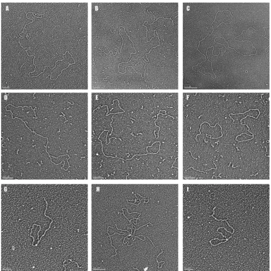
potentially contribute to the stability of the t-loop. We ad-dressed this by generating two different mutant sequences. The first contains a single base substitution in which the middle G is replaced by thymidine. The second mutant has the same base composition as the wild-type repeat but in a scrambled order. To produce such dsDNAs with lengths that approach that of human telomeres and contain a T7 RNA polymerase promoter at one end, we used rolling cir-cle replication of a small circular template with29 DNA polymerase and generated three different ssDNA species as long as 15 000 nt (Figure6A), which were then converted to dsDNA (Figure 6B) with DNA polymerase I (Materi-als and Methods). The three ssDNAs consisted of repeating TTAGGGn, TTAGTGn in which the run of 3 G’s that can form G quadruplexes is disrupted, and TGAGTGn, which has the same ratio of bases as TTAGGG but no runs of G’s. The resulting DNAs ranged from a few hundred bp to some molecules approaching 40 kb, with a significant proportion in the range of 3 to 10 kb (Figure7A–C). The 3end of the ds DNA opposite the T7 promoter may be blunt or may have a short 3G-rich ss tail depending on where the most
terminal C-rich oligonucleotide annealed prior to synthe-sis of the complementary strand. This was verified by SSB staining and EM (not shown.)
Transcription of human-sized telomeric DNA generates t-loops and HR products
lin-Figure 6. Agarose gel electrophoresis of long telomeric DNA and TERRA. (A) Schematic of the rolling circle synthesis of the long telom-eric ssDNA and dsDNA. (B) Agarose gel electrophoresis of long G-rich ss telomeric DNA, (C) telomeric dsDNA and (D) TERRA. P1 refers to TERRA generated using the Megascript kit (Supplementary Materials) and P2 refers to TERRA generated using T7 RNA polymerase purified in this laboratory. The marker ladder consists of 1 kb steps with highest molecular weight band at 10 kb.
ear DNAs with loops at one end (Figure 7D–G and I) as well as molecules with one or more branches along their length, representing HR events between two or more DNAs (Figure7H). In three separate experiments, 28% (57/211, 58/205, 64/223) of the long DNAs were arranged into t-loops and 23% (50/211, 47/205, 52/223) of the molecules contained one or more branches along their length (Table 1B). When the DNA was incubated for 2 h under transcrip-tion conditranscrip-tions but without RNA polymerase, in 3 separate experiments the percentage of DNAs with a loop at one end was∼4% (7/169, 5/111, 9/219). When T7 RNA polymerase was included but the incubation lacked NTPs, 5% (13/197, 9/174, 11/239) of the DNA were scored as t-loop forms and 3% (7/197, 5/174, 8/239) showed branches in three differ-ent experimdiffer-ents (Table1B).
When the quadruplex mutant DNA (TTAGTG)n was
transcribed, the looping frequency dropped to 11% (n = 385) and 8% (n=385) of the total molecules were branched. Similarly, in reactions with DNA containing the scrambled sequence (TGAGTG)n,14% (n=368) were looped and 10%
(n=368) were branched (Table1B). Thus while DNAs con-sisting of the two mutant sequences tested here can gener-ate looped or branched molecules upon transcription, they do so at a 2- to 3-fold lower frequency than DNA with the wild-type telomeric repeat.
In previous studies of t-loops isolated from human cells, there was no correlation between the size of the circular por-tion and the length of the tail, with small loops being found at the ends of long telomeric DNAs, and other, shorter telomeric fragments being mostly circular with only a short tail. This was the case here, as in several experiments em-ploying the long DNA with the wild-type repeat the looped portions varied from 0.3 kb to 2.5 kb, with the majority be-ing between 1 and 1.5 kb (Supplementary Figure S2). There was no clear relation between the size of the loop and the total length of the t-loop molecule.
To ask if a ss tail is required for loop formation, the long wild type telomeric DNA was digested with S1 nuclease. Agarose gel electrophoresis revealed only a modest shift to smaller lengths pointing to a relative lack of nicks or gaps in the DNA. Following S1 treatment, the long DNA was gel purified and transcribed by T7 RNA polymerase. In 3 dif-ferent experiments, the percentage of DNA in t-loops was 27% (n = 243), 26% (n= 208) and 26% (n =223), and branched forms were 25% (n =212), 25%(n =173) and 25%(n=187) (Table1B). Thus, blunt ended molecules will form t-loops upon transcription and nicks or gaps along the DNA are not required for looping.
Examination of the loop junctions formed by the long telomeric DNA revealed a fraction with small beaded par-ticles similar to those observed with the pRST5 template (Figure7G and I). In one experiment, 21% (n=54) of the loop junctions showed such a bead following extensive pu-rification with RNase A, Proteinase K and SDS. In addi-tion, chicken foot stems were also present (Figure7I). This observation further points to the conclusion that the loop junctions may involve a complex structure, likely containing residual TERRA.
Since the loops formed by the long (wild type) telomeric DNA were much larger than those formed on the pRST5 template, it was of interest to examine their stability. The long telomeric DNA was transcribed with T7 RNA poly-merase as above but not photo crosslinked. One aliquot was then treated with only Proteinase K and SDS while another was treated with RNase A, Proteinase K and SDS. Both samples were chromatographed over agarose beads and ei-ther mounted for EM within 1 h of chromatography or al-lowed to remain at 4◦C for 72 h before EM preparation.The sample treated with Proteinase K and RNase A at 1 h con-tained 26% loops (n=226), whereas after 72 h at 4◦C 17% (n=219) of the molecules were in t-loops. For the aliquot not treated with RNase A, 29% (n=253) of the DNA was in t-loops 1 h after chromatography, and after 72 h at 4◦C, 21% (n=233) of the DNA was in t-loops. Thus, the large sized transcription mediated t-loops are highly stable.
DISCUSSION
Figure 7. Visualization of long telomeric DNA and transcription products.Long telomeric dsDNA was generated (Figure6, Supplementary Material) and prepared for (A–C) EM. The sizes of these very large telomeric DNAs are 21.6, 26.9 and 41.0 kb. (D–G,I) Following transcription with T7 RNA polymerase DNAs arranged into loops were common as well as (H) high branched structures. (G–I) Samples were prepared by surface spreading with cytochrome C or by adsorption to carbon supports and tungsten shadow casting. Lengths of the DNAs in D-G and I respectively are: 11.1, 8.6, 7.4, 2.2 and 2.3 kb. Magnification bars are shown in each panel.
including t-loops. Indeed, Horard and Gilson suggested that t-loops might be stabilized by TERRA (28) and Az-zalin and Lingner have discussed how TERRA might be involved in telomere remodelling (29). In agreement with these authors, we observed TERRA tightly associated with the loop junctions. The work reported here describes a path-way of t-loop formation that is transcription driven rather than being dependent on TRF2.
We employed two RNA polymerases (T7, T3), which function as single polypeptides and clear their promoters at high efficiency. They are robust, readily available, and eas-ily purified. Future studies will test reconstituted eukaryotic RNA polymerase II systems. However, this will require de-veloping different model telomere DNAs, multiple factors will be required, and promoter clearance would be less ef-ficient. The size of transcription bubbles is similar between the phage RNA polymerases and the eukaryotic RNA poly-merase II complex (30,31). Thus, the basic changes in DNA structure induced by transcription, including formation of an R loop and/or G quartets in the displaced G-rich strand are likely to be dictated by sequence features of the DNA
and not the particular RNA polymerase. Initial experiments in which the telomeric DNA was preincubated with puri-fied TRF1 or TRF2 did not show any significant effects on t-loop formation. They were likely displaced by T7 RNA polymerase and, once removed, the subsequent events oc-curred as if in their absence. More detailed experiments will be warranted in the future using a eukaryotic RNA poly-merase II transcription system, and the shelterins, ideally in the context of a chromatinized template.
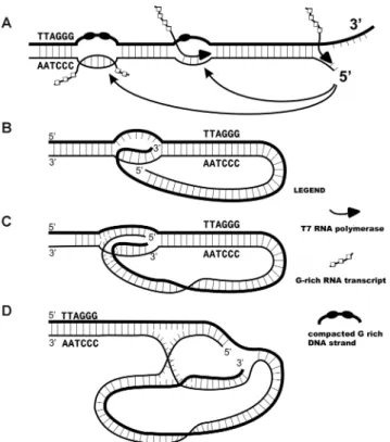
Figure 8. Models of transcription mediated telomere looping and archi-tecture of the t-loop junctions. (A) A telomere undergoing transcription by RNA polymerase generates G-rich RNA arranged into chains of 24 nt beads (19). Here, a segment on the DNA following transit of the poly-merase remains in an R loop with the rich strand compacted into G-quartet stabilized particles. When the polymerase reaches the end of the telomere the DNA strands are split apart and can pair either at the R loop or at the site of transcription. (B) Classic diagram of a t-loop junction in which just the long 3overhang has inserted into the preceding DNA to form a D-loop. (C) A more stable joint is generated if both DNA strands are inserted. (D) Illustration of the t-loop junction as a hybrid replication fork and Holliday junction. The joint in C has been drawn to illustrate its architecture in which the right side has features of a replication fork where the G-rich strand (dark strand) represents the leading strand and the C rich strand, the lagging strand. On the left, the strands cross over in man-ner suggestive of a Holliday junction. Migration of this crossover along the telomere repeats to the left together with pairing of the ‘leading and lag-ging strand termini’ on the right could lead to the extrusion of a regressed chicken foot structure (Figure5I, J, Supplementary Figure S2E and F).
lead to their disruption. Given that telomere loops presum-ably open to allow transcription and replication, there may be multiple means for reforming and stabilizing loops.
In the cell, looping might occur between the terminus of the telomere bound by the shelterin complex includ-ing TRF2, and an internal R loop containinclud-ing an extended TERRA tail which would engage the TRF2 component of the shelterin complex and bring the two sites together. Here, the primary function of the 3 ss overhang is to position the shelterin complex at the telomere terminus poised to loop back and engage an internal R loop (Figure8). Addi-tional shelterin complexes bound at the R loops could fur-ther facilitate looping. For cells maintaining their telomeres by ALT, the higher levels of TERRA may allow the telom-eric DNA to remain in a more open configuration peppered with R loops which would promote HR with extrachromo-somal t-circles or other chromosomes.
In the diagram shown in Figure8, a junction in which both terminal strands have invaded the preceding duplex is drawn to illustrate that these junctions have features of both a replication fork (right side) and a Holliday junction (left side). Fork regression and movement of the Holliday junction to the left would result in an extruded chicken foot stem, a possibility supported by the images shown in Fig-ure5I, J and Supplementary Figure S2. This complex junc-tion could be a target for juncjunc-tion resolving factors, which in cells employing ALT might release t-circles. Replicative extension of the inserted 3terminus could extend telomere length in a telomerase-independent fashion. TRF2 and the TRF2-Rap1 complex bind Holliday junctions, suggesting a possible role in stabilizing these structures (26,32).
An unexpected feature of the loop junctions was the fre-quent presence of small∼11×13 nm beads. Extensive de-proteinization along with RNase A and RNase T treatment did not eliminate them; however, they were reduced to some extent in number by treatment with RNase H. Localization of biotin-tagged TERRA with streptavidin–iron particles showed that some residual TERRA remains at the t-loop junctions in spite of extensive RNase treatments. Thus, the beads may consist of a mixture of ss G-rich or C-rich DNA and TERRA, possibly stabilized by G-quadruplexes in a form resistant to the RNases.
Further evidence for the involvement of G quadruplexes came from the experiments with two long DNAs com-posed of mutants of the wild type repeat. Their transcrip-tion resulted in a 2- to 3-fold lower fractranscrip-tion of t-loops when compared with the wild-type repeat. The sequence changes in both disrupted the run of 3 G’s capable of forming G quadruplexes. Thus, this motif appears important for either formation of loops or stabilizing them once formed. Future experiments will explore the relative stability of the loops formed by these different repeats.
Inherent in the repetitive nature of telomeric DNA, to-gether with one strand that can form G quartets is the abil-ity to loop back on itself upon opening of the helix by tran-scription. If the looping provided some benefit(s) for the sta-bilization and maintenance of the terminal parts of a linear genome, the ability to form t-loops may have been one of the selection forces accompanying the evolution of eukaryotic telomeric sequences and may have preceded the origin of the proteins that protect, resolve or generate t-loops them-selves.
SUPPLEMENTARY DATA
Supplementary Dataare available at NAR Online.
ACKNOWLEDGEMENTS
FUNDING
National Institutes of Health [GM 31819; ES 13773 to J.D.G]. Funding for open access charge: National Institutes of Health [GM 31819; ES 13773 to J.D.G].
Conflict of interest statement.None declared.
REFERENCES
1. Palm,W. and de Lange,T. (2008) How shelterin protects mammalian telomeres.Annu. Rev. Genet.,42, 301–334.
2. Griffith,J.D. (2013) Many ways to loop DNA.J. Biol. Chem.,288, 29724–29735.
3. Griffith,J.D., Comeau,L., Rosenfield,S. and Stansel,R.M. (1999) Mammalian telomeres end in a large duplex loop.Cell,97, 503–514. 4. Doksani,Y., Wu,J.Y., De Lange,T. and Zhuang,X. (2013)
Super-resolution fluorescence imaging of telomeres reveals TRF2-dependent t-loop formation.Cell,155, 345–356. 5. Cesare,A.J., Quinney,N., Willcox,S., Subramanian,D. and
Griffith,J.D. (2003) Telomere looping in P. sativum (common garden pea).Plant J.,36, 271–279.
6. Wright,W.E., Tesmer,V.M., Huffman,K.E., Levene,S.D. and Shay,J.W. (1997) Normal human chromosomes have long G-rich telomeric overhangs at one end.Genes Dev.,11, 2801–2809. 7. Stansel,R.M., De Lange,T. and Griffith,J.D. (2001) T-loop assembly
in vitro involves binding of TRF2 near the 3telomeric overhang.
EMBO J.,20, 5532–5540.
8. Azzalin,C.M., Reichenbach,P., Khoriauli,L., Giulotto,E. and Lingner,J. (2007) Telomeric repeat containing RNA and RNA surveillance factors at mammalian chromosome ends.Science,318, 798–801.
9. Blasco,M. and Schoeftner,S. (2008) Developmentally regulated transcription of mammalian telomeres by DNA-dependent RNA polymerase II.Nat. Cell Biol.,10, 228–236.
10. Porro,A., Feuerhahn,S., Reichenbach,P. and Lingner,J. (2010) Molecular dissection of telomeric repeat-containing RNA biogenesis unveils the presence of distinct and multiple regulatory pathways.
Mol. Cell. Biol.,30, 4808–4817.
11. Deng,Z., Norseen,J., Wiedmer,A., Riethman,H. and Lieberman,P.M. (2009) TERRA RNA binding to TRF2 facilitates heterochromatin formation and ORC recruitment at telomeres.Mol. Cell,35, 403–413. 12. Redon,S., Zemp,I. and Lingner,J. (2013) A three-state model for the
regulation of telomerase by TERRA and hnRNPA1.Nucleic Acids Res.,41, 9117–9128.
13. Deng,Z., Wang,Z., Stong,N., Plasschaert,R., Moczan,A., Chen,H.S., Hu,S., Wikramasinghe,P., Davuluri,R.V., Bartolomei,M.S.et al.
(2012) A role for CTCF and cohesin in subtelomere chromatin organization, TERRA transcription, and telomere end protection.
EMBO J.,31, 4165–4178.
14. Arora,R., Lee,Y., Wischnewski,H., Brun,C.M., Schwarz,T. and Azzalin,C.M. (2014) RNaseH1 regulates TERRA-telomeric DNA hybrids and telomere maintenance in ALT tumour cells.Nat. Commun.,5, 5220.
15. Episkopou,H., Draskovic,I., Van Beneden,A., Tilman,G., Mattiussi,M., Gobin,M., Arnoult,N., Londono-Vallejo,A. and Decottignies,A. (2014) Alternative lengthening of telomeres is
characterized by reduced compaction of telomeric chromatin.Nucleic Acids Res.,42, 4391–4405.
16. Ng,L.J., Cropley,J.E., Pickett,H.A., Reddel,R.R. and Suter,C.M. (2009) Telomerase activity is associated with an increase in DNA methylation at the proximal subtelomere and a reduction in telomeric transcription.Nucleic Acids Res.,37, 1152–1159.
17. Cusanelli,E., Perez Romero,C.A. and Chartrand,P. (2013) Telomeric noncoding RNA TERRA is induced by telomere shortening to nucleate telomerase molecules at short telomeres.Mol. Cell,51, 780–791.
18. Porro,A., Feuerhahn,S., Delafontaine,J., Riethman,H., Rougemont,J. and Lingner,J. (2014) Functional characterization of the TERRA transcriptome at damaged telomeres.Nat. Commun.,5, 5379. 19. Randall,A. and Griffith,J.D. (2009) Structure of long telomeric RNA
transcripts: The G-rich RNA forms a compact repeating structure containing G-quartets.J. Biol. Chem.,284, 13980–13986.
20. Rippe,K. and Luke,B. (2015) TERRA and the state of the telomere.
Nat. Struct. Mol. Biol.,22, 853–858.
21. Bower,B.D. and Griffith,J.D. (2014) TRF1 and TRF2 differentially modulate Rad51-mediated telomeric and nontelomeric displacement loop formation in vitro.Biochemistry,53, 5485–5495.
22. Thresher,R. and Griffith,J. (1992) Electron microscopic visualization of DNA and DNA-protein complexes as adjunct to biochemical studies.Methods Enzymol.,211, 481–490.
23. Griffith,J.D. and Christiansen,G. (1978) Electron microscope visualization of chromatin and other DNA-protein complexes.Annu. Rev. Biophys. Bioeng.,7, 19–35.
24. Cech,T.R. and Pardue,M.L. (1976) Electron microscopy of DNA crosslinked with trimethylpsoralen: test of the secondary structure of eukaryotic inverted repeat sequences.Proc. Natl. Acad. Sci. U.S.A., 73, 2644–2648.
25. Cordeiro-Stone,M., Makhov,A.M., Zaritskaya,L.S. and Griffith,J.D. (1999) Analysis of DNA replication forks encountering a pyrimidine dimer in the template to the leading strand.J. Mol. Biol.,289, 1207–1218.
26. Fouche,N., Cesare,A.J., Willcox,S., Ozgur,S., Compton,S.A. and Griffith,J.D. (2006) The basic domain of TRF2 directs binding to DNA junctions irrespective of the presence of TTAGGG repeats.J. Biol. Chem.,281, 37486–37495.
27. Griffith,J., Bianchi,A. and de Lange,T. (1998) TRF1 promotes parallel pairing of telomeric tracts in vitro.J. Mol. Biol.,278, 79–88. 28. Horard,B. and Gilson,E. (2008) Telomeric RNA enters the game.
Nat. Cell Biol.,10, 113–115.
29. Azzalin,C.M. and Lingner,J. (2015) Telomere functions grounding on TERRA firma.Trends Cell Biol.,25, 29–36.
30. Gnatt,A.L., Cramer,P., Fu,J., Bushnell,D.A., Kornberg,R.D., Cramer,P., Zhang,G., Cramer,P., Bushnell,D.A., Kornberg,R.D.et al.
(2001) Structural basis of transcription: An RNA Polymerase II elongation complex at 3.3 A resolution.Science,292, 1876–1882. 31. Jiang,M., Ma,N., Vassylyev,D.G. and McAllister,W.T. (2004) RNA
displacement and resolution of the transcription bubble during transcription by R7 RNA polymerase.Mol. Cell,15, 777–788. 32. Arat,N.O. and Griffith,J.D. (2012) Human Rap1 Interacts Directly