Introduction
Inherited metabolic disorders present great challenges for patients, families, and healthcare professionals. In amino acidopathies, treatment is for life and dietary management consists of attempting to provide nutrition for growth and development while keeping serum concentrations of potential toxins to a minimum. Medical nutrition therapy for inherited metabolic disorders requires attention to more than just protein, calories, and micronutrient recommendations, as will be seen in this case study.
Maple syrup disease (MSUD), or branched-chain alpha-ketoaciduria, is a genetic disorder
inherited in an autosomal recessive manner; both the mother and father of an affected infant must have passed on a defective gene for the branched-chain keto-acid dehydrogenase (BCKAD) enzyme1. They will each carry one copy of the defective gene but will nevertheless be unaffected1. Enzyme activity has to be reduced to perhaps less than five percent of "normal," requiring two defective copies, before toxic metabolites of MSUD can accumulate to dangerous concentrations (Joseph Muenzer, MD, PhD, personal communication, October 18, 2013). Therefore, many parents have no knowledge of their carrier status until an affected child is born. As approximately 1 in 215 persons carries a defective gene, MSUD has an estimated incidence ranging from 1 in 185,000 to 1 in 200,000 in the general population2,6,8.
For treatment of Maple Syrup Disease (MSUD), metabolic dietitians must adjust relative amounts of protein, calories, and a special metabolic food containing all amino acids except for the branched-chain amino acids (BCAAs) in order to compensate for changes in weight or serum
therefore, that a patient of UNC Department of Pediatrics, Division of Genetics and Metabolism came to present with an acrodermatitis enteropathica-like rash most likely due to a deficiency of isoleucine - one of the very amino acids whose elevation is a hallmark of MSUD.
MSUD is one of many inherited metabolic disorders in which impaired enzyme activity leads to accumulation of toxic substrates in untreated individuals1. The mitochondrial branched-chain ketoacid dehydrogenase complex (BCKAD) is responsible for irreversible oxidative decarboxylation of the three branched chain amino acids, leucine, isoleucine, and valine1. BCAAs are not metabolized properly and both branched-chain ketoacids and the BCAAs themselves accumulate in abnormally high amounts (Figure 1)1. MSUD is named for an accumulation of a volatile and odorous metabolite of branched-chain amino acid hydroxylation called sotolone that occurs only with impaired decarboxylation, and can be detected in an infant's cerumen and urine3. MSUD's consequences can be as severe as death in the first one to two weeks of life if untreated1,6. Damage occurs through leucinosis, the accumulation in the brain of leucine and its corresponding ketoacid alpha-ketoisocaproate4. The result is a complex
Figure 1: MSUD Biochemical Pathway and Metabolite Accumulations
Isoleucine
Leucine
Valine
Alpha-keto-beta methylvaleric
acid
Alpha-ketoisocaproic acid
Alpha-ketoisovaleric
acid
Isovaleryl-CoA Alpha-
methylbutyrl-CoA
Isobutyrl-CoA
Branched-chain amino acids
Branched-chain keto-acids Transaminase
Branched-chain keto-acid dehydrogenase
NH2
CO2 NADH + H+ CoA-SH
NADH+ TPP
Reports of the disorder first appeared in scientific literature in 1954 when four cases of a rapidly progressing lethal encephalopathy during the first week of life was noted to be accompanied by a peculiar odor of maple syrup in the urine6. By 1960, the enzymatic cause of MSUD was known and patients of the disorder were able to be treated before the disorder could result in death6. Today, because newborn screening can now implicate MSUD before clinical symptoms appear, proper treatment of MSUD can allow a benign neonatal course, normal childhood growth and development, and a low rate of hospitalization, although normal growth is not guaranteed1,7. There are many adult patients of MSUD currently followed and treated by metabolic clinics across the country that live full and healthy lives8.
decompensation actually arise from catabolism of body tissues at times of fasting, injury, excessive exercise, surgery, or physical stress9. Best practices include immediate action taken during illness to minimize catabolism of body protein through intravenous provision of glucose and enteral or parenteral BCAA-free protein depending on the patient's ability to use the gastrointestinal tract10. Insulin may also be provided to stimulate anabolism10. Even when well, and especially during infancy when growth is rapid and illnesses can be common, medical nutrition therapy for the patient of MSUD will require regular alterations to nutrient content and amino acid balance.
Description of the Case
In early July 2013, the UNC Department of Pediatrics, Division of Genetics and Metabolism first saw a 22 day old boy diagnosed with MSUD in the state of Florida after an abnormal newborn screen with very elevated leucine and leucine-to-phenylalanine ratio, as well as elevated valine. When
newborn screening results were available at 6 days of life, the patient had been admitted to the hospital and begun on IV glucose, BCAA-free formula, and supplemental isoleucine and valine. Isoleucine and valine are known to decrease in concentration at a much greater rate than leucine, thus
supplementation is necessary for the protein anabolism that will lower leucine concentration.
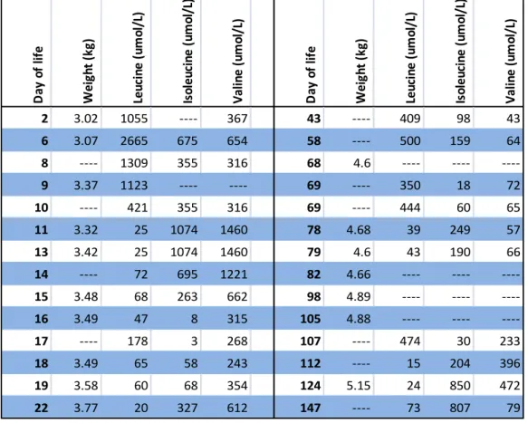
Table 1. Serum concentrations of BCAAs over time
At his first clinic visit he was found to have gained weight, prompting a change in the most current formula prescription from Florida to increase calories and protein, both intact and BCAA-free (Table 2). His formula included BCAA-free formula, regular infant formula, and supplemental isoleucine and valine. His analysis of amino acids from day 19 of life showed leucine to be slightly low (Table 1), but otherwise the patient's BCAAs were within normal reference range and he clinically appeared healthy. The patient's mother was told that the prescribed volume of metabolic formula should be consumed in its entirety each day and that if more or less volume was required for two days in a row, contact with the UNC metabolic team would be necessary for a dietary adjustment. Metabolic formula prescriptions involve weighing by gram all of the components of the formula and it is important, especially in an outpatient setting, to know whether or not a patient like this one is receiving more or less of the calories, protein, and BCAAs that his team believes him to be receiving. Before he left his first
D ay o f lif e W ei gh t (k g) Le ucin e (u m ol /L ) Is ol eu cin e (u m ol /L ) V al in e (u m ol /L ) D ay o f lif e W ei gh t (k g) Le ucin e (u m ol /L ) Is ol eu cin e (u m ol /L ) V al in e (u m ol /L )
2 3.02 1055 ---- 367 43 ---- 409 98 43
6 3.07 2665 675 654 58 ---- 500 159 64
8 ---- 1309 355 316 68 4.6 ---- ----
----9 3.37 1123 ---- ---- 69 ---- 350 18 72
10 ---- 421 355 316 69 ---- 444 60 65
11 3.32 25 1074 1460 78 4.68 39 249 57
13 3.42 25 1074 1460 79 4.6 43 190 66
14 ---- 72 695 1221 82 4.66 ---- ----
----15 3.48 68 263 662 98 4.89 ---- ----
----16 3.49 47 8 315 105 4.88 ---- ----
----17 ---- 178 3 268 107 ---- 474 30 233
18 3.49 65 58 243 112 ---- 15 204 396
19 3.58 60 68 354 124 5.15 24 850 472
clinic visit, blood was taken for an amino acid analysis and the patient's mother was instructed to take a blood spot each week and mail it to the clinic for analysis. His next clinic visit was scheduled for three weeks later.
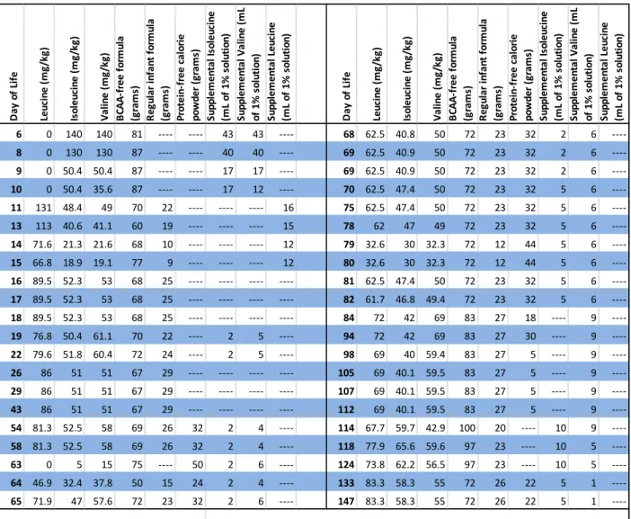
Table 2. Dietary Prescriptions with BCAAs Supplied as Compared with Recommendations
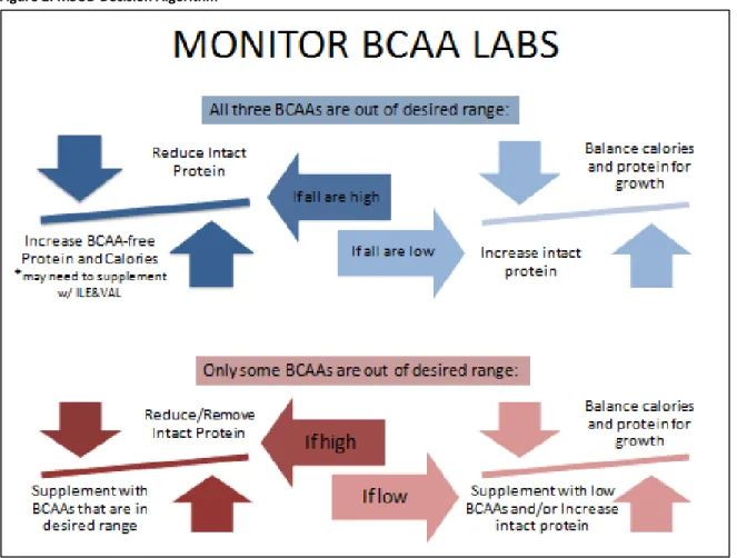
When BCAAs are outside normal range, metabolic dietitians must make decisions to increase or decrease intact protein (protein containing all amino acids) and other diet components to ensure growth and development but bring potentially toxic compounds under control (Figure 2). Ten days after his visit, this patient's amino acid analysis from clinic was available, showing leucine was low but
isoleucine and valine were elevated, as were several other amino acids. The patient's isoleucine and
D ay o f Li fe Le ucin e (m g/ kg ) Is ol eu cin e (m g/ kg ) V al in e (m g/ kg ) B CA A -f re e fo rm ul a (g ra m s) R eg ul ar in fa nt f or m ul a (g ra m s) Pr ot ei n-fr ee cal or ie po wd er ( gr am s) Su pp le m en ta l I so le ucin e (m L of 1 % s ol ut io n) Su pp le m en ta l V al in e (m L of 1 % s ol ut io n) Su pp le m en ta l L eu cin e (m L of 1 % s ol ut io n) D ay o f Li fe Le ucin e (m g/ kg ) Is ol eu cin e (m g/ kg ) V al in e (m g/ kg ) B CA A -f re e fo rm ul a (g ra m s) R eg ul ar in fa nt f or m ul a (g ra m s) Pr ot ei n-fr ee cal or ie po wd er ( gr am s) Su pp le m en ta l I so le ucin e (m L of 1 % s ol ut io n) Su pp le m en ta l V al in e (m L of 1 % s ol ut io n) Su pp le m en ta l L eu cin e (m L of 1 % s ol ut io n)
6 0 140 140 81 ---- ---- 43 43 ---- 68 62.5 40.8 50 72 23 32 2 6 ----8 0 130 130 87 ---- ---- 40 40 ---- 69 62.5 40.9 50 72 23 32 2 6 ----9 0 50.4 50.4 87 ---- ---- 17 17 ---- 69 62.5 40.9 50 72 23 32 2 6 ----10 0 50.4 35.6 87 ---- ---- 17 12 ---- 70 62.5 47.4 50 72 23 32 5 6 ----11 131 48.4 49 70 22 ---- ---- ---- 16 75 62.5 47.4 50 72 23 32 5 6 ----13 113 40.6 41.1 60 19 ---- ---- ---- 15 78 62 47 49 72 23 32 5 6 ----14 71.6 21.3 21.6 68 10 ---- ---- ---- 12 79 32.6 30 32.3 72 12 44 5 6 ----15 66.8 18.9 19.1 77 9 ---- ---- ---- 12 80 32.6 30 32.3 72 12 44 5 6 ----16 89.5 52.3 53 68 25 ---- ---- ---- ---- 81 62.5 47.4 50 72 23 32 5 6 ----17 89.5 52.3 53 68 25 ---- ---- ---- ---- 82 61.7 46.8 49.4 72 23 32 5 6 ----18 89.5 52.3 53 68 25 ---- ---- ---- ---- 84 72 42 69 83 27 18 ---- 9 ----19 76.8 50.4 61.1 70 22 ---- 2 5 ---- 94 72 42 69 83 27 30 ---- 9 ----22 79.6 51.8 60.4 72 24 ---- 2 5 ---- 98 69 40 59.4 83 27 5 ---- 9 ----26 86 51 51 67 29 ---- ---- ---- ---- 105 69 40.1 59.5 83 27 5 ---- 9 ----29 86 51 51 67 29 ---- ---- ---- ---- 107 69 40.1 59.5 83 27 5 ---- 9 ----43 86 51 51 67 29 ---- ---- ---- ---- 112 69 40.1 59.5 83 27 5 ---- 9 ----54 81.3 52.5 58 69 26 32 2 4 ---- 114 67.7 59.7 42.9 100 20 ---- 10 9 ----58 81.3 52.5 58 69 26 32 2 4 ---- 118 77.9 65.6 59.6 97 23 ---- 10 5
----63 0 5 15 75 ---- 50 2 6 ---- 124 73.8 62.2 56.5 97 23 ---- 10 5
----64 46.9 32.4 37.8 50 15 24 2 4 ---- 133 83.3 58.3 55 72 26 22 5 1
----65 71.9 47 57.6 72 23 32 2 6 ---- 147 83.3 58.3 55 72 26 22 5 1
valine supplementation were eliminated, but intact protein was increased in his diet to provide greater amounts of all three BCAAs.
Figure 2: MSUD Decision Algorithm
Blood spots sent by the patients' mother between clinic visits were never received, so the next time blood was taken for analysis of amino acids came eleven days after the adjustment to his diet at his clinic visit at 43 days of age. It was learned at this visit that he had been consuming 960 mL of formula, rather than the 720 mL he was prescribed, which would provide 165 kcal/kg if prepared as
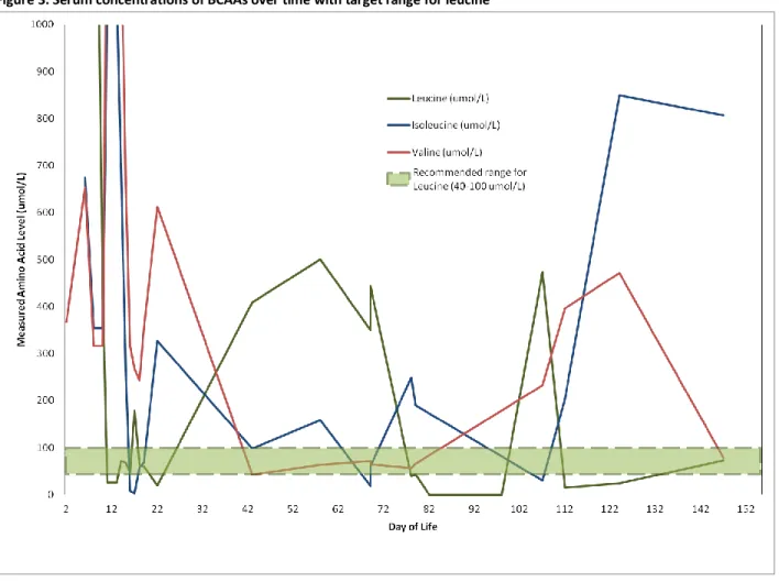
The next update on serum concentrations of amino acids came when the clinic amino acid analysis was returned at 54 days of age. His leucine was elevated and isoleucine slightly elevated, but valine was now low (Figure 3). Adjustments made by the metabolic dietitian involved reintroducing isoleucine and valine supplementation while decreasing intact protein (Table 2). Nine days later, results of a between-clinic blood spot amino acid analysis revealed that the patient's leucine had since gone higher as had isoleucine, and though valine had increased somewhat, it was still well outside a desirable range. As his leucine had remained high despite decreasing intact protein in his formula, a three-day correction regimen was attempted (Table 2). Over the three days, a removal and gradual reintroduction of leucine in the presence of adequate calories intended to reduce serum concentrations of leucine with protein turnover.
Figure 3. Serum concentrations of BCAAs over time with target range for leucine
On day 78 of life, the patient was once again seen in the UNC metabolic outpatient clinic as well as in general pediatric clinic, with whom his primary care had recently been established. His general pediatric team reported that he was acting normally, feeding appropriately, and afebrile but having frequent, watery diarrhea. The general pediatric team also noted the patient had an erythematous macular rash within his neck folds that appeared to be caused by yeast, a systolic murmur, and small fingernail scratches on his right cheek and nose. The patient's mother reported that the neck rash had previously resolved but had returned with recent illness and antibiotic use. Antifungal cream was again prescribed for his rash to be applied until two days after the rash cleared. His metabolic dietitian reported the patient was adequately receiving his metabolic formula, so blood for plasma amino analysis was drawn and he was scheduled to return three weeks later.
Unfortunately, just three days later he was again admitted to the hospital due to vomiting at the on-call metabolic physician's recommendation. He had been able to consume only half of his prescribed formula, was unable to tolerate liquids, had continuing diarrhea, and the rash in his diaper area was worsening. He was found to have multiple small sores on his face, right thumb, and left great toe. His mother reported that the diarrhea had not resolved since discharge from his last hospital stay. His stool was Clostridium difficile negative but his urine grew the same bacteria present on his last admission. Due to normocytic anemia, he was given a transfusion of red blood cells. His previously noted systolic murmur resolved post-transfusion. He was again provided intravenous glucose to avoid catabolism with his illness and after three days was discharged with a diagnosis of urinary tract infection and diarrhea possibly secondary to gastroenteritis due to antibiotic use. The rash was considered by his inpatient medical team to be consistent with fungal infection. He was again prescribed antifungal and barrier creams for the rashes.
was made: intact protein and BCAA-free protein were increased, non-protein calories decreased, valine supplementation increased, and isoleucine supplementation discontinued (Table 2). Ten days later, the patient's mother reported spitting up to the metabolic dietitian, hence the formula was concentrated with the addition of non-protein calories.
Figure 5. Peeling erythematous rash on patient's ear and neck
Seven days later he had not yet been seen by a dermatologist and the patient was admitted for his worsening rash (Figures 4,5). As the rash had the appearance of acrodermatitis enteropathica, serum concentrations of zinc were analyzed and the zinc content of his metabolic formula was
were also noted and suspected to be due to GI losses from chronic diarrhea. At discharge, the patient was prescribed a new dietary formula with increased isoleucine and valine supplementation, increased BCAA-free protein, and a reduced amount of a complete, but elemental, amino acid formula (Table 2). His dietary management continues to struggle to stabilize all three BCAAs in desirable range, but increased supplemental isoleucine is provided to ensure that he will not become deficient again.
Discussion
MSUD itself is rare, so infrequently encountered rashes similar to acrodermatitis enteropathica in this population warrant reporting. In 1993, Giacoia and Berry described the presentation of a severe rash in an infant that evolved over three weeks of dietary treatment consisting of protein only from BCAA-free formula11. Concurrent with the rash were loose stools and depressed hemoglobin and hematocrit, as were seen in this case study patient before his transfusion at day 81 of life. Zinc sulfate therapy failed to resolve Giacoia and Berry's patient's rash, but within seven days of isoleucine
supplementation the rash had healed. Koch et al. reported on recurrent eruptive dermatitis in two patients with MSUD who were fed only BCAA-free formulas after diagnosis until low amino acid
concentrations of leucine, isoleucine, or both were noted and supplementation initiated12. These rashes also healed with increased dietary BCAAs. De Raeve et al., Bosch et al, and Tabanlioglu et al. all
described patients of various metabolic disorders with acrodermatitis enteropathica-like rashes corresponding with low concentrations of BCAAs13,14,15. Tabalioglu et al. stated that the mechanism for the rash is as yet unknown but suggest it to be most likely related to a deficiency of one or more amino acids and proposed the name "acrodermatitis dysmetabolica" for the condition15.
that in an attempt to ensure that BCAAs are not in a toxic range, dietitians may cause a deficiency12. Getting treatment "just right" can be quite difficult. Though the exact mechanism of the development of "acrodermatitis dysmetabolica" rashes seen in patients of inherited metabolic disorders has not been fully elucidated, the correlation between manifestation of rash and deficiency of amino acids, especially isoleucine, is strong enough to necessitate special attention from metabolic dietitians.
Might the medical team have taken different steps that would have prevented this patient's experience? There are guidelines for medical nutrition therapy for MSUD set forth this year by GMDI and SERC to ensure that MSUD patients receive adequate but not excessive BCAAs for growth and development10. Discussion of a few of these guidelines compared with this patient's management are pertinent. GMDI/SERC recommendations are that infants have daily amino acid analysis until
concentrations stabilize, once to twice weekly until the infant is six months of age, and weekly from the age of six months to one year. The guidelines recommend providing 30-90 mg/kg of isoleucine for infants. The guidelines also suggest managing serum concentrations of BCAAs differently from following the reference ranges for individuals without the disorder. "Normal" serum concentrations of isoleucine should range between 40-90 umol/L while GMDI/SERC recommend concentrations of 200-400 umol/L for a patient of MSUD10.
The suspicion is that this patient's rash developed due to isoleucine deficiency. Discharge documentation from his last inpatient visit made note of the fact that supplemental isoleucine was discontinued four weeks prior when his plasma amino acid analysis indicated elevated isoleucine. At the time, he was receiving 50 mg of supplemental isoleucine as 5 mL of a 1% isoleucine solution added to his metabolic formula. However, the same dietary prescription that discontinued his supplemental
never without isoleucine from diet or supplement before the development of his rash, in contrast with some previously described patients11-14. Additionally, as compared to GMDI/SERC recommendations, this patient's dietary prescription for isoleucine intake was below recommendations for only a single day during his time in North Carolina, regardless of having to change his diet over a dozen times in less than four months (Table 2, Figure 5).
Figure 5. Serum Isoleucine versus Dietary Isoleucine over time
Analyses of this patient's amino acid concentrations reveal that all three of the BCAAs have fluctuated from below normal to well above normal (Figure 2). Before his admission for his
the medical team might have had the patient come in for a blood draw before so much time elapsed. Opportunities for amino acid analysis were missed on days 82 and 98 of life, when the patient was either in the hospital or in clinic. An additional complication with regular, serial amino acid analysis occurred when machine regularly used by the UNC metabolic team needed repair on two separate occasions during the time span covered by this case.
Obtaining the frequency of amino acid analysis recommended by GDMI/SERC is certainly possible in an inpatient scenario but becomes much more complicated when the patient is managed through an outpatient clinic. Following the protocol from the recommendations might have allowed an isoleucine deficiency to be avoided, but this patient did not have a primary care provider for an
extended period of time after moving to North Carolina and coming to clinic involved hours of travel. The metabolic team opted to try to avoid hardship for the family by having the patient's mother provide blood spots by mail for analysis, though when blood spots were not received it is unknown whether they were lost in the mail or never truly taken by the patient's mother.
This patient's serum concentrations of isoleucine were infrequently below the "normal" reference range but were below GMDI guidelines for 45% of the measurements (10 of 22 analyses). The metabolic dietitians have had a very difficult time attempting to stabilize amino acid concentrations within recommended ranges for this patient. MSUD presents a particularly great challenge because serum concentrations of three BCAAs must be balanced and the priority is often stabilization of leucine, since it and its ketoacid can lead to toxicity if elevated while elevations of isoleucine and valine are not known to be toxic4.
Though this patient was not monitored as regularly as recommended (sometimes for reasons outside the medical team's control) and serum concentrations of isoleucine were not able to be
was likely a strong contributor to development of isoleucine deficiency. Irritation of the gastrointestinal tract over time can result in a diminished ability to absorb nutrients16. Therefore, though his dietary prescriptions were thought to have supplied ample isoleucine, the amount absorbed was possibly less than adequate to avoid deficiency. It is also possible that the great fluctuations in his amino acid concentrations over time (Figure 3) were contributory to the development of rash.
The metabolic dietitians and his metabolic physicians considered a deficiency-induced rash to be a possibility for this patient well before his diagnosis. However, as he was assumed to be receiving adequate dietary isoleucine and his most recent labs at the time showed elevated isoleucine
concentrations, the team discounted the idea and believed his rash to have been induced by diarrhea and yeast infection, as had been diagnosed in the hospital. Their assumptions were faulty, however, partly because the information available was outdated or incomplete.
Perhaps the most important lesson that can be drawn from the experience with this patient is the reminder that the care a metabolic dietitian can provide is only as good as the information available to the dietitian. In an outpatient setting, considerations of a family's resources and social situation might lead a medical team to forgo an amino acid analysis or be understanding of a missed clinic appointment. Surekha Pendyal, MSc, MEd, RD, a member of his metabolic team, states, "The over restriction [of isoleucine] can occur intentionally or unintentionally due to various problems: difficult social situation, communication difficulty, discrepancy between what is recommended and how much the child has consumed, and inadequate monitoring" (personal communication November 2, 2013). Most of those concerns, however, can be ameliorated with frequent patient-dietitian communication. This patient's experience demonstrates that the need for regular follow-up is critical and social support should have been implemented.
have changed the outcome for this patient. In an outpatient setting, though, strict adherence may be improbable or even impossible. Regardless, the experience with this patient can teach that any time deviation from nutrition management guidelines is considered, the risk and benefit of different actions should be weighed against one another and the ultimate decision should be the one that will most likely result in getting medical nutrition therapy for the patient "just right."
References:
1. Acosta, P. Nutrition management of patients with inherited metabolic disorders. Sudbury, Mass.: Jones and Bartlett Publishers.2010.
2. Chuang DT, Shih VE (2000) Disorders of branched chain amino acid and keto acid metabolism. In: Scriver CR, Beaudet A, Sly WL, Valle D (eds) The metabolic and molecular bases of inherited disease, 8th ed. McGraw-Hill, New York, pp 1971–2006.
3. Korman SH, Cohen E, Preminger A. Pseudo-maple syrup urine disease due to maternal prenatal ingestion of fenugreek. J Paediatr Child Health. 2001 Aug;37(4):403-4.
4. Strauss KA, Puffenberger EG, Morton DH. Maple Syrup Urine Disease. 2006 Jan 30 [Updated 2013 May 9]. In: Pagon RA, Adam MP, Bird TD, et al., editors. GeneReviews™ [Internet]. Seattle (WA): University of Washington, Seattle; 1993-2013. Available from: http://www.ncbi.nlm.nih.gov/books/NBK1319/
5. Muelly ER, Moore GJ, Bunce SC, Mack J, Bigler DC, Morton DH, Strauss KA. Biochemical correlates of neuropsychiatric illness in maple syrup urine disease. J Clin Invest. 2013 Apr 1;123(4):1809-20.
6. Zinnanti WJ, Lazovic J. Interrupting the mechanisms of brain injury in a model of maple syrup urine disease encephalopathy. J Inherit Metab Dis. 2012 Jan;35(1):71-9.
7. Strauss KA, Morton DH. Branched-chain Ketoacyl Dehydrogenase Deficiency: Maple Syrup Disease. Curr Treat Options Neurol. 2003 Jul;5(4):329-341.
8. le Roux C, Murphy E, Hallam P, Lilburn M, Orlowska D, Lee P. Neuropsychometric outcome predictors for adults with maple syrup urine disease. J Inherit Metab Dis. 2006 Feb;29(1):201-2.
9. Morton DH, Strauss KA, Robinson DL, Puffenberger EG, Kelley RI. Diagnosis and treatment of maple syrup disease: a study of 36 patients. Pediatrics. 2002 Jun;109(6):999-1008.
10. GMDI/SERC. MSUD Nutrition Management Guidelines. Available from:
https://southeastgenetics.org/ngp/guidelines.php/45/MSUD%20Nutrition%20Guidelines/Version%201.37
11. Giacoia GP, Berry GT. Acrodermatitis enteropathica-like syndrome secondary to isoleucine deficiency during treatment of maple syrup urine disease. Am J Dis Child. 1993 Sep;147(9):954-6.
12. Koch SE, Packman S, Koch TK, Williams ML. Dermatitis in treated maple syrup urine disease. J Am Acad Dermatol. 1993 Feb;28(2 Pt 2):289-92.
14. Bosch AM, Sillevis Smitt JH, Van Gennip AH, Abeling NG, Schutgens RB, Bakker HD, Wijburg FA. Iatrogenic isolated isoleucine deficiency as the cause of an acrodermatitis enteropathica-like syndrome. Br J Dermatol. 1998 Sep;139(3):488-91.
15. Tabanlioğlu D, Ersoy-Evans S, Karaduman A. Acrodermatitis enteropathica-like eruption in metabolic disorders: acrodermatitis dysmetabolica is proposed as a better term. Pediatr Dermatol. 2009 Mar-Apr;26(2):150-4.