University of Warwick institutional repository: http://go.warwick.ac.uk/wrap
A Thesis Submitted for the Degree of PhD at the University of Warwick
http://go.warwick.ac.uk/wrap/38168
This thesis is made available online and is protected by original copyright. Please scroll down to view the document itself.
The Molecular Determinants and Consequences
of Recombination in the Evolution of Human
Enteroviruses
Kym Sheree Lowry
A thesis submitted for the degree of Doctor of Philosophy
University of Warwick
School of Life Sciences
Table of Contents
List of Tables... 3
List of Figures... 4
Acknowledgements... 6
Declaration... 8
Summary... 9
Abbreviations... 10
CHAPTER ONE: Introduction... 14
1.1 Classification... 14
1.2 Virus Structure and Life Cycle ... 21
1.3 Enterovirus Evolution ... 35
1.4 Aims ... 47
CHAPTER TWO: Materials and Methods... 48
2.1 Cell Culture and Virological Methods ... 48
2.2 Molecular Genetic Techniques ... 52
2.3 Stock Solutions and Buffers... 58
2.4 List of DNA plasmids ... 60
2.5 Restriction Enzymes to Linearise Plasmids ... 61
2.6 List of Oligonucleotides... 62
CHAPTER THREE: RNA-mediated Interference to Enrich Recombinants.... 63
3.1 Introduction... 63
3.2 Construction of anti-PV miRNA Expressing Plasmid ... 70
3.3 Transient Expression of miRNA to Reduce Virus Replication ... 76
3.4 Transient Expression of siRNA to Reduce Virus Replication... 80
3.5 Discussion ... 93
CHAPTER FOUR: In vitro Recombination... 99
4.1 Introduction... 99
4.2 Proposed Methodology ... 102
4.3 Recovery of Intratypic Recombinants... 105
4.4 Recovery of Intertypic Recombinants... 108
4.5 Recovery of Intraspecies Recombinants ... 109
4.6 Co-transfections with RNA Partners Belonging to Species B... 115
4.7 Discussion ... 121
CHAPTER FIVE: Characterisation of Recombinants... 124
5.1 Introduction... 124
5.2 PV3/PV1 Recombinants ... 125
5.3 Intraspecies Recombinants... 155
5.4 Comparative Bioinformatic Analysis of RNA Partner Sequences ... 164
5.5 Discussion ... 185
CHAPTER SIX: Construction of RNA Partners for Co-transfections... 190
6.1 Introduction... 190
6.2 Generation of a Full-length Infectious E30 cDNA ... 193
6.3 Generation of a Full-length EV7 CRE Mutant cDNA ... 206
6.4 Discussion ... 212
CHAPTER SEVEN: General Discussion... 215
List of Tables
Table 1.1 Classification of Picornavirus family ... 16
Table 1.2 Clinical symptoms associated with certain enteroviruses ... 19
Table 1.3 Some enterovirus serotypes and their receptors... 23
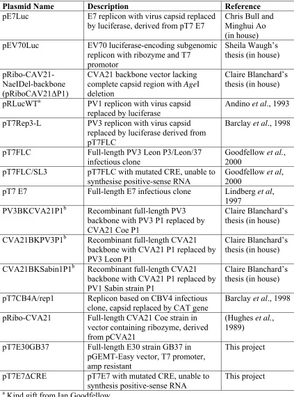
Table 2.1 List of subgenomic replicon and full-length clones used in this study ... 60
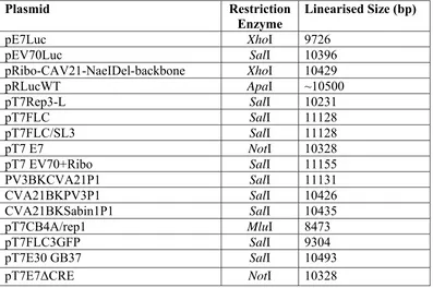
Table 2.2 Restriction sites used to linearise plasmids for RNA transcriptions ... 61
Table 2.3 Oligonucleotides used throughout this study ... 62
Table 3.1 Summary of RNAi and delivery methods ... 66
Table 3.2 Synthesised miRNA oligonucleotides (DNA) and corresponding nucleotide positions in PV1 and PV3 genome ... 71
Table 3.3 Synthesised siRNA oligonucleotides (RNA) and corresponding nucleotide positions in PV1 and PV3 genome... 82
Table 4.1 Combinations of interspecies co-transfections performed ... 118
Table 5.1 Characterisation of PV3/PV1 recombinants by crossover region from single co-transfection... 130
Table 5.2 Characterisation of PV3/PV1 recombinants by crossover region from an independent co-transfection in L929 cells... 138
Table 5.3 Characterisation of PV3/PV1 recombinants by crossover region after continuous passage through HeLa cells ... 150
List of Figures
Figure 1.1 Genome structure of enteroviruses. ... 25
Figure 1.2 Summary of the PV life cycle... 29
Figure 1.3 Poliovirus negative-strand synthesis... 31
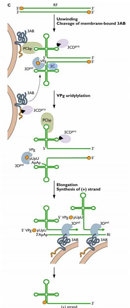
Figure 1.4 Poliovirus positive-strand RNA synthesis. ... 33
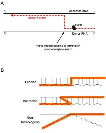
Figure 1.5 Current replication-dependent template switching models for RNA recombination. ... 43
Figure 3.1 RNAi silencing of mRNA in cells. ... 68
Figure 3.2 RNAi regimes to enrich recombinants in a virus population... 69
Figure 3.3 The expression vector kit used to deliver virus specific miRNA to cells.75 Figure 3.4 Immunofluorescence of GFP expressing plasmids in cells. ... 77
Figure 3.5 Effects of miRNA on PV replication... 79
Figure 3.6 Intra-assay variation using miRNA. ... 81
Figure 3.7 Effects of siRNA on PV replication. ... 84
Figure 3.8 Effects of MET-2C siRNA dose-response on PV replication. ... 87
Figure 3.9 Effects of si-6243PV3 siRNA dose-response on PV3 replication... 88
Figure 3.10 Effects of combinations of siRNA on PV replication... 90
Figure 3.11 Optimisation of siRNA transfection and infection time points. ... 92
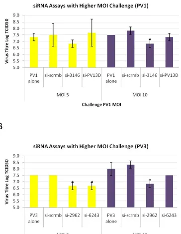
Figure 3.12 Challenging siRNA transfected HeLa cells at higher MOIs of PV. ... 94
Figure 4.1 Generating recombinant virus in the absence of infectious parental virus. ... 103
Figure 4.2 Method used to generate recombinant viruses by co-transfection... 104
Figure 4.3 Virus recovered from L929 cells co-transfected with PV3 RNA molecules. ... 107
Figure 4.4 Virus recovered from BsrT7 cells co-transfected with PV1 and PV3 RNA partners. ... 110
Figure 4.5 Intraspecies C recombinants constructed by Dr. Claire Blanchard. ... 112
Figure 4.6 Virus recovered from intraspecies RNA co-transfections. ... 114
Figure 4.7 Virus recovered from pT7EV7luc + pT7EV7ΔCRE co-transfections. . 116
Figure 4.8 Plaque assays of interspecies co-transfection supernatant... 120
Figure 5.1 Identification and biological cloning of 100 PV3/PV1 recombinant viruses. ... 126
Figure 5.2 An example of a non-clonal recombinant virus mixture. ... 129
Figure 5.3 Chromatogram of sequenced recombinant virus 105B... 131
Figure 5.4 Sixteen distinct crossover junctions identified from 100 cloned PV3/PV1 recombinants... 133
Figure 5.5 Sites of crossover junctions in recovered PV3/PV1 recombinants. ... 135
Figure 5.6 Origin of the inserted 15 nt sequence in recombinant 25A at the crossover site... 137
Figure 5.7 Four distinct crossovers identified from 31 cloned PV3/PV1 recombinants in a repeated independent experiment... 139
Figure 5.8 Single-step growth curves... 141
Figure 5.9 Plaque phenotypes of parent and two selected recombinant viruses. .... 143
Figure 5.11 Changes in crossover regions from passaged PV3/PV1 recombinants.
... 147
Figure 5.12 Crossover regions of PV3/PV1 recombinants after serial passage through HeLa cells... 149
Figure 5.13 Sites of the recombination junctions in the passaged recombinants 51G and 36B... 152
Figure 5.14 New crossover sites for serially passaged recombinants... 153
Figure 5.15 RT-PCR product of biologically cloned intraspecies C recombinants.156 Figure 5.16 Single intraspecies recombinant formed from RNA partners... 159
Figure 5.17 Crossover junctions of intraspecies C recombinants formed from replicons and Dr. Blanchard’s constructed recombinants. ... 160
Figure 5.18 Changes in crossover region of serially passaged intraspecies recombinants... 163
Figure 5.19 Imprecise and precise recombination junctions and correlation to sequence identity between RNA partners... 166
Figure 5.20 Mononucleotide composition of RNA partners... 168
Figure 5.21 Comparison of junction sites and homopolymers located in recombination regions of PV1 and PV3. ... 169
Figure 5.22 Palindromic sequences identified within targeted recombination region of PV1 and PV3. ... 171
Figure 5.23 Local secondary structures using MFED analysis for PV1 and PV3 regions of recombination. ... 173
Figure 5.24 Amino acid similarity scan between PV1 and PV3... 175
Figure 5.25 Comparing 2Apro domain structures to precise junction sites... 178
Figure 5.26 Amino acid similarity scans comparing PV1, PV3, and CVA21 protein sequences across the recombination region. ... 180
Figure 5.27 Crossover sites in PV3 and PV1 recombinants from this study and from the literature. ... 181
Figure 5.28 Correlation coefficients for locations of junction sites in recombinants recovered... 184
Figure 5.29 Distance of template switching from original displacement from donor template... 187
Figure 6.1 Overview of proposed E30 full-length infectious clone construction. .. 192
Figure 6.2 Amplification and restriction digests of E30 cDNA... 195
Figure 6.3 Construction of pT7E30 full-length infection clone for E30... 197
Figure 6.4 Phylogenetic analysis of cloned E30 strain GB37... 200
Figure 6.5 Recovery of infectious E30 from RNA transfections... 201
Figure 6.6 Growth characteristics of infectious full-length pT7E30 GB37 clone. . 203
Figure 6.7 Overview of construction of E30 subgenomic replicon encoding luciferase... 205
Figure 6.8 Predicted secondary structure of wild-type and mutagenised EV7 CRE stem loop... 207
Figure 6.9 Strategy for mutagenising CRE in full-length infectious clone pT7EV7. ... 209
Figure 6.10 Overlapping PCR of EV7 CRE fragments. ... 210
List of Appendices
Acknowledgements
This work was funded by a University of Warwick Studentship, to which I am extremely grateful for making this research project possible. I have appreciated the opportunity to gain valuable experience in such a rich research environment and have enjoyed all that Warwick University (including an outstanding virology section) has to offer.
I would like to thank my supervisor Professor David J. Evans for awarding me a scholarship – thank you for taking that leap of faith having never met the potential student on the other side of the world. I appreciate the endless time, help, and support throughout my PhD and realise how lucky I was to have such an approachable
supervisor.
I need to thank the members of the Evan’s lab for making it such an enjoyable work environment. I’ll miss the cheeky sports banter, the constant laughs, and the wealth of knowledge and experience that made it so enriching.
Thanks must go to my family and friends who have offered constant support
throughout and always provided help or a sympathetic ear. I can’t wait to spend more time with you all.
I would like to dedicate this thesis to my mother, Judy, who overcame serious illness during the course of my study and taught me what it was to ‘knuckle down’ and keep going when the times were tough. You are brave and always look to the future.
Declaration
This work was completed at the University of Warwick between July 2007 and November 2010 and has not been submitted for another degree. The work is original and unless otherwise stated in the text, has been completed by the author.
Signed
Summary
Recombination is an important biological process in a diverse range of viruses, particularly those with single-stranded ribonucleic acid (RNA) genomes including the enteroviruses. Mutations caused by the error-prone RNA-dependent RNA polymerase (RdRp) and the vast population size of these virus populations are evolutionary mechanisms that generate genetic diversity – this allows viruses to survive under changing environmental pressures (e.g. adaptive host immunity). Ribonucleic acid recombination has been identified as another contributing
mechanism involved in diversification, by removing interfering or lethal mutations from a virus genome, and by establishing new viruses.
Virus RNA recombination is well documented and is identified in several virus families including picornaviruses. However, recombination is a rare event and the study of the molecular mechanisms behind virus recombination is complicated by our ability to isolate and analyse recombinants from a mixed virus population including the parental viruses.
The objectives of this study were to firstly devise a method for generating populations of natural recombinant viruses, and secondly, to study the molecular processes that determine where and when recombination occurred in the enteroviral genome.
During this project, an in vitro system was developed to allow the recovery of recombinant enteroviruses in the absence of their parental viruses. Two virus RNA molecules containing “lesions” rendering them unable to generate viable virus on their own were co-transfected into mouse L929 cells. The method required two parental virus RNA molecules to be present in a single cell to produce a viable recombinant virus. Reverse Transcription-Polymerase Chain Reaction (RT-PCR) and sequencing analysis confirmed the recombinant nature of progeny virus genomes. Experimental data confirmed the effectiveness of the method and provided evidence that recombination occurs in at least two phases. Initial template switching, referring to the transfer of RdRp from one RNA template to another mid-replication, occurred apparently indiscriminately and with the addition of extra virus and non-virus
Abbreviations
ANOVA Analysis of variance
aVDPV Ambiguous vaccine-derived poliovirus BLAST basic local alignment search tool
bp(s) base pair(s)
CD cluster of differentiation
cDNA complementary DNA
CI confidence interval
CMV cytomegalovirus
CO2 carbon dioxide
CPE cytopathic effect
CRE cis-acting replication element
cVDPV circulating vaccine-derived poliovirus
D redundant nucleotide for adenine, guanine, or thymine DAPI 4’,6-diamidino-2-phenylindole
DEPC diethylpyrocarbonate
dH2O distilled water
DI defective interfering
DMEM Dulbecco’s modified Eagle’s medium
DNA deoxyribonucleic acid
DNase deoxyribonuclease
ds double stranded
DTT dithiothreitol
dNTP deoxyribonucleotide triphosphate
E. coli Escherichia coli
EDTA ethylenediaminetetraacetic acid
EMEM minimum essential medium with Earle’s salts
FBS foetal bovine serum
FITC fluoroscein isothiocyanate
FLC full-length clone
g gram GFP green fluorescent protein GI gastrointestinal
GMEM Glasgow minimum essential medium GuHCl guanidine hydrochloride
HeLa human cervical cancer cell line
HI heat inactivated
hr hour
ICAM-1 intracellular adhesion molecule type - 1 IF immunofluorescence
IPV inactivated poliovirus vaccine
IRES internal ribosome entry site
iVDPV immunodeficiency-associated vaccine-derived poliovirus K redundant nucleotide for guanine or thymine
kbp kilobase pair
LB Luria Bertani
MCS multiple cloning site MFED minimal free energy differences mg milligram
min minute miRNA microRNA ml millilitre mM millimolar
MOI multiplicity of infection
mRNA messenger RNA
NCBI National Center for Biotechnology Information
NCR non-coding region
ng nanogram nm nanometre nt nucleotide OPV oral poliovirus vaccine
ORF open reading frame
PABP poly(A) binding protein PBS phosphate buffered saline PCBP2 poly(rC) binding protein 2 PCR polymerase chain reaction
PFU plaque forming unit
pH power of hydrogen
pmol picomole
PS Penicillin and streptomycin
PTB polypyrimidine tract binding protein PTGS post-transcriptional gene silencing PVR poliovirus type 3 receptor (CD155)
R redundant nucleotide for adenine or guanine
RD rhabdomyosarcoma cells
RD-ICAM rhabdomyosarcoma cells expressing ICAM-1
RdRp RNA-dependent RNA polymerase
RF replicative form
RI replication intermediate
RISC RNA-induced silencing complex
RNA ribonucleic acid
RNAi RNA-mediated interference RNase ribonuclease
rpm revolutions per minute
RT-PCR reverse transcription polymerase chain reaction
SDS sodium dodecyl sulphate
s second
SG stress granule
shRNA short hairpin RNA
siRNA short interfering RNA
TCID50 50% tissue culture infective dose
U unit
μg microgram
μM micromolar
UV ultraviolet
VAPP vaccine-associated paralytic poliovirus
VDPV vaccine-derived poliovirus
VPg virus protein genome linked
v/v volume per volume total
W redundant nucleotide for adenine or thymine
w/v weight per volume total
WHO World Health Organisation
Y redundant nucleotide for cytosine or thymine
°C degrees Celsius
Virus abbreviations (enterovirus species where appropriate)
BVDV bovine viral diarrhoea virus CVA coxsackievirus A serotypes CVA21 coxsackievirus A21 (species C) CVA16 coxsackievirus A16 (species A) CVB coxsackievirus B serotypes CVB3 coxsackievirus B3 (species B) CVB4 coxsackievirus B4 (species B) E30 echovirus 30 (species B)
E7 echovirus 7 (species B)
EV70 enterovirus 70 (species D) EV71 enterovirus 71 (species A) FMDV foot-and-mouth disease virus
HAV hepatitis A virus
HCV hepatitis C virus
HEV human enterovirus
HEVC human enterovirus C serotypes
HRV human rhinovirus
Protein abbreviations
VP0 precursor protein before cleavage to make VP2 and VP4
VP1 capsid protein
VP2 capsid protein
VP3 capsid protein
VP4 capsid protein
2Apro virus protease
2BC precursor protein, interacts with cellular membranes 2B interacts with cellular proteins, induces vesicles in cells 2C interacts with cellular proteins, induces vesicles in cells 3AB precursor protein, binds to one or more CREs
3A component of virus replication complex
3B protein VPg
3CDpro precursor protein with protease activity, binds to one or more CREs 3Cpro virus protease
3Dpol RNA-dependent RNA polymerase (RdRp)
VPg virion protein genome linked covalently to 5’ NCR of virus genome CRE cis-acting replication element in 2C encoding region
p200 large subunit of eIF-4 degraded by 2Apro
G3BP regulated effecter of stress granule assembly p53 tumour suppressor protein that regulates cell cycle
CHAPTER ONE: Introduction
Evolution requires genetic variation. Positive-sense single stranded RNA viruses including important human pathogens like poliovirus (PV) and rhinovirus have error prone polymerases, short replication cycles, and high yields that together contribute to genetic diversity. Far greater genetic variation is achieved by chance
recombination events than is possible in a single round of genome replication, and this is an important biological process that accelerates virus evolution further.
Although the result of many separate recombination events have been observed and characterised in nature, other than an understanding of the underlying principle, little is known about the mechanisms (viral and host) that establish the location of
recombination breakpoints across the virus genome. Recombination is a rare event (possibly only one per million progeny), so this complicates isolation and analysis of recombinants from a mixed population. To this end, a method has been developed to recover recombinant viruses in vitro in the absence of infectious parental viruses.
This review will briefly consider the mechanisms of enteroviral evolution, methods of RNA recombination and evidence of recombination in enteroviruses both
naturally and in the laboratory. Poliovirus, being the prototype strain of
picornaviruses, is one of the best studied viruses and is used extensively during this project. As such, the majority of this literature review will focus on this virus.
1.1 Classification
Virus classification and the species concept
Classifying viruses into a taxonomic system is primarily performed by the
species. A group of species, in turn, comprise a genus. The term virus species implies a single genotype whereas RNA viruses, due to their large population sizes and high mutation rates, are in fact a diverse population of genotype variants or quasispecies (Lauring & Andino, 2010). Virus species are further subdivided into serotypes, which are classically defined as antigenically distinct viruses that are incapable of being neutralised to a significant extent by other serotype antibodies from the same species. Genetic variation within serotypes leads to the identification of virus strains, which are the limit to virus classification.
The picornavirus family
The picornaviruses are small (pico is a very small unit of measure), single stranded positive-sense RNA viruses belonging to the family Picornaviridae. Poliovirus is the type species (Flint et al., 2004) and the family includes 12 genera that are responsible for morbidity in both humans and animals (summarised in table 1.1). After
poliomyelitis, probably the most notable disease-causing picornavirus is foot-and-mouth disease virus (FMDV) (genus aphthovirus), which infects cloven-hoofed animals such as cattle and swine (Bachrach, 1968). This highly infectious virus has devastating financial effects on the agricultural industry through the necessary elimination of millions of animals and containment/sanitation measures. Other picornaviruses that greatly impact public health comprise enteroviruses (including PV and echoviruses); rhinoviruses (causing the majority of cases of the common cold); and hepatoviruses (hepatitis A virus [HAV]).
The enterovirus genus
Human enteroviruses are currently subgrouped into seven species, Human
Enterovirus (HEV) A-D and Human Rhinovirus (HRV) A-C (Arden & Mackay,
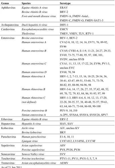
Table 1.1 Classification of Picornavirus family
Genus Species Serotype
Aphthovirus Equine rhinitis A virus ERAV Bovine rhinitis B virus BRV-2
Foot-and-mouth disease virus FMDV-A, FMDV-Asia1,
FMDV-C, FMDV-O, FMDV-SAT1-3
Avihepatovirus Duck hepatitis A virus DHV-1
Cardiovirus Encephalomyocarditis virus EMCV
Theilovirus TMEV, VHEV, TLV, RTV-1
Enterovirus Bovine enterovirus BEV-1, BEV-2
Human enterovirus A CVA2-8, 10, 12, 14, 16, EV71, 76, 89-92,
SV46
Human enterovirus B CVA9, CVB1-6, E-1-9, 11-21, 24-27, 29-33,
EV69, 73-75, 77-88, 95, 97, 100, 101, SVDV, unclass HVB
Human enterovirus C CVA1, 11, 13, 15, 17-22, 24, EV96, PV1-3,
unclass EVC
Human enterovirus D EV68, 70, 94
Human rhinovirus A HRV-1, 2, 7-13, 15, 16, 18-25, 28-34, 36,
38-41, 43-47, 49-51, 53-68, 71, 73-78, 80-82, 85, 88-90, 94-96, 98
Human rhinovirus B HRV-3-6, 14, 17, 26, 27, 35, 37,42, 48, 52,
69, 70, 72, 79, 83, 84, 86, 91-93, 97, 99
Human rhinovirus C HRV-1-3, HRV-4-6, 8, 10, 12, 13, 17-20, (not defined) 22-28, 30-35, 37, 38, 40-48, 51-57, 59-61,
63, 64, 66-71, 73-84, 86-88, 90-100
Porcine enterovirus B PEV-9, 10, J10
Simian enterovirus A A-2PV, SVSA4, SVSV4, SVSV28, SPV7
Erbovirus Equine rhinitis B virus ERV-2
Hepatovirus Hepatitis A virus HAV, SAV
Kobuvirus Aichi virus AiV, unclass KV
Bovine kobuvirus BKV
Parechovirus Human parechovirus E1-8, 10, 11
Ljungan virus LV87-012, LV145SL, LV174F
Sapelovirus Avian sapelovirus ASV
Porcine sapelovirus PV8, PVJ4, PVJ6
Senecavirus Seneca Valley virus SVV
Teschovirus Porcine teschovirus PTV1-11, PV11, PVJ1-3, 5, 7, 9
Tremovirus Avian encephalomyelitis virus AEMV
As of 21 Jan 2011
enterovirus species through partial or complete genome sequencing and phylogenetic analysis (Lukashev, 2005; Oberste et al., 2004a; Oberste et al., 2004b). Molecular typing has been adopted for the identification of enterovirus isolates, and this is based on comparisons of a portion of the VP1 capsid gene (Oberste et al., 1999). Enterovirus serotypes within a species typically share > 75% nucleotide sequence identity (> 85% amino acid identity) (Oberste et al., 2007).
Poliovirus was recently reclassified as a member of HEV-C as a result of phylogenetic analysis which showed that members of this species were closely related in the non-capsid coding region (Benschop et al., 2008; Brown et al., 2003). Previous separation of PVs from HEV-C was based on disease manifestations, specifically the ability of PV to cause poliomyelitis, and to bind to the specific poliovirus receptor (PVR; CD[cluster differentiation]155) (Lukashev, 2005; Minor, 2004). There is generally little correlation between the virus serotype and the resulting disease in the host, as in virus serotypes belonging to different enterovirus species can cause similar symptoms.
Transmission
Enteroviruses infect the host by two main routes namely; (1) faecal-oral transmission (such as PV) and (2) respiratory routes (most predominantly with rhinoviruses). Faecal-orally transmitted viruses such as PV are acid-stable (pH 3.0 to 5.0) for 1 to 3 hours (hrs), and replicate at 37°C which makes them suited to enteric infection. These faecal-orally transmitted enteroviruses are also resistant to inactivation by many common disinfectants and soaps (Sutter et al., 2008). In contrast, rhinoviruses are acid-labile and replicate more efficiently at 33°C (hence their preference for the upper respiratory tract) (Kistler et al., 2007; Pallansch & Roos, 2001).
Diseases associated with enterovirus infection
Table 1.2 outlines a number of enteroviruses and their clinical symptoms (adapted from Flint et al., 2004). The patient age also influences the severity of disease experienced after infection by enterovirus serotypes. For example, children tend to have mild or asymptomatic symptoms following PV infection while older children and adults experience more severe symptoms. Neonates are at higher risk of disease caused by non-poliovirus enteroviruses (44% of total infections by age group) (Khetsuriani et al., 2006) presumably due to underdeveloped immunity, while all ages are at risk of rhinovirus infection (causing predominantly upper and lower respiratory illness).
Overview of PV pathogenesis and eradication strategy
Poliovirus was discovered by Landsteiner and Popper in 1909 and was the cause of extensive poliomyelitis disease outbreaks up to the 1950s (Wimmer et al., 1993). Infection with PV can lead to one of the following outcomes: (1) asymptomatic infection which is the most common outcome (72%); (2) minor illness such as fever, vomiting, constipation, or sore throat; (3) non-paralytic poliomyelitis (aseptic
meningitis); or (4) paralytic poliomyelitis which is a rare outcome (usually <1% of cases) (reviewed in Sutter et al., 2008). Poliovirus progressively penetrates other tissues after escaping the tonsils and gut following initial infection and can cause symptoms elsewhere, e.g. myalgia, vomiting, and pain in the limbs. Studies in the 1940s and 1950s determined that this form of disease lasted from 5 to 25 days, and resulted in complete recovery (reviewed in Nathanson & Kew, 2010). Progression to severe symptoms occurs if the virus penetrates the blood brain barrier of the central nervous system (CNS) by infecting motor neurons (Minor, 2004), as the name suggests – (polio = grey, myelitis/myelo = marrow). Virus replicates and destroys motor neurons in the grey matter of the spinal cord, brain stem, or motor cortex, and can ultimately lead to paralysis or even death (reviewed in Sutter et al., 2008).
Table 1.2 Clinical symptoms associated with certain enteroviruses
Virus Paralytic Encephalitis, Carditis Neonatal Hand-foot- Rash Acute Respiratory Fever Diarrhea, Diabetes, Orchitis Congenital Disease Meningitis disease and-mouth hemorrhagic Tract GI disease pancreatitis anomalies disease conjunctivitis infections
Poliovirus 1-3 + + + +
Coxsackieviruses A1-24 + + + + + + + + + Coxsackieviruses B1-6 + + + + + + + + +
Echovirus 1-33 + + + + + + + +
Enterovirus 70 + +
Enterovirus 71 + + +
Parechoviruses 1-3 + + + + +
(Bodian, 1951; Toyoda et al., 1984). The introduction of trivalent, inactivated polio vaccine (IPV) (Salk) and trivalent, live attenuated (Sabin) oral poliovirus vaccine (OPV) (Wimmer et al., 1993) reduced the incidence of poliomyelitis between 1950 and 1970. The former was a formalin trivalent IPV developed by Dr. Jonas Salk and licensed in 1955 (Salk, 1953). In 1963, an attenuated oral vaccine containing all three serotypes was developed and licensed by Dr. Albert Sabin (Sabin & Boulger, 1973). Oral polio vaccine contained attenuated strains of PV and was developed by rapidly passaging neurovirulent strains at suboptimal temperatures in simian cells and tissues. Numerous studies have since identified the multiple genetic determinants of Sabin vaccine strain attenuation, and Kew et al have summarised the critical
mutations responsible in all three strains (Kew et al., 2005). Briefly, Sabin-1 vaccine strain was distinguishable from its neurovirulent progenitor by 55 nucleotide (nt) substitutions which were located throughout the genome (McGoldrick et al., 1995). Through reverse genetics, it was determined that the most important attenuating substitution occurred in the internal ribosome entry site (IRES) at position 480 (Kawamura et al., 1989) while four other substitutions in the capsid region
contributed to the attenuating phenotype. Sabin-2 contained only two substitutions (in the IRES and VP1 coding region) from the original P712 strain that contributed to the attenuated phenotype (Macadam et al., 1993). Lastly, the attenuated Sabin-3 strain differed from the neurovirulent Leon strain by 10 nt substitutions, with the principal attenuating nts located in the IRES, VP3, and VP1 coding regions (Stanway
et al., 1984). The attenuation of the neurovirulent phenotype in PV vaccine strains
provides one of the best model for rapid virus evolution and how we exploited it for the generation of highly effective vaccines.
Poliovirus vaccines are included in the national paediatric vaccine schedules of most developed countries. In 1988, the World Health Organisation (WHO) implemented the global eradication target to improve vaccination efforts in developing countries where PV infections resulted in annual cases of poliomyelitis in sub-optimally vaccinated or other vulnerable populations (WHO, 1988). To date, only four
vaccine strains are inherently unstable and occasionally mutate to cause PV infections in vaccinated individuals or close contacts.
Cases of vaccine-associated paralytic poliomyelitis (VAPP) are rare independent events that occur following administration of OPV (Kew et al., 2005). The risk is low - approximately one case per 2.4 million doses delivered - and is a direct consequence of genetic mutation of OPV strains (CDC, 1997; Kew et al., 2005). During virus replication in a vaccinee, Sabin vaccine strains can spontaneously revert to enhanced virulence by key nt substitutions that conferred attenuation in the first place (outlined above). Sabin-3 contributes to the highest rates of VAPP post-vaccination, with low genetic stability of critical substitutions the main cause
(Chumakov et al., 1992; Kew et al., 2005). Such vaccine-derived outbreaks of VAPP arise as a direct result of OPV replication in vaccinees, and highlights the role of rapid genetic evolution within these virus populations.
Several outbreaks caused by vaccine-derived polioviruses (VDPVs) have been identified in recent years due to more modern sequencing methods. Vaccine-derived PVs differ from 1% to 15% of VP1 sequence from the original Sabin vaccine strains indicating longer circulation post-vaccination (Kew et al., 2005). These VDPVs may circulate within a population, especially in areas where wild PV no longer occurs and where there is low vaccine coverage (CDC, 2006; Rousset et al., 2003). There are several categories of VDPVs including circulating-VDPVs (cVDPVs) that arise from recombination events between VDPVs and species C enteroviruses (Kew et al., 2002; Rousset et al., 2003; Shimizu et al., 2004; Yang et al., 2003),
immunodeficiency-associated VDPVs (iVDPVs) which are excreted from immunodeficient recipients usually over the long term, and ambiguous-VDPVs (aVDPVs) which do not fall into either of the previous categories and have no defined origin (CDC, 2006).
1.2 Virus Structure and Life Cycle
Virus morphology
viral capsid proteins (VP4, VP2, VP3, and VP1) and forms a virion with a single virus protein genome (VPg)-linked positive-sense RNA (Hogle et al., 1985; Lee et
al., 1977; Rossmann et al., 1985). Each heteromeric structural unit comprises one
copy of VP1, VP2, and VP3, while one copy of VP4 lies on the inner surface of the viral capsid, purportedly to support the capsid interior and interact with the RNA genome (Guttman & Baltimore, 1977; Lee et al., 1993). Each of the 12 five-fold axes of the capsid (with prominent peaks) is surrounded by a surface depression, or
“canyon”, where the host receptor CD155 docks (Bernhardt et al., 1994; Colston & Racaniello, 1994).
Tissue tropism
Pathogenesis of enteroviruses is influenced by extracellular factors including the presence of cellular receptors required for virus attachment and cell infection, and this largely determines tissue tropism. As well as requirements for cellular entry, viruses must also be able to replicate within the cell by utilising the cell’s own machinary.
Experimental studies suggest that many enteroviruses exploit more than one cell surface protein in order to infect host cells (Evans & Almond, 1998). However, PV and the major group of HRV are exceptions in that they only require one receptor, CD155 and the intracellular adhesion molecule type-1 (ICAM-1) respectively (Greve
et al., 1989; Mendelsohn et al., 1989). Other enteroviruses use different
immunoglobulin-like molecules and adhesion proteins for attachment, and a current list of identified receptors used by a selection of enteroviruses is outlined in table 1.3.
Table 1.3 Some enterovirus serotypes and their receptors
Serotype Receptor Reference
Poliovirus 1-3 CD155 (PVR) (Mendelsohn et al., 1989)
Coxsackievirus A21 ICAM-1, DAF (Shafren et al., 1997)
Rhinovirus (major group) ICAM-1 (Greve et al., 1989)
Coxsackievirus B1, B3, B5 DAF (Shafren et al., 1995)
(Bergelson et al., 1994)
(Powell et al., 1998)
Echovirus 3, 6, 7, 11-13, 20, 21, 24, 29, 33 DAF
(Ward et al., 1994)
Echovirus 22 αvβ3 vitronectin (Roivainen et al., 1994)
(Nishimura et al., 2009)
Enterovirus 71
SCARB2, PSGL-1 (Yamayoshi et al., 2009)
(L cells) which ordinarily do not support PV infection (Mendelsohn et al., 1989). As a result, L cells also became susceptible to PV infection, and not just permissive to PV replication. In further experiments, transgenic mice containing the human PVR gene in the germ line were established for PV studies (Koike et al., 1991; Ren et al., 1990). Both groups observed that transgenic mice were susceptible to either infection by PV1 (Mahoney) or all PV serotypes, and presented with the same clinical
symptoms identified in humans and monkeys. PVR transcripts were shown to be expressed in a wide range of transgenic mouse tissues, and this was also observed in humans (e.g. CNS) (Koike et al., 1991; Mendelsohn et al., 1989).
Genome organisation
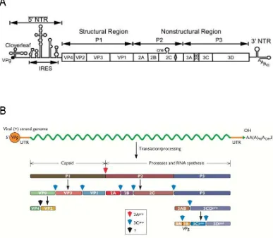
The enterovirus genome consists of a single stranded, non-segmented positive-sense RNA molecule approximately 7500 nts long. The genome encodes a long single open reading frame preceded by a highly structured 5’ non-coding region (NCR) and followed by a short 3’ NCR with a poly(A)-tail. The polyprotein is cleaved co- and post-translationally by the virus’s own encoded proteases (2Apro and 3Cpro/3CDpro). The enterovirus genome encodes four structural (capsid) proteins and seven non-structural proteins (figure 1.1A). The P1 region encodes the proteins necessary for the virion capsid, proteins from the P2 region are involved in host cell membrane rearrangement, protein processing and RNA replication, and P3 proteins are required for immune response interference and virus RNA replication. Figure 1.1B
demonstrates how the cleavage products from P1, P2, and P3 regions are processed from the polyprotein.
Figure 1.1 Genome structure of enteroviruses. (A) The virus RNA genome contains the essential signals for viral translation, replication, virion assembly and release of viral progeny from the host cell and is divided into 3 regions, P1-P3 (Source: Jiang et al., 2007). (B) Genome structure and cleavage products of
[image:26.595.126.513.161.498.2](Basavappa et al., 1994). Not only do virus proteases cleave the polyprotein, they are also involved in cleaving host cell proteins in order to inactivate cellular targets and inhibit host defences. Virus proteinase 2Apro plays an essential role in disrupting cap-dependent translation in the cell, by cleaving subunit p220 of the eukaryotic
translation initiation factor 4 (eIF-4) (Kräusslich et al., 1987). Likewise, PV 3Cpro has been implicated in many host cell processes. In one instance, 3Cpro cleaves a cellular factor (G3BP), which is involved in the formation of cellular messenger (m)RNA stress granules (SGs) (White et al., 2007). Stress granules are thought to regulate mRNA metabolism during stress and favour the translation of stress
response proteins. Additionally, 3Cpro mediates cleavage of transcriptional activator p53 with the help of cellular factors, thereby regulating the transcription of cellular genes (Weidman et al., 2001).
The enterovirus genome also consists of cis-acting features required for translation and replication, and are discussed in further detail in the following section.
Regulatory elements in enterovirus/picornavirus genomes
The picornavirus replication cycle relies not only on virally encoded proteins, but is also mediated by cis-acting elements within the genome that drive translation and replication.
The 5’ NCR contains cis-acting features required for translation and replication including the IRES, responsible for recruiting ribosomal subunits to translate the single polyprotein from which mature virus cleavage products are derived – these are essential for subsequent RNA replication and virion packaging (Pelletier &
Sonenberg, 1988). An 88 nt cloverleaf structure is also located in the 5’ NCR
upstream of the IRES. This is crucial for RNA replication and plays a role in forming a ternary complex with virus protein 3CD, cellular poly(rC)-binding protein (PCBP), and poly(A)-binding protein (PABP), which circularises the genome and initiates replication from the 3’ poly(A)-tail (Andino et al., 1990; Herold & Andino, 2001; Parsley et al., 1997).
been identified in this region (Melchers et al., 1997; Mirmomeni et al., 1997; Rohll
et al., 1995), and although the role of the 3’ NCR in general is not clear, it may
provide a role in stabilising spatial interactions during replication (Pilipenko et al., 1996). Rohll and colleagues found that extensive mutagenesis of the single stem-loop structure formed by the HRV-14 3’ NCR and replaced in a PV3 replicon, severely debilitated virus replication, indicating that this structure was essential for
replication. Herold and Andino suggest that the 3’ NCR plays a regulatory role, but is not an origin of replication, as PVs are still viable if this region is interchanged with another enterovirus (HRV14) or if the region is deleted (Herold & Andino, 2001; Rohll et al., 1995; Todd et al., 1997). Similarly, the poly(A)-tail is an important cis-acting element for RNA replication, since removal or significant shortening results in defects in replication (Herold & Andino, 2001; Spector et al., 1975).
Within the coding region itself, a further cis-acting replication element (CRE) is located in the 2C coding region of PV and all other enteroviruses, and is a conserved 61 nt stem-loop structure (Goodfellow et al., 2000). The CRE acts as a template for VPg uridylylation and VPg-pUpU functions as a primer for RNA synthesis by 3Dpol. Uridylylation is the process of covalently linking two uridine nts to a tyrosine residue (Crawford & Baltimore, 1983; Kuhn et al., 1988) and this process relies on an
A1A2A3CA motif in the terminal loop of the CRE structure (Goodfellow et al., 2003) . It has been demonstrated using a cell-free in vitro translation and replication
reaction with PV that 2CCRE is not required for negative-strand synthesis, even though both positive- and negative-strand synthesis are primed by VPg (Goodfellow et al., 2003). In this case it is possible that the poly(A)-tail functions to template VPg uridylylation prior to negative-strand synthesis, as shown in in vitro assays (Paul et
al., 1998).
Similar CRE structures have been identified in various regions of other non-enterovirus genomes, including the VP1 encoding region of HRV14 (McKnight & Lemon, 1998), the 5’ NCR of FMDV (Mason et al., 2002), and 2A of HRV2 (Gerber
et al., 2001), indicating that the CRE location is not conserved. Goodfellow et al
Likewise, the FMDV CRE can be moved to the 3’ end of the genome without affecting replication (Mason et al., 2002).
In a competitive intracellular environment, virus genomes contain cis-acting
elements, including the cloverleaf, CRE, and componants of the 3’ NCR, to allow 3D polymerase (3Dpol) to differentiate between viral and cellular RNAs (reviewed in Bedard & Semler, 2004).
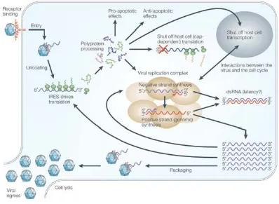
Overview of the enterovirus life cycle
Enteroviruses share similar genome structure and morphology, and as a result, have a similar life cycle. The following overview describes the PV life cycle as this is well studied and the subject of research presented in this thesis. Many aspects of PV infection have been mentioned, therefore will be shortened for brevity. A diagrammatic summary of the life cycle is shown in figure 1.2.
Poliovirus infection is initiated by the attachment of virus particles to PVR, and this triggers conformational changes in the virus particle. Upon cell attachment, both VP4 and the amino terminus of VP1 are externalised and are subsequently inserted into the host cell membrane (Levy et al., 2010). This results in the formation of
RNA genomes before mature virions are released from the cell following lysis (Novak & Kirkegaard, 1991). In all, PVs have a short growth cycle which takes approximately 8 hrs from cell attachment to virus release (Mueller et al., 2005).
Poliovirus RNA replication
Although PV RNA synthesis is an intricate process, it has been examined extensively to provide a clear understanding of the viral and cellular proteins involved. Once the virus genome is released into the cytoplasm, VPg is cleaved by a cellular enzyme (Ambros et al., 1978). The genome is translated and virus proteins necessary for RNA replication and for inhibiting host cell processes are produced. Virus RNA replication proceeds in a replication complex, formed by the rearrangement of cellular membranes, and involves virus and cellular proteins that initiate negative-strand RNA synthesis by genome circularisation (Ansardi et al., 1996; Herold & Andino, 2001).
Replication complexes have been studied extensively, including their role in virus replication (Bienz et al., 1980; Bienz et al., 1990). The P2 virus proteins are found to be associated with the endoplasmic reticulum from 3 to 3.5 hrs post-infection, and RNA synthesis begins to decline approximately 1 hr later (Ansardi et al., 1996, Bienz et al., 1987). These virus-induced vesicles are described as closed ‘entities’ which limit or prevent the exchange of viral protein, RNA, or membranes, in order to concentrate necessary components (Egger et al., 2000; Miller & Krijnse-Locker, 2008). They may also protect newly synthesised virus RNA from cellular factors and competing processes (Ahlquist, 2002).
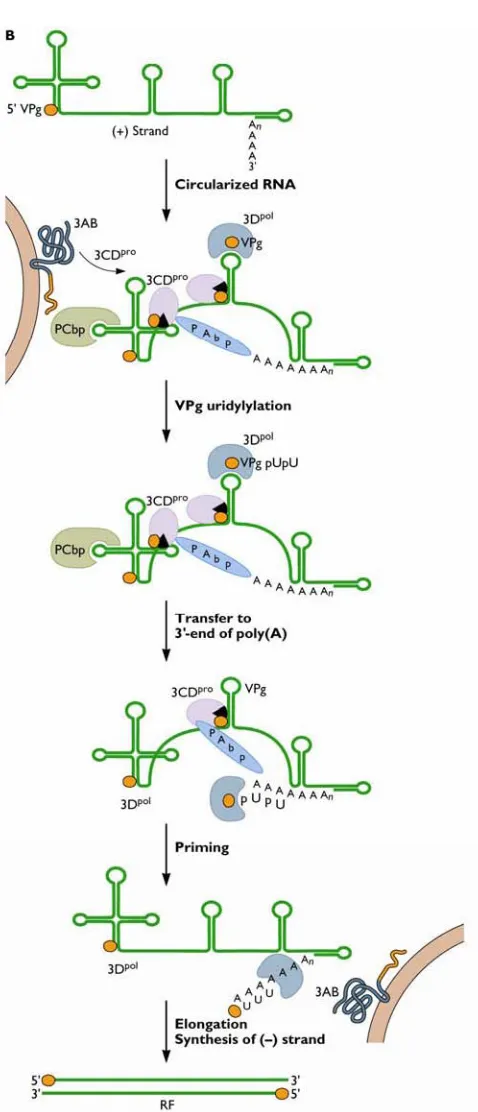
Virus proteins 2C and 3AB are responsible for binding virus RNA to membranes during replication, as 3Dpol cannot associate with membranes (Plotch & Palant, 1995; Semler et al., 1982; Teterina et al., 1997). Figure 1.3 outlines PV negative-strand RNA synthesis. Virus precursor protein 3CDpro, together with poly(rC)-binding protein 2 (PCBP2) facilitates the formation of a ribonucleprotein complex as both bind to the 5’ terminal cloverleaf structure on the positive-strand RNA (Andino et
al., 1990; Andino et al., 1993; Gamarnik & Andino, 1997). Poly(A)-binding protein
1 (PABP1), which is bound to the 3’ poly(A)-tail also interacts with the
Figure 1.3 Poliovirus negative-strand synthesis. Once the virus genome is released into the cytoplasm, VPg, a virus protein covalently linked to the 5’ terminal of RNA, is cleaved by a cellular enzyme. The genome is translated and virus proteins
necessary for RNA replication are produced. Virus RNA replication proceeds in a replication complex, which incorporates cellular membranes associated with initial translation, and virus and cellular proteins that initiate negative-strand RNA
structure also binds 3Dpol, 3CDpro, and VPg in which the latter serves as a protein primer for RNA synthesis. Negative-strand RNA synthesis proceeds by 3Dpol
elongation once the complex is transferred to the 3’ end of the genome. As a result, a double stranded replicative form (RF) is produced, containing a positive- and
negative-strand virus genome.
Following synthesis of negative RNA strands, membrane-associated virus protein 2C anchors this RNA strand to replication membranes (Banerjee & Dasgupta, 2001) (figure 1.4). It has been demonstrated that guanidine inhibits initiation of negative-strand synthesis and mutants resistant to guanidine contain mutations mapped to the 2C region (Barton & Flanegan, 1997). Binding to protein 2C might aid recruitment of uridylylated VPg-containing complexes to the 3’ end of the negative-strand to initiate positive-strand RNA synthesis. VPg-pUpU (bound to the 3’ end of negative-strand) is uridylylated by 3Dpol using 3’ terminal A residues, and serves as the primer
for positive-sense RNA synthesis. VPg-pUpU is elongated by 3Dpol to synthesise multiple copies of positive-sense strands (Barton & Flanegan, 1997; Richards et al., 1984) and allows for suitable genome packaging with structural proteins prior to virion release.
The use of reverse genetics (infectious virus clones and subgenomic replicons)
permissive cells that are able to support infection. This was first achieved by Taniguchi and colleagueswith the recovery of an RNA bacteriophage, Qβ, from cloned cDNA (Taniguchi et al., 1978). The cloning of the first animal RNA virus, PV, occurred only a few years later. Racaniello and Baltimore initially cloned three fragments spanning the PV1 genome into plasmids before combining these into one final clone to make the first infectious PV clone (Racaniello & Baltimore, 1981a; b).
Likewise, the HRV14 genome was cloned in its entirety and infectious virus was recovered from RNA transcripts transfected in HeLa cells (Mizutani & Colonno, 1985). Subsequently, the coxsackievirus B3(CVB3) genome was cloned directly into pBR322, and the entire coxsackievirus B4 (CVB4) (strain JVB) genome was cloned also into pBR322, both by the cDNA:RNA hybrid method to construct a first full-length infectious clone for sequence comparison (Jenkins et al., 1987; Tracy et al., 1985). Since then, molecular techniques have improved to construct complete enterovirus cDNA plasmids, especially using amplification by polymerase chain reaction (PCR) and cloning of entire genomes (Lindberg et al., 1997).
Infectious cDNA in plasmid form allows for genetic manipulations that cannot be performed on an RNA genome alone. Mutations or modifications can be introduced into the cDNA in order to characterise the properties of subsequently recovered virus
in vitro. One such example was the disruption of the CRE structure by site-directed
mutagenesis, which led to the inhibition of positive-strand RNA synthesis and ultimately a lack of recovered virus in cell culture (Goodfellow et al., 2000).
Virus cDNA plasmids are also useful in order to map spontaneous mutations that arise when wild-type viruses are able to grow under restrictive conditions (selective pressures). Mutations that allow viruses to escape such restrictive conditions can therefore be identified by sequencing and used for recombination studies. One such example was the mapping of PV mutants that were resistant to 2 millimolar (mM) guanidine, which usually inhibits replication (Emini et al., 1984). Resistance to guanidine was mapped to mutations in the 2C region, and similar studies have identified genetic markers linked to selection by temperature, escape from
monoclonal antibody neutralisation, and actinomycin D (Dewalt et al., 1990; Kean et
Subgenomic replicons emerged as a powerful tool in the mid 1980s when researchers became interested in the mechanism of PV replication and the role of defective interfering (DI) particles during virus infection. Defective interfering particles are shown to replicate in cells but due to missing portions of their genome, are not able to propagate virus unless in the presence of helper virus (Kuge et al., 1986). Minimal genome elements required for replication and signals necessary for virion packaging can be determined as a result of such particles. Poliovirus DI particles have been isolated from virus populations propagated in cell culture and results indicate that the locations of deletions were limited to the capsid coding region (Hagino-Yamagishi & Nomoto, 1989; Kajigaya et al., 1985). This suggests that such incomplete genomes retain the ability to replicate and package due to cis-acting non-structural proteins, in the presence of infectious virus. This knowledge was applied to the construction of subgenomic replicons in which large fragments of the P1 region were deleted from infectious PV cDNA clones (Kaplan & Racaniello, 1988). Enterovirus cDNA genomes can be altered by the replacement of the capsid region with a reporter gene (e.g. luciferase) to create subgenomic replicons. The capsid can be removed without any consequences to RNA synthesis and allows for accurate quantitative detection of RNA, replication, and translation by in vitro assay. A subgenomic replicon for PV3 was constructed by the removal of the capsid region and replacement by a reporter gene encoding chloramphenicol acetyltransferase (CAT) (Percy et al., 1992). A luciferase-expressing Mahoney strain of PV1 replicon was also engineered to study the ribonucleoprotein complex during virus replication (Andino et al., 1993). These are a few of the several virus clones that exist for picornaviruses.
1.3 Enterovirus Evolution
Virus evolution
Evolution requires genetic variation. Positive-sense single-stranded RNA viruses including important human pathogens like PV and hepatitis C virus (HCV) have error prone polymerases, short replication cycles, and high virus yields that together contribute to genetic diversity. The small genome of PV ultimately allows for it to replicate quickly so that more progeny RNA can be generated, and this has
Virus genome adaptability in a host depends on three broad factors; (1) the genetic heterogeneity of the population (or quasispecies) or the number of stable mutations in individual genomes compared to the consensus sequence; (2) the population size; and (3) the complexity of the viral genome (Domingo & Holland, 1997).
Consequently, a viral population can contain genomes with one or several mutations that make mutants dissimilar from the type species or reference genomes (Bouslama
et al., 2007). This high level of variation allows RNA viruses to exist as a
heterogeneous population and is the reason why viruses are constantly changing, with the continuous emergence of new strains from a quasispecies population. Mutations within viral populations lead to variations in virulence and possibly modifications in host cell tropism as a result of selective pressures imposed by the host.
Mechanisms of evolution in enteroviruses
Most genetic diversity in positive-sense RNA virus populations arise from mutations caused by the error-prone nature of the virus’s own RdRp (Domingo & Holland, 1997, Steinhauer et al., 1992). As a result, enteroviruses (and indeed all positive-sense RNA viruses), exist as quasispecies; a collection of non-identical but related genomes (Lauring and Andino, 2010; Domingo & Holland, 1997). This is significant given the size of such quasispecies genome populations. Virally encoded RdRps lack exonucleolytic editing ability (Ishihama et al., 1986), and it has been shown that PV polymerases have an estimated mis-insertion error rate ranging from 1.2 x 10⁻⁴ to 1 x 10⁻⁶ for transition mutations and 3.2 x 10⁻⁵ to 4.3 x 10⁻⁷ for transversion
mutations (Freistadt et al., 2007). As a result, repeated passages of large populations of virus over time enrich those mutant genomes that replicate more efficiently, and can also alter virulence by modifying host cell tropism (Domingo & Holland, 1997). The best example of this is the attenuation of the three serotypes of PV to create the OPV. Strains of PV were repeatedly passaged through monkey cells (in vivo and ex
vivo) to eventually select viruses with multiple spontaneous point mutations that led
to the inability of these strains to replicate as efficiently in human neurons while retaining the ability to replicate in the human gut cells where they were capable of inducing the desired protective immune response (Kew et al., 2005, Sabin &
and was demonstrated by Westrop et al., when they showed that attenuation of PV3 Sabin vaccine strain from its neurovirulent wild-type parent only required two point mutations at positions 472 and 2034 of the genome (Westrop et al., 1989).
The natural replication of all three PV serotypes can be followed easily due to rapid genome evolution, and this can be calculated with high precision. Nucleotide
substitutions (of which > 80% are in the coding region) accumulate at an average rate of ~ 1% per year at each position, and this rate increases to ~ 3% per year when calculated for synonymous sites only (Kew et al., 1995; Martín et al., 2000). Rapid evolution of vaccine viruses, such as PV, also accounts for the adverse reactions associated with the use of live-attenuated vaccines. The level by which VDPVs differ from the vaccine Sabin strains can help determine when such viruses were originally transmitted. Isolated VDPVs can differ from OPV strains at 1% to 15% of VP1 nts when resequenced, which provides useful timelines for tracing origins of outbreaks (Shimizu et al., 2004).
Over a decade ago, extensive sequence and phylogenetic analysis of enterovirus genomes revealed that evolutionary rates differed depending on the genomic region studied (Hyypiä et al., 1997). Different selection pressures are most likely applied to different regions; the capsid region is directly linked to host immune responses and over a significant amount of time, has diversified and evolved due to genetic drift, and this is clear from the wide range of receptors recognised for cell attachment. The 5’ NCR shows the slowest rate of evolution (Hyypiä et al., 1997; McWilliam Leitch
et al., 2009). Only two distinct phylogenetic groups exists for 5’ NCR in human
enteroviruses, those belonging to species A and B, and the remaining serotypes in species C and D (Santti et al., 1999).
Even though the RdRp of RNA viruses are usually cited as the reason for high sequence variability, the evolutionary rates observed in PV sequences are in fact higher than those attributed to RdRp fidelity alone (Freistadt et al., 2007). Genetic recombination may well provide the opportunity for evolutionary advancement that may be greater than that seen by the steady accumulation of point mutations.
on the process, the causes and outcomes of recombination were evaluated as part of this project.
Evidence of virus recombination
Recombination is a way for viruses to achieve dramatic alterations of a significant part of the genome. Evolution therefore occurs quicker than by the gradual
acquisition and accumulation of natural mutations during replication (genetic drift). As a general rule, recombinants arise and evolve from either initial dual infections in the same cell, or as a result of superinfections. Superinfection is the process by which a cell that has previously been infected with a virus is subsequently co-infected by a different virus strain or type altogether.
The phenomenon of recombination in non-segmented RNA viruses has been demonstrated previously and is an important evolutionary mechanism. Interspecies recombination in alphaviruses between an eastern equine encephalitis-like virus (EEEV) and a Sindbis-like virus gave rise to a related new virus – western equine encephalitis virus (WEEV) (Hahn et al., 1988). This virus combined the
encephalogenic properties of EEEV but maintained the antigenic properties of Sindbis virus.
In a further example, a recombination event between virus and host RNA enhanced the cytopathic properties of the virus, bovine viral diarrhoea virus (BVDV).
Cytopathic and non-cytopathic forms of BVDV are classified by culture through cells, and it was determined that the cytopathic form contained host-cellular RNA encoding ubiquitin (Meyers et al., 1989). It was suggested that in persistently infected cattle, non-cytopathic BVDV evolved to the cytopathic form, triggered by recombination events that inserted cellular RNA into the virus genome.
Recombination in enteroviruses
Recombination in RNA viruses was first described in PV in the early 1960s (Hirst, 1962; Ledinko, 1963) and it has been identified since then in many enteroviruses, and studied extensively in PV (Agol et al., 1985; Minor et al., 1986; Romanova et
al., 1980; Savolainen-Kopra et al., 2009b). In the early PV experiments, both groups
were able to characterise recombinants generated in vitro by mapping different selection markers passed onto recombinant progeny virus. Since then, recombination between PV serotypes has been shown to occur as soon as 2.5 hrs post co-infection in replication complexes within HeLa cells (Egger & Bienz, 2002). Once cells were co-infected with PV1 (Mahoney strain) and PV2 (Sabin strain), type-specific fluorescent riboprobes were used to visualise the genome regions within replication complexes by fluorescent in situ hybridisation (FISH). Recombination was
subsequently demonstrated by RT-PCR and sequencing.
Multiple levels of recombination have been identified in natural PVs outside the laboratory: intraserotypic recombinants (within each PV serotype) and interserotypic recombinants (combining PV1, PV2, or PV3) have been isolated from OPV
vaccinees (Cammack et al., 1988; Cuervo et al., 2001; Furione et al., 1993; Yang et
al., 2005); wild-type PV serotypes 1, 2, and 3 have been shown to recombine with
each other (Dahourou et al., 2002); wild-type and OPV strains have recombined (Georgescu et al., 1995; Guillot et al., 2000), and lastly, vaccine-derived PVs have recombined with unknown HEV-C strains (Kew et al., 2002; Rakoto-Andrianarivelo
et al., 2007; Shimizu et al., 2004). As yet, there have been no reported cases of
interspecies recombinants involving PV and other HEV species. Interspecies recombination is rare (Simmonds & Welch, 2006), and so far only one event has been suggested between HEV-B and HEV-C viruses (Bolanaki et al., 2007).
resulted in more cases of intraspecies recombinants being identified from PV outbreaks. For example, phylogenetic analysis revealed that the source of the 2000-2001 outbreak of poliomyelitis in Hispaniola was due to recombinants formed between OPV derived PVs and the non-structural regions derived from an unknown enterovirus strain (species C) (Kew et al., 2002). Two other reports of cVDPV in Madagascar and the Philippines, implicated vaccine/non-polio recombinants
(Rousset et al., 2003; Shimizu et al., 2004). These and other reports have confirmed that live attenuated strains of PV originating from vaccinees are able to recombine with circulating non-polio species C enteroviruses creating neurovirulent
recombinant viruses capable of causing serious disease. Outbreaks attributed to PV or recombinant forms of PV are more likely to occur in regions where vaccine
coverage is sub-optimal and the circulation of other enteroviruses is high (Kew et al., 2005).
Certain enteroviruses are thought to have emerged as a result of early recombination events. This includes evidence of speciation of PV from a coxsackievirus A (species C) ancestor that occurred through mutation of the capsid region. This speciation event involved a cellular receptor switch in order to possibly escape selective pressure imposed by the host (Jiang et al., 2007). Extensive analysis of HEV B species has also indicated recombination events involving other unknown species B virus sequences and this has occurred within the HEV B species group many times to create a complex mosaic of RNA genomes in a short, eight year period (Simmonds & Welch, 2006). Surprisingly, recombination events were observed not only between VP1 and 3Dpol, but between VP1 and VP4 virus sequences. Frequencies of
recombination (between VP1 and 3D regions) were also related to time periods (i.e. virus isolation dates). Approximately 40% of species B isolates from the same year were recombinant, and this rose to 70% for isolates collected two to three years apart. Almost all isolates were recombinant in nature when the collection period exceeded three years. It was also noted that enterovirus diversity occurred
asymmetrically between structural and non-structural coding regions, particularly in echovirus (E30) strains (McWilliam Leitch et al., 2009). Phylogenetic analysis shows that while VP1 sequences diverged by natural genetic drift, 3Dpol sequences clustered in groups and interspersed with other species B serotypes, further
regions. This illustrates the dynamic nature of species B evolution, where fluctuating outbreaks of E30 activity have been associated with new genomic lineages that have replaced previously circulating genotypes (Oberste et al., 1999).
The location of recombination sites in enteroviruses appear to occur throughout the genome with most crossover events occurring in the 5’ NCR and in the 2A, 2B, 2C, and 3D protein encoding regions (Savolainen-Kopra et al., 2009). Recombination sites in the capsid region predominantly occur between PV serotypes 2 and 3, and usually at the C-terminal end of the protein region (Blomqvist et al., 2010; Tao et al., 2010). Interserotypic recombination in the capsid region is rare, possibly due to the sensitivity of structural constraints that are important in order to maintain the correct capsid shell.
Although recombination has been responsible for diverging viruses from ancestral lineages, the role of recombination in allowing virus populations to respond to selective pressures is unclear. At a genetic level this would include recombination in the genome that is ‘selected for’ by specific events, such as immunological responses from the host, differing temperature ranges of host cells and the influence of
environmental chemicals.
Methods of RNA virus recombination
recombination in PV may occur as the joining of broken RNA molecules by transesterification (Gmyl et al., 1999), although this would occur less often. Homologous recombination by contrast, is the result of crossovers between two similar or closely related RNA molecules containing extensive sequence similarity. This form of recombination is known as ‘copy-choice’ and is the consequence of template switching by the RdRp during negative-strand synthesis as demonstrated by Kirkegaard and Baltimore (1986). ‘Copy choice’ was first proposed as the
mechanism for recombination in PV (Copper et al., 1974), and to date, the majority of studies of recombination in RNA viruses have supported this model (reviewed in Agol, 1997).
RdRp ‘copy choice’ by template switching
Replication-dependent recombination in plant and animal viruses requires RNA templates and viral replicase for successful template switching to occur. The simplest model suggests that two RNA templates are necessary, including the primary RNA template (donor) which binds RdRp to initiate replication, and the acceptor RNA template that receives the RdRp mid-elongation (figure 1.5A). The crossover sites or ‘breakpoints’ occur at positions similarly matched in sequence so that the resultant recombinant RNA templates retain the same sequence length and structural integrity as the parental RNA molecules (Lai, 1992). This is commonly referred to as precise recombination. Imprecise recombination, on the other hand, results in changes including nucleotide mismatches, deletions, or insertions at the junction site, or close to it (figure 1.5B) (Lai, 1992; Nagy & Simon, 1997). Depending on the modifications that have occurred, a viable virus may still result from this form of genetic exchange. ‘Copy choice’ template switching may not always occur between homologous RNA strands, but crossover sites in coding regions are somewhat constrained by reading frame, recognition sequences for proteolytic processing, and protein function, which could conceal such events (Wimmer et al., 1993).
Figure 1.5 Current replication-dependent template switching models for RNA recombination. (A) Template switching during replication. The RdRp exchanges one template (donor) for another (acceptor) at corresponding positions in the genome. There are several possibilities as to why the RdRp is removed from the donor template (see text). (B) Diagrams depicting precise and imprecise
al., 1985;(Cole et al., 1971; Kajigaya et al., 1985; Kuge et al., 1986; Wimmer et al.,
1993). It was observed that all deletions in isolated DI particles of PV occurred in frame (Kuge et al., 1986), suggesting that some viral proteins were required to be encoded incis for genome replication. Poliovirus DI particles can therefore initiate their own replication but cannot produce progeny virions as they do not synthesise capsid proteins – these must be provided by a homologous helper virus.
Since the same mechanisms for RNA recombination are thought to be responsible for the generation of DI particles, Kuge and colleagues performed comparative sequence analysis of PV1 DI particles to determine the underlying genetic mechanisms behind their generation (Kuge et al., 1986). Selection pressures may have influenced the location of deletions, as these were limited to between nucleotide positions 1226 and 2705 of the capsid coding region. There was no evidence of sequence homology between immediate donor and acceptor template sites, and it was proposed that deleted regions were ‘looped out’ by secondary RNA structures, meaning that the RdRp skipped these regions during replication. As PV DI particles are rarely formed or identified, further study of the mechanisms behind their formation are limited.
What mechanisms influence recombination?
At the genomic level, intrinsic signals present on the donor or acceptor RNA templates may influence switching of the RdRp between templates. At present, it is suggested that RNA signals either pause or terminate the RdRp, both of which cause the release of RdRp from the RNA template (King, 1988; Nagy & Simon, 1997). Possible signals include: A/U-rich and U-rich sequences that promote RdRp
slippage, local strong hairpin structures present on either template, the formation of stable heteroduplexes between templates, and mis-incorporations (Carpenter et al., 1995; Makino et al., 1986; Pilipenko et al., 1995; Tolskaya et al., 1987). If local RNA signals are essential for template switching of the RdRp, then it is unclear why there is no consistency of crossover sites for the same PV strains reported in the literature.
Researchers have succeeded in achieving genetic recombination of enteroviruses in
vitro (in particular in PV) in an attempt to define the preferred region/s (but not