Abstract — A key feature of single-cell marine organisms called diatoms is their silica exoskeleton with nanoporous morphology. These naturally grown structures provide a promising basis for new biomimetic structural designs, but may also be used directly in their native form.
Microscale diaphragms found in nature often can withstand large deformations. They also show vibration eigenfrequencies in the high MHz to GHz range. These structural properties open up the possibilities for the development of production technologies based on micromanipulation of bio-inspired or bio-derived microscale structures. In this work we report the results of FEM simulations aimed at investigating the effects of stiffness, pore diameter, and thickness on the vibrational characteristics of diatom frustule structures.
Index Terms—Biomaterials, Coscinodiscus sp., Diatom Frustules, Eigenfrequency, Finite Element Method (FEM) simulation
I. INTRODUCTION
ANOMETRE scale manipulation of material volumes has become a major scientific theme in recent decades, since it opens the possibilities of obtaining substances and structures with hitherto unprecedented combinations of functional and deformation properties. [1] One of the major challenges in nanotechnology lies in the stringent demands that arise in terms of the high complexity and cost of the processing equipment, and associated significant environmental impact. In view of these considerations, significant efforts have been directed at the search for envi -
Manuscript received April 9, 2019; revised April 12, 2019. This work was supported in part by the Royal Society, UK (IEC/R2/170223), EPSRC UK (EP/P005381/1), and the Russian Foundation for Basic Research (RFBR 18-44-920012).
Bakhodur Abdusatorov is a doctoral student of Skolkovo Institute of Science and Technology, Center for Energy Science and Technology Moscow, 121205, Russia, email: [email protected]
Joris Everaerts is with MBLEM, the University of Oxford, Department of Engineering Science, Oxford OX1 3PJ; e-mail:
Alexei I. Salimon is Senior Research Engineer at the Hierarchically Structured Materials (HSM) lab, Skoltech Center for Energy Science at Technology (CEST), Skolkovo Institute of Science and Technology, Moscow 121205, Russia, e-mail: [email protected]
Alexander M. Korsunsky is Head of the Multi-Beam Laboratory for Engineering Microscopy (MBLEM), and Professor of Engineering Science, Department of Engineering Science, University of Oxford, Parks Road, Oxford OX1 3PJ, UK (tel. +44 1865 273043, e-mail:
[email protected]), and Visiting Professor, Hierarchically Structured Materials (HSM) lab, SM) lab, Skoltech Center for Energy Science at Technology (CEST), Skolkovo Institute of Science and Technology, Moscow 121205, Russia
-ronmentally friendly, nature-like processes for nanofabrication.
In Nature, nanostructuration arises in many instances within living systems, not only soft matter, but also in mineralized tissues containing ceramics such as hydroxyapatite (mammalian bone and teeth), calcite (crustacean nacre), and silica (diatom algae).
Manipulating materials to obtain nanostructured objects and components offer a route to significant advances towards new technology [2]. Whilst the development of nature-like (biomimetic) approaches is still at its early stages, some fundamental insights are beginning to be collected into ‘soft’ processes and mechanisms that underlie the transport and deposition of matter in a controlled and precise fashion used by Nature in its complex architectures. For example, the dissolution of amorphous glassy germania (GeO2) and its re-precipitation in crystalline form were
observed using in situ liquid cell within TEM [3]. This study represents a significant step towards elucidating the possible mechanisms that make use of aqueous solution to produce solid structures with precisely defined properties. This is of obvious relevance for understanding the formation of marine crustacean and algal skeletons that involves atomic species present in the world ocean and freshwater water bodies. Imitating these natural processes is likely to provide a route towards water-based, environmentally friendly fabrication of intricately nanostructured elements.
However, many questions remain unanswered. How does nanostructuration occur? What templates underlie the formation of specific structural elements, such as micron-sized pores containing finer, nano-scale sub-channels? How and why did these intricate architectures evolve through natural selection and optimization? What vital functions do they fulfil in the host organism, through the combination of their photonic, chemical, micro-fluidic, strength, and vibrational properties? [4]
In the present study we focus attention on the numerical study of the mechanical vibration behavior of the mineralized structural shell (frustule) of an important group of microscopic algae: diatoms. Diatoms obtain their name from the asexual reproductive mechanism, in which cells divide into two. They are one of the most wide-spread microorganisms on the planet. Diatom algae produce about 1/2 of the world's ocean biomass, 2/3 of the oxygen produced by the World's oceans, and about 1/4 of all oxygen and organic matter release on the planet Earth. There are around 200,000 species of diatom algae known to science. They have a unique porous architecture with dimensions ranging from 2 µm to 2000 µm [5]. The walls cells mainly composed of amorphous hydrated silica SiO2*2H2O with a
On the prospects of using Biogenic Silica for
MEMS (Micro-Electro-Mechanical Systems)
Bakhodur Abdusatorov, Joris Everaerts, Alexei I. Salimon, and Alexander M. Korsunsky,
Member, IAENG
small proportion of organic macromolecules. About 60% of the dried weight is made up of silica.
The unique structure and diversity of diatoms motivated researchers to consider their unique properties, and propose potential use of these in optical devices [6], drug delivery [7-8], bio-sensing [9], molecular or particle separation [10], nano-photonics [11], micro, and nanodevices [12]. This warrants further exploration of the relationship between diatom frustule composition, structure, and function.
There are two general types of diatoms depending on symmetry, frustule shape, ribs, as shown in Figure 1. The shell of the centric diatom has a form of Petri dish (Fig. 1(a) one half (cover) slightly more than the other and radial symmetry. Pennate diatoms display bilateral symmetry.
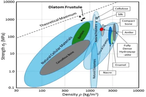
Within the family of biomaterials, diatom frustules are some of the most lightweight, stiff, and strong. Fig.2 shows that the diatom frustules demonstrate an excellent combination of high strength and low density (black dots) compared to other natural materials, approaching the theoretical maximum.
Fig. 2. Ashby plot of strength and density of natural biological materials [15]
Computer simulation of the mechanics of diatom frustules can help understand the deformation response of these structures through the analysis of their load bearing capacity and vibration dynamics.
II. THE FORMULATION OF PARAMETRIC HIERARCHICAL CAD MODEL
The present study focuses on exploring the vibrational properties of centric and pennate diatom frustules. Centric frustule shape, e.g. Coscinodiscus sp., was chosen for the first part of this study, because these frustules are
abundantly widespread in nature, and also display hierarchical morphology. As the second object of this study, pennate frustule was studies that is representative of another large class of diatom algae.
The requirement of optimal consumption of computing effort imply that some simplification of the structure provides efficient route to structural analysis. 3D models were created on the basis of Scanning Electron Microscopy images and modeled using Solidworks (CAD). At first, 2D models of the contour of diatoms were created with the correct dimensions based on SEM images. The result was extruded numerically to obtain 3D model. Then circular through pores were added. The porosity was calculated by using equation (1), determined by Aempty (empty area per
pores, top of diatom model) and Atotal (total area, top of
diatom model):
(1)
Hereinafter, d is the pore diameter, D is the diatom frustule diameter, N is the number of pores, and t is the shell thickness [16]. Complete 3D CAD diatom models of the top of the frustule assembly were created to carry out eigenfrequency analysis using the appropriately adapted mesh. 3D CAD diatom models were imported into FEM software (COMSOL Multiphysics 5.4) and some simulations performed.
Fig. 3. CAD model of a dome-shaped diatom Coscinodiscus sp. frustule (right) with details of the generatrix curve (left) . Dimensions of various frustule models are listed in Tables 1 and 2 depending on simulation.
III. FEM SIMULATION
Computer simulation provides extensive possibilities for the study of structural and material response to a range of multi-physics loading conditions. The high cost and complexity of performing experiments makes computer simulation an attractive alternative and supporting analytical framework for parametric investigation and design of complex multi-material structures.
In this section we report the results of simulating the mechanical response of diatom frustules under various loading conditions that are not easily accessible via experiment.
Enhanced dome-shaped 3D frustule models were created using shell elements for computational efficiency, in order to investigate the role of hierarchical structure on the mechanical properties of the diatom. The initial properties used in the simulations were taken from the literature data. Young’s modulus was set to 73 GPa, Poisson’s ratio of 0.17, and density 2300 kg/m3 [17]. The parametric dependence of eigenfrequency was studied on the pore diameter, shell thickness, and material stiffness. The pore diameter was varied from 0.8 µm to 2.6 µm. The frustule thickness varied Fig. 1. SEM images of Diatom Frustules: (a) of centric diatom
(Coscinodiscus sp.) [13] (a) and (b) pennate diatom (Fragilariopsis kerguelensis)[14].
[image:2.595.48.257.227.318.2] [image:2.595.307.535.384.465.2] [image:2.595.47.283.450.606.2]from 0.1 µm to 0.5 µm. and Young’s modulus values were chosen in the range from 10 Pa to 100 GPa.
IV. RESULTS AND DISCUSSION
One-layer truncated cone model was used in this study to compute eigenfrequency of vibrational response. As seen from Fig. 4, as the wall thickness increases, the eigenfrequency increases gradually. The effect of the layer thickness on the eigenfrequency is seen to be moderate.
In this study the shell thickness was assumed to be uniform. However, it is also possible to investigate the response of shells of non-uniform thickness.
Fig. 4. The effect of wall thickness on the base eigenfrequency of the hemispherical shell (d of pores=1.5 µm)
Fig. 5. The correlation between the pore size d of the Coscinodiscus sp.
frustule model and eigenfrequency of the hemispherical shell (t=0.3 µm)
Fig. 6. The effect of material stiffness on the eigenfrequency of the hemispherical shell (d pores=1.5 µm). The results are shown for shell thickness of t = 1.5 µm (lower curve) and t = 0.3 µm (upper curve).
Fig. 7. The effect of inverse frustule diameter 1/D on the base eigenfrequency of the hemispherical shell (d of pores=1.5 µm).
Fig. 8. The effect of inverse root of frustule density on the base eigenfrequency of the hemispherical shell (d of pores=1.5 µm).
Fig. 5. illustrates the decrease in frustule eigenfrequency as the pore diameter increases. For the pore diameter value of ~3.5 µm, the eigenfrequency drops to 0. This reflects the fact that this pore size corresponds to the merging of the pores, thus destroying the continuity of the frustule structure.
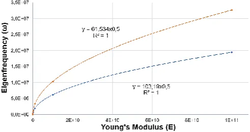
Result in Fig. 6. show that the eigenfrequency increases with material stiffness (Young’s modulus) following a parabolic law.
The effects of the frustule diameter on eigenfrequency is presented in Fig. 7. It shows that as the diameter of diatom frustule decreases, the eigenfrequency increases, respectively. The ratio of density shows (Fig. 8.) the increase of density leads to a decrease eigenfrequency.
Overall, we find that the parametric study results are consistent with the theoretical formula for vibration eigenfrequency of thin circular plates, given by [18, 19]:
. (2)
Here Y is the geometric parameter that has the units of inverse length, and depends on the ratio of the shell thickness t to the diameter D, but also on the diameter d and the arrangement of pores.
[image:3.595.306.539.50.161.2] [image:3.595.305.539.200.316.2] [image:3.595.48.289.206.322.2] [image:3.595.49.290.367.496.2] [image:3.595.46.292.541.671.2]Fig. 9. FEM natural frequency simulation results for Coscinodiscus sp.
[image:4.595.47.288.47.337.2](centric) diatom frustule models. Frequencies of individual modes are indicated. The diatomite frustule diameter was D = 50 µm, and the pore size was set to d = 1.6 µm.
Fig. 10. SEM image of Staurosira diatom frustule valve halves, with the overall diameter of about 2 µm.
A full 3D model would be useful to obtain additional information about the material and structural response, i.e. the details of local stress values. The correlations between the eigenfrequency and the frustule morphology and properties (pore diameter, wall thickness, material modulus of elasticity) allows identifying the optimal choice of diatom frustules for MEMS applications.
[image:4.595.305.540.161.425.2]In [20], the phase transition from amorphous silica to crystalline quartz was observed to occur due to heating the sample of diatomic earth to 1100oC. Given that Young’s modulus of crystalline quartz is 102.4 GPa and exceeds that of amorphous silica by almost 50%, and that the eigenfrequency increases with Young’s modulus as illustrated in Fig.6, conversion from amorphous to crystalline quartz provides a means of increasing the vibrational frequency response of the structure.
Fig. 11. SEM of image Corethron diatom frustule (diameter ~26 µm).
(a) (b)
(c) (d)
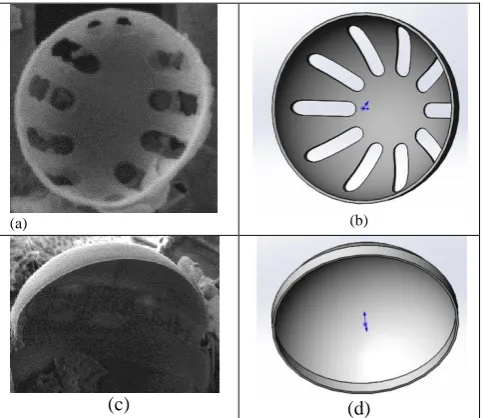
Fig. 11. (a) SEM micrograph of Staurosira diatom frustule exoskeleton half, and (b) its CAD model for FEM simulations. (c) SEM image of
Corethron diatom frustule half, and (d) CAD model based on the micrograph used for the simulation, with the dimensions given in Table 1.
[image:4.595.46.281.395.659.2] [image:4.595.300.542.455.664.2]contains large aspect ratio radial slits, whilst the latter is larger in diameter, with the half-shell of a shape close to a semi-sphere.
[image:5.595.63.271.140.232.2]The results of FEM calculations are summarized in Table 1, illustrating the fact that the resulting change in the shell eigenfrequency spans almost a decade of values.
TABLE I
THE DIMENSIONS AND PROPERTIES OF STAUROSIRA AND
CORETHRON DIATOM FRUSTULES
Staurosira Corethron
d 2 [µm] 26 [µm]
t 0.03 [µm] 0.3 [µm]
E 73 [GPa] 73 [GPa]
ρ 2300 [kg/m3] 2300 [kg/m3]
ω 360 [MHz] 59 [MHz]
Where, pore diameter d, wall thickness t, Young’s modulus E, density ρ) and the eigenfrequency (ω) results from FEM analysis. where and eigenfrequency, respectively.
[image:5.595.42.285.340.457.2]In the final part of the present study, the vibration response of pennate diatom frustules was considered. Fig.11 illustrates the shape of Louboutensis Kolski (var. Diploneis interrupta), along with the simplified numerical model constructed.
Fig. 11. SEM micrograph (left, shown with false colour) of external valve of Louboutensis Kolski (var. Diploneis interrupta) [4], and the CAD model (right) created for the purpose of FEM simulation.
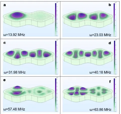
Fig. 14. FEM results of natural vibration mode simulation of Diploneis interrupta diatom frustule model. The eigenfrequencies of different modes are indicated. The diatomite frustule size was l = 42 µm, and the pore size of d = 0.08 µm was used.
Fig.14 shows the normal mode shapes for these diatom shells, along with eigenfrequencies indicated. This finding demonstrates the rich variety of vibration modes displayed by pennate diatom frustules. This type of diatoms had recently been studied by our group [4]. A diatom colony was grown and imaged on silicon substrate. This provided an established route towards obtaining MEMS-like structures for microelectronic integration. Provided a suitable excitation mode is found and activated, frustules mounted on silicon wafers can serve as sensors or actuators e.g. for high frequency vibration detector, energy harvesting, radiofrequency tagging, etc.
Further advancements may be made by controlled arrangement of frustules on the substrate, e.g. by guided diatom colonization; control over structural stiffness, e.g. by amorphous silica crystallization, reduction to silicon, etc.
V. CONCLUSION
The findings of the present study show that morphological features such as pore size, wall thickness, and material stiffness have a significant effect on the vibrational behavior of diatom frustules. The ‘natural’ (unprocessed) frustules are expected to have vibration eigenfrequencies in the range 10 MHz to 100 MHz, making them a promising candidate for MEMS applications. Diatom frustules with thin silica walls would be preferred for high frequency applications. The presence of large diameter or high density of pores reduces the eigenfrequency of response.
ACKNOWLEDGMENT
Alexander M. Korsunsky wishes to acknowledge the support from Trinity College, Oxford, UK.
REFERENCES
[1] Colton, R. J. Nanoscale measurements and manipulation. Journal of Vacuum Science & Technology B: Microelectronics and Nanometer Structures, 22(4), 1609, 2004
[2] Mann, D. G., & Droop, S. J. M. (1996). 3. Biodiversity, biogeography and conservation of diatoms. Hydrobiologia, 336(1-3), 19–32, October 1996.
[3] Xiao, Z.H., Sun, X.Y., Li, X.Y., et al. Phase Transformation of GeO2
Glass to Nanocrystals under Ambient Conditions. Nano Lett., 18, 3290-96, 2018.
[4] A.M. Korsunsky, P. Sapozhnikov, J. Everaerts, A.I. Salimon, “Nature’s neat nanostructuration: fascinating frustules of diatomic algae”, Materials Today, 22C, 169-170, 2019.
[5] Yu Wang & Jun Cai & Yonggang Jiang & Xinggang Jiang, Preparation of biosilica structures from frustules of diatoms and their applications: Current state and perspectives, Applied Microbiology and Biotechnology, Vol.95, number 2 November 2012
[6] De Stefano, L. Rendina, De Stefano, M. Bismuto A., Maddalena P., Marine diatoms as optical chemical sensors, Applied Physics letters, 87(23), 2005
[7] Ryan W. Drum and Richard Gordon, Star Trek replicators and diatom nanotechnology, TRENDS in Biotechnology, Vol.21 No.8, August 2003.
[8] Moom Sinn Aw, Spomenka Simovic, Yang Yu, Porous silica microshells from diatoms as biocarrier for drug delivery applications, Powder Technology, 52–58, 2012.
[9] De Tommasi, E., Rendina, I., Rea, I., De Stefano, M., Lamberti, A., & De Stefano, L. Intrinsic photoluminescence of diatom shells in sensing applications, Optical Sensors, Vol.7356, 2009.
[10] Losic, D., Rosengarten, G., Mitchell, J. G., & Voelcker, N. H. Pore Architecture of Diatom Frustules: Potential Nanostructured Membranes for Molecular and Particle Separations. Journal of Nanoscience and Nanotechnology, 6(4), 982–989, 2006.
[11] L. De Stefanoa, P. Maddalena, L. Moretti, I. Rea, I. Rendina, Nano-biosilica from marine diatoms: A brand new material for photonic applications, Superlattices and Microstructures, 84–89, 2009.
[image:5.595.48.290.503.730.2][12] Rea I, Terracciano M, De Stefano, L.Synthetic vs Natural: Diatoms Bioderived Porous Materials for the Next Generation of Healthcare Nanodevices, Adv Healthc Mater., 6(3), Feb 2017
[13] F. Round, R. Crawford and D. Mann, The Diatoms, Biology & Morphology of the Genera, Cambridge University Press, Cambridge, 1990.
[14] VictorSmetacek, Bacteria and silica cycling, Nature 397, 475-476, 1999.
[15] Zachary H. Aitken, Shi Luo, Stephanie N. Reynolds, Christian Thaulow, and Julia R. Greer, Microstructure provides insights into evolutionary design and resilience of Coscinodiscus sp. Frustule, National Academy of Science, 113 (8), 2017-2022, February 2016. [16] Miguel Diaz Moreno, KakaMab, Julie Schoenung, Lilian P.Dávilaa
An integrated approach for probing the structure and mechanical properties of diatoms: Toward engineered nanotemplates, Acta Biomaterialia, Volume 25, Pages 313-324, October 2015.
[17] Yao, S., Subhash, G., & Maiti, S. (2007). Analysis of nanoindentation Response of Diatom Frustules. Journal of Nanoscience and Nanotechnology, 7(12), 4465–4472, 2007.
[18] Sader, J.E. Frequency response of cantilever beams immersed in viscous fluids with applications to the atomic force microscope. Journal of Applied Physics 84, 64-76, 1998.
[19] Senjanovic, I., Hadzic, N., Vladimir, N. et al. Natural vibrations of thick circular plate based on the modified Mindlin theory. Arch. Mech., 66, 389–409, 2014.