N
-Acetyl-
N
-{2-[(
Z
)-2-chloro-3,3,3-tri-fluoroprop-1-enyl]phenyl}acetamide
Jia-Jia Niu,a,bZhi-Gang Lia,band Jing-Wei Xua*
aNational Analytical Research Center of Electrochemistry and Spectroscopy, Changchun Institute of Applied Chemistry, Chinese Academy of Sciences, Changchun 130022, People’s Republic of China, andbGraduate School of the Chinese Academy of Sciences, Beijing 100039, People’s Republic of China Correspondence e-mail: [email protected]
Received 14 March 2009; accepted 21 April 2009
Key indicators: single-crystal X-ray study;T= 293 K; mean(C–C) = 0.004 A˚;
Rfactor = 0.052;wRfactor = 0.136; data-to-parameter ratio = 14.5.
The title compound, C13H11ClF3NO2, adopts a Z conforma-tion. Halogen oxygen interactions [Cl O = 2.967 (3) A˚ ] in the crystal packing lead to the formation of a dimer joined by two Cl O bonds.
Related literature
The title compound is an important medical intermediate, see: Zhouet al.(2009). For the van der Waals radii of chlorine and oxygen, see: Politzeret al.(2007).
Crystal data C13H11ClF3NO2
Mr= 305.68
Triclinic,P1
a= 8.4408 (19) A˚
b= 9.385 (2) A˚
c= 9.455 (2) A˚
= 64.599 (3)
= 80.727 (4)
= 89.756 (3)
V= 666.0 (3) A˚3
Z= 2
MoKradiation
= 0.32 mm 1
T= 293 K
0.130.130.07 mm
Data collection
Bruker SMART APEX CCD area-detector diffractometer Absorption correction: multi-scan
(SADABS; Bruker, 2003)
Tmin= 0.958,Tmax= 0.978
3747 measured reflections 2562 independent reflections 2155 reflections withI> 2(I)
Rint= 0.010
Refinement
R[F2> 2(F2)] = 0.052
wR(F2) = 0.136
S= 1.03 2562 reflections
177 parameters
H-atom parameters constrained
max= 0.37 e A˚ 3
min= 0.38 e A˚ 3
Data collection:SMART(Bruker, 1998); cell refinement: SAINT-Plus(Bruker, 1998); data reduction:SAINT-Plus; program(s) used to solve structure: SHELXS97(Sheldrick, 2008); program(s) used to refine structure:SHELXL97(Sheldrick, 2008); molecular graphics:
SHELXTL(Sheldrick, 2008); software used to prepare material for publication:SHELXTL.
This work was supported by the Changchun Institute of Applied Chemistry, Chinese Academy of Sciences.
Supplementary data and figures for this paper are available from the IUCr electronic archives (Reference: KJ2121).
References
Bruker (1998).SMARTandSADABS. Bruker AXS Inc., Madison, Wisconsin, USA.
Bruker (2003).SAINT-Plus. Bruker AXS Inc., Madison, Wisconsin, USA. Politzer, P., Lane, P., Concha, M. C., Ma, Y. & Murray, J. S. (2007).J. Mol.
Model.13, 305–311.
Sheldrick, G. M. (2008).Acta Cryst.A64, 112–122.
Zhou, H., Niu, J.-J., Xu, J.-W. & Hu, S.-J. (2009).Synth. Commun.39, 716–732.
Structure Reports
Online
supporting information
Acta Cryst. (2009). E65, o1305 [doi:10.1107/S1600536809014779]
N
-Acetyl-
N
-{2-[(
Z
)-2-chloro-3,3,3-trifluoroprop-1-enyl]phenyl}acetamide
Jia-Jia Niu, Zhi-Gang Li and Jing-Wei Xu
S1. Comment
The title compoud is an important medical intermediate (Zhou et al., 2009). The conformation of the C=C bond (Z or E)
is usually determined by 1H and 19F NMR. Here we report the crystal structure of title compound to establish the
conformation.
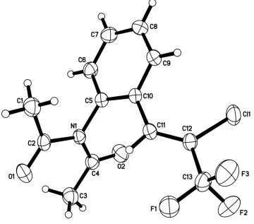
The title compound, as shown in Fig. 1, a has Z conformation, with the benzene ring and the Cl atom on the same side
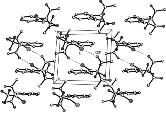
of C=C bond. In the crystal packing a distance between Cl and O3[-x,-y,1-z) of 2.967 (3) Å is observed, which is
obviously shorter than the sum of van der Waals radii of chlorine and oxygen (3.27 Å, Politzer et al., 2007), showing the
strong Cl···O interaction indicative of a halogen bond with a nearly linear C12—Cl1···O3[-x,-y,1-z] angle of 173.5 (4)°.
Two monomers, related by a centre of symmetry, are linked into a dimer by two Cl···O halogen bonds (Fig. 2). There are
no distinct interactions between the dimers in the crystal packing.
S2. Experimental
N-(2-formylphenyl)acetamide (1 mmol) and zinc powder (5 mmol) were added to DMF (5 ml, distilled from CaH2), the
flash was then evacuated and backfilled with argon (3 cycles). Acetic anhydride (3 mmol) was added by syringe at room
temperature, and then 3 mmol 1,1,1-trichloro-2,2,2-trifluoro-ethane was added to the reaction mixture slowly in 10
minutes. The reaction mixture was stirred at room temperature for 3 h. The mixture was partitioned between ethyl acetate
and water. The organic layer was separated, and the aqueous layer was extracted with ethyl acetate. The combined
organic layers were washed with brine, dried over Na2SO4, and concentrated in vacuo. The residual oil was loaded on a
silica gel column and eluted with ethyl acetate/petroleum ether (1/9) to afford the product (55%). The purified product
was recrystallized from petroleum ether to get colorless block crystals
S3. Refinement
H atoms were placed geometrically and refined with fixed individual displacement parameters [Uiso(H) = 1.2Ueq(C,N)],
using a riding model, with C—H distances of 0.93 Å for Csp2 [U
iso(H) = 1.2Ueq(C)] and 0.96 Å for methyl C [Uiso(H) =
Figure 1
The structure of the title compound, showing the atomic numbering scheme. Displacement ellipsoids are drawn at the
Figure 2
Perspective view of the packing structure of the title compound along the c axis. Dashed lines indicate Cl···O interactions.
N-Acetyl-N-{2-[(Z)-2-chloro-3,3,3-trifluoroprop-1-enyl]phenyl}acetamide
Crystal data C13H11ClF3NO2 Mr = 305.68 Triclinic, P1 Hall symbol: -P 1 a = 8.4408 (19) Å b = 9.385 (2) Å c = 9.455 (2) Å α = 64.599 (3)° β = 80.727 (4)° γ = 89.756 (3)° V = 666.0 (3) Å3
Z = 2 F(000) = 312 Dx = 1.524 Mg m−3
Mo Kα radiation, λ = 0.71073 Å Cell parameters from 1359 reflections θ = 2.3–24.8°
µ = 0.32 mm−1 T = 293 K Block, colorless 0.13 × 0.13 × 0.07 mm
Data collection
Bruker APEX CCD area-detector diffractometer
Radiation source: fine-focus sealed tube Graphite monochromator
φ and ω scans
Absorption correction: multi-scan (SAINT-Plus; Bruker, 2003) Tmin = 0.958, Tmax = 0.978
3747 measured reflections 2562 independent reflections 2155 reflections with I > 2σ(I) Rint = 0.010
θmax = 26.0°, θmin = 2.4° h = −10→7
Refinement on F2
Least-squares matrix: full R[F2 > 2σ(F2)] = 0.052 wR(F2) = 0.136 S = 1.03 2562 reflections 177 parameters 0 restraints
Primary atom site location: structure-invariant direct methods
Secondary atom site location: difference Fourier map
Hydrogen site location: inferred from neighbouring sites
H-atom parameters constrained w = 1/[σ2(F
o2) + (0.06P)2 + 0.4882P]
where P = (Fo2 + 2Fc2)/3
(Δ/σ)max < 0.001
Δρmax = 0.37 e Å−3
Δρmin = −0.38 e Å−3
Special details
Geometry. All e.s.d.'s (except the e.s.d. in the dihedral angle between two l.s. planes) are estimated using the full covariance matrix. The cell e.s.d.'s are taken into account individually in the estimation of e.s.d.'s in distances, angles and torsion angles; correlations between e.s.d.'s in cell parameters are only used when they are defined by crystal symmetry. An approximate (isotropic) treatment of cell e.s.d.'s is used for estimating e.s.d.'s involving l.s. planes.
Refinement. Refinement of F2 against ALL reflections. The weighted R-factor wR and goodness of fit S are based on F2,
conventional R-factors R are based on F, with F set to zero for negative F2. The threshold expression of F2 > σ(F2) is used
only for calculating R-factors(gt) etc. and is not relevant to the choice of reflections for refinement. R-factors based on F2
are statistically about twice as large as those based on F, and R- factors based on ALL data will be even larger.
Fractional atomic coordinates and isotropic or equivalent isotropic displacement parameters (Å2)
x y z Uiso*/Ueq
Cl1 0.34597 (9) 0.48032 (9) 1.17904 (9) 0.0624 (3)
F1 0.1487 (3) 0.0570 (2) 1.3535 (2) 0.0876 (7)
F2 0.3488 (2) 0.1468 (2) 1.4086 (2) 0.0839 (6)
F3 0.1179 (3) 0.2263 (3) 1.4482 (2) 0.0849 (6)
N1 0.2505 (2) 0.2173 (2) 0.8013 (2) 0.0395 (5)
O1 0.1014 (3) −0.0027 (2) 0.8367 (3) 0.0615 (5)
O2 0.5147 (2) 0.2397 (3) 0.8026 (3) 0.0669 (6)
C1 −0.0380 (4) 0.2291 (4) 0.7833 (4) 0.0625 (6)
H1A −0.1288 0.1624 0.7927 0.094*
H1B −0.0184 0.3158 0.6790 0.094*
H1C −0.0600 0.2692 0.8617 0.094*
C2 0.1066 (3) 0.1358 (3) 0.8087 (3) 0.0439 (6)
C3 0.4252 (4) −0.0064 (4) 0.8221 (4) 0.0625 (6)
H3A 0.5363 −0.0280 0.8237 0.094*
H3B 0.3932 −0.0141 0.7325 0.094*
H3C 0.3602 −0.0820 0.9185 0.094*
C4 0.4031 (3) 0.1564 (3) 0.8091 (3) 0.0462 (6)
C5 0.2485 (3) 0.3838 (3) 0.7692 (3) 0.0387 (5)
C6 0.2754 (3) 0.4964 (3) 0.6127 (3) 0.0501 (6)
H6 0.2997 0.4655 0.5310 0.060*
H9 0.1863 0.6208 0.9356 0.058*
C10 0.2205 (3) 0.4277 (3) 0.8945 (3) 0.0376 (5)
C11 0.2003 (3) 0.3065 (3) 1.0601 (3) 0.0401 (5)
H11 0.1495 0.2099 1.0813 0.048*
C12 0.2460 (3) 0.3182 (3) 1.1831 (3) 0.0419 (5)
C13 0.2142 (4) 0.1867 (4) 1.3472 (3) 0.0559 (7)
Atomic displacement parameters (Å2)
U11 U22 U33 U12 U13 U23
Cl1 0.0723 (5) 0.0633 (5) 0.0603 (5) −0.0114 (3) −0.0172 (3) −0.0328 (4) F1 0.1321 (18) 0.0637 (11) 0.0503 (10) −0.0312 (11) −0.0168 (11) −0.0086 (9) F2 0.0850 (13) 0.0953 (15) 0.0632 (12) 0.0159 (11) −0.0323 (10) −0.0202 (11) F3 0.0907 (14) 0.1127 (16) 0.0463 (10) 0.0056 (12) 0.0090 (9) −0.0369 (10) N1 0.0450 (11) 0.0410 (10) 0.0386 (11) 0.0010 (8) −0.0083 (8) −0.0226 (9) O1 0.0679 (12) 0.0511 (11) 0.0724 (14) −0.0081 (9) −0.0115 (10) −0.0334 (10) O2 0.0480 (11) 0.0663 (13) 0.0952 (17) 0.0048 (9) −0.0180 (11) −0.0414 (12) C1 0.0555 (12) 0.0646 (13) 0.0786 (15) 0.0087 (10) −0.0172 (10) −0.0398 (12) C2 0.0502 (14) 0.0494 (14) 0.0369 (13) −0.0043 (11) −0.0062 (10) −0.0235 (11) C3 0.0555 (12) 0.0646 (13) 0.0786 (15) 0.0087 (10) −0.0172 (10) −0.0398 (12) C4 0.0487 (14) 0.0510 (14) 0.0452 (14) 0.0062 (11) −0.0096 (11) −0.0263 (12) C5 0.0398 (12) 0.0399 (12) 0.0389 (12) 0.0010 (9) −0.0073 (10) −0.0193 (10) C6 0.0584 (16) 0.0553 (15) 0.0371 (13) 0.0008 (12) −0.0074 (11) −0.0208 (12) C7 0.0726 (19) 0.0469 (15) 0.0438 (15) −0.0042 (13) −0.0144 (13) −0.0067 (12) C8 0.0687 (18) 0.0388 (14) 0.0620 (18) 0.0044 (12) −0.0221 (14) −0.0184 (13) C9 0.0547 (15) 0.0457 (14) 0.0532 (15) 0.0074 (11) −0.0149 (12) −0.0277 (12) C10 0.0375 (12) 0.0399 (12) 0.0391 (12) 0.0006 (9) −0.0080 (9) −0.0202 (10) C11 0.0433 (12) 0.0404 (12) 0.0404 (13) −0.0009 (10) −0.0045 (10) −0.0219 (11) C12 0.0416 (12) 0.0485 (13) 0.0402 (13) 0.0009 (10) −0.0040 (10) −0.0247 (11) C13 0.0649 (17) 0.0642 (18) 0.0407 (14) −0.0006 (14) −0.0095 (13) −0.0246 (13)
Geometric parameters (Å, º)
Cl1—C12 1.726 (2) C3—H3C 0.9600
F1—C13 1.312 (3) C5—C6 1.382 (3)
F2—C13 1.338 (3) C5—C10 1.399 (3)
F3—C13 1.328 (3) C6—C7 1.378 (4)
N1—C4 1.407 (3) C6—H6 0.9300
N1—C2 1.414 (3) C7—C8 1.378 (4)
N1—C5 1.458 (3) C7—H7 0.9300
O1—C2 1.209 (3) C8—C9 1.377 (4)
O2—C4 1.204 (3) C8—H8 0.9300
C1—C2 1.489 (4) C9—C10 1.396 (3)
C1—H1A 0.9600 C9—H9 0.9300
C1—H1B 0.9600 C10—C11 1.471 (3)
C1—H1C 0.9600 C11—C12 1.329 (3)
C3—C4 1.493 (4) C11—H11 0.9300
C4—N1—C2 125.8 (2) C5—C6—H6 119.9
C4—N1—C5 115.01 (19) C8—C7—C6 119.7 (3)
C2—N1—C5 119.0 (2) C8—C7—H7 120.2
C2—C1—H1A 109.5 C6—C7—H7 120.2
C2—C1—H1B 109.5 C9—C8—C7 120.1 (2)
H1A—C1—H1B 109.5 C9—C8—H8 119.9
C2—C1—H1C 109.5 C7—C8—H8 119.9
H1A—C1—H1C 109.5 C8—C9—C10 121.5 (2)
H1B—C1—H1C 109.5 C8—C9—H9 119.3
O1—C2—N1 121.5 (2) C10—C9—H9 119.3
O1—C2—C1 122.1 (2) C9—C10—C5 117.2 (2)
N1—C2—C1 116.3 (2) C9—C10—C11 122.6 (2)
C4—C3—H3A 109.5 C5—C10—C11 120.2 (2)
C4—C3—H3B 109.5 C12—C11—C10 127.9 (2)
H3A—C3—H3B 109.5 C12—C11—H11 116.1
C4—C3—H3C 109.5 C10—C11—H11 116.1
H3A—C3—H3C 109.5 C11—C12—C13 122.5 (2)
H3B—C3—H3C 109.5 C11—C12—Cl1 126.5 (2)
O2—C4—N1 118.3 (2) C13—C12—Cl1 111.00 (18)
O2—C4—C3 121.3 (2) F1—C13—F3 107.5 (2)
N1—C4—C3 120.4 (2) F1—C13—F2 106.4 (3)
C6—C5—C10 121.0 (2) F3—C13—F2 105.2 (2)
C6—C5—N1 118.5 (2) F1—C13—C12 113.0 (2)
C10—C5—N1 120.5 (2) F3—C13—C12 112.0 (2)
C7—C6—C5 120.3 (2) F2—C13—C12 112.2 (2)