RESEARCH ARTICLE
Embryonic hypoxia programmes postprandial cardiovascular
function in adult common snapping turtles (
Chelydra serpentina
)
Oliver H. Wearing1,*, Justin Conner2, Derek Nelson2, Janna Crossley2and Dane A. Crossley, II2
ABSTRACT
Reduced oxygen availability (hypoxia) is a potent stressor during embryonic development, altering the trajectory of trait maturation and organismal phenotype. We previously documented that chronic embryonic hypoxia has a lasting impact on the metabolic response to feeding in juvenile snapping turtles (Chelydra serpentina). Turtles exposed to hypoxia as embryos [10% O2(H10)] exhibited an earlier and increased peak postprandial oxygen consumption rate, compared with control turtles [21% O2(N21)]. In the current study, we measured central blood flow patterns to determine whether the elevated postprandial metabolic response in H10 turtles is linked to lasting impacts on convective transport. Five years after hatching, turtles were instrumented to quantify systemic (Q_sys) and pulmonary (Q_pul) blood flows and heart rate (fH) before and after a∼5% body mass meal. In adult N21 and H10 turtles, fH was increased significantly by feeding. Although total stroke volume (VS,tot) remained at fasted values, this tachycardia contributed to an elevation in total cardiac output (Q_tot). However, there was a postprandial reduction in a net left–right (L–R) shunt in N21 snapping turtles only. Relative to N21 turtles, H10 animals exhibited higherQ_sysdue to increased blood flow through the right systemic outflow vessels of the heart. This effect of hypoxic embryonic development, reducing a net L–R cardiac shunt, may support the increased postprandial metabolic rate we previously reported in H10 turtles, and is further demonstration of adult reptile cardiovascular physiology being programmed by embryonic hypoxia.
KEY WORDS: Cardiac output, Cardiac shunting, Developmental programming, Chronic hypoxia, Phenotypic plasticity, Reptile
INTRODUCTION
Chronic hypoxia during embryonic development has a profound effect on reptiles, changing the trajectory of phenotype maturation
of multiple systems, with a pronounced impact on the
cardiovascular system (Kam, 1993; Crossley et al., 2003; Crossley and Altimiras, 2005; Owerkowicz et al., 2009; Eme et al., 2011, 2013, 2014; Enok et al., 2013; Tate et al., 2015, 2016; Wearing et al., 2016). Over the past decade, our understanding of embryonic cardiovascular development in these species has improved markedly. However, very few studies have determined lasting
impacts of morphological and physiological phenotypic
modifications on juveniles and adults (Owerkowicz et al., 2009;
Galli et al., 2016; Wearing et al., 2016). Much of the work that has been conducted so far has illustrated that embryonic reductions in oxygen availability restrict body mass at hatching (Kam, 1993; Crossley and Altimiras, 2005; Owerkowicz et al., 2009; Eme et al., 2011, 2013, 2014; Marks et al., 2013; Tate et al., 2015, 2016; Crossley et al., 2017b), and this persists into the first years of post-hatching life (Owerkowicz et al., 2009; Galli et al., 2016; Wearing et al., 2016). In addition, heart mass is relatively enlarged in embryonic reptiles chronically exposed to low oxygen (Kam, 1993; Crossley and Altimiras, 2005; Eme et al., 2013, 2014; Marks et al., 2013; Tate et al., 2015, 2016), which again persists at least into the first years after hatching in American alligators (Owerkowicz et al., 2009; Galli et al., 2016), indicating that developmental hypoxia has a lasting impact on the cardiovascular phenotype of this species. If cardiac enlargement is a common juvenile characteristic of reptiles exposed to embryonic hypoxia, functional parameters dictated by
cardiac structure, such as stroke volume (VS) and cardiac output (Q_),
may be altered in adult life.
The capacity to meet tissue oxygen demand during periods of elevated aerobic activity is an important metric of cardiovascular capacity (Hicks and Bennett, 2004). For reptiles, feeding elicits
marked elevations in oxygen consumption rate (V_O2) that last for
several hours (Secor and Diamond, 1999; Hicks et al., 2000; Wang et al., 2001a; Overgaard et al., 2002a; Hicks and Bennett, 2004; Enok et al., 2013; Wearing et al., 2016). This postprandial increase
in oxygen demand necessitates increasedQ_ to sufficiently supply
tissues with oxygen (Hicks et al., 2000; Secor et al., 2000; Wang et al., 2001a; Hicks and Bennett, 2004; Andersen et al., 2005; Secor and White, 2010; Enok et al., 2012, 2013, 2016; Wearing et al.,
2016). In terrestrial vertebrates,Q_ can be modulated by changes in
heart rate (fH) and/orVS. The relative size of the adult reptile heart,
as dictated by the embryonic environment, may therefore have
important implications for maximal VS(and therefore Q_) during
periods of increased oxygen demand.
Our recent study of the common snapping turtle (Chelydra
serpentina) was the first to demonstrate the lasting impacts of developmental hypoxia on the post-hatching functional phenotype of reptiles (Wearing et al., 2016). After feeding 3-year-old juvenile snapping turtles that had been incubated in hypoxia during
embryonic development, we observed postprandial increases infH
(up to∼25–35%) similar to those elicited in normoxic controls.
However, previously hypoxic animals exhibited a relatively large
postprandialV_O2while maintaining a markedly lowerfHcompared
with normoxic controls, both before and after an∼5% body mass
meal. These findings suggest that hypoxic development allows
juvenile snapping turtles to maintain or elevateQ_ while exhibiting a
lowerfH, probably through an increase inVS. However, we did not
quantify aspects of blood flow during this study.
In the current study, we determined central blood flow patterns to assess whether embryonic hypoxia has lasting impacts on convective transport in adult common snapping turtles. We
Received 1 April 2017; Accepted 3 May 2017
1Department of Biology, McMaster University, Hamilton, Ontario, Canada, L8S 4K1. 2Department of Biological Sciences, University of North Texas, Denton, TX 76203-5017, USA.
*Author for correspondence ([email protected])
O.H.W., 0000-0002-1866-0416
Journal
of
Experimental
previously speculated that hypoxic juvenile turtles achieve
increased postprandialV_O2 relative to normoxic turtles through a
greater postprandial increase inVS(Wearing et al., 2016). In light of
these findings, we hypothesized that H10 turtles have a greater
capacity to increaseVS, allowing for reducedfHduring periods of
elevated oxygen demand, e.g. after feeding. To test this hypothesis, we measured total systemic and pulmonary blood flow of 5-year-old snapping turtles both before and after feeding.
MATERIALS AND METHODS
Turtle embryo acquisition and incubation
Ten clutches of common snapping turtle eggs [Chelydra serpentina
(Linnaeus 1758)] were collected as previously described (Wearing et al., 2016). Briefly, eggs were collected from nests in north central Minnesota, USA (Minnesota Department of Natural Resources Permit No. 18337 to D.A.C.) and transported to the University of North Texas. All eggs were incubated in plastic containers, with the eggs being placed in a bed of moist vermiculite mixed in a 1:1 ratio of vermiculite:water. Water content of the vermiculite was determined by box mass and maintained by weighing the box twice weekly, with water added as needed. Egg boxes were held at 30°C in a walk-in Percival environmental room (model IR-912L5; Percival Scientific, Perry, IA, USA), ensuring that all embryos were
female (Yntema et al., 1968; Andersen et al., 2005). At∼20% of
development, i.e. ∼9–12 days post-laying (Yntema et al., 1968;
Eme et al., 2013), 20 eggs from each clutch were randomly moved in
vermiculite boxes to one of two 75 l Ziploc® bags (SC Johnson,
Racine, WI, USA) so that at least 20 eggs from each clutch was in each
bag. Each bag was continuously flushed with either room air (21% O2;
normoxia) or 10% O2 (hypoxia) at a rate of 2–3 l min−1. For
normoxia, room air was pumped into the Ziploc®bag. For the hypoxic
bag, room air was mixed with compressed N2, controlled by a
rotameter (Sho-Rate Brooks Instruments Division, Hatfield, PA,
USA) to maintain 10% O2. Each gas mixture was passed through a
H2O-bubbler so that water saturation remained above 80% relative
humidity. Oxygen and CO2percentages were continuously monitored
(S-3AI, Applied Electrochemistry, IL, USA) and recorded
(PowerLab®16/35 data recorder, 10 Hz; LabChart Pro® software,
v7.2, ADInstruments, CO, USA). Thus, at least 20 eggs from each clutch were incubated in normoxia (N21) or hypoxia (H10) during the
last ∼80% of embryonic development. Hatching began at
approximately 55 days after the eggs were laid, after which hatchlings were relocated to 10 l holding tanks until all eggs hatched.
Post-hatching turtle maintenance
All hatchling turtles were maintained in normoxic room air at 24°C such that turtles were only exposed to hypoxia during embryonic
development. Holding tanks (50–500 l) were tilted and partially
filled with fresh water, allowing animals to voluntarily fully
submerge. All animals were fed (Mazuri® Crocodilian Diet,
Mazuri®, PMI Nutrition International, Brentwood, MO, USA)ad
libitumfour times per week, and kept on a constant 12 h:12 h light:
dark (LD) cycle (light=08:00 h–20:00 h).
Surgical procedure
Five years after hatching, size-matched snapping turtles from each egg incubation condition (N21 or H10) were selected for use in this study. Animals were fasted for 1 week before being weighed
(to ±0.01 kg) (mean masses±s.e.m.: N21, 3.53±0.37 kg, N=6;
H10, 3.29±0.35 kg, N=5; Mettler Toledo MS3001S; P=0.6510).
Turtles were then induced into anaesthesia in a sealed plastic box
containing isoflurane-saturated (Isoflo®, Abbott Laboratories,
North Chicago, IL, USA) cotton gauze. Once pedal reflexes were no longer present, turtles were removed from the box, placed ventral
side up and intubated with a section of flexible Tygon® tubing.
To maintain anaesthesia and gas exchange, animals were ventilated
with 20 ml kg−1 of 3% isoflurane/room air gas mix at a rate of
3–4 breaths min−1 (Harvard Apparatus 665 ventilator, Harvard
Apparatus, Holliston, MA, USA; FluTec vaporizer, FluTec, Ohmeda, OH).
After a surgical plane had been established, a 5 cm×5 cm square of the plastron, immediately ventral to the major outflow vessels, was carefully cut out using a cast saw (941 Cast Cutter, Stryker Instruments, Kalamazoo, MI, USA). Pectoral muscles and connective tissue were blunt-dissected away from the internal surface of the plastron, preventing loss of blood. The excised section of plastron was wrapped in sterile 0.9% saline-soaked cotton gauze to prevent desiccation. The major outflow vessels of the heart were accessed by blunt dissection of surrounding connective tissue, ensuring that the pericardium was not punctured. A blood flow probe (PS Series Flowprobes, Transonic Systems Inc., NY, USA) was then placed around each of the following outflow vessels: the
left pulmonary artery (LPA; 2.5–3 mm); left aortic arch (LAo;
3 mm); and all primary branches of the brachiocephalic artery (RAo; 2 mm), i.e. the right aorta as well as both subclavian and both carotid arteries. Leads from the probes were externalized through an incision made medial to each front limb, through the pectoral girdle and between the carapace and plastron. These incisions were sutured (4.0 silk with tapered needle) closed, and the positioning of the probes around the vessels was checked before the exiting leads were carefully sutured to the skin. Once in place, flow probes were bathed in acoustic coupling gel and secured in position using dental silicone (Delikit Light Body, UC Dental Products, Santa Fe Springs, CA, USA). The excised plastron was then replaced and dental
silicone applied to seal the body cavity. Fast-setting epoxy (Loctite®
Epoxy Instant Mix™1 Minute, Loctite®, Henkel Corporation, OH,
USA) was then used externally to further secure the plastron. Animals were injected intramuscularly with 0.2 ml of antibiotic List of symbols and abbreviations
fH heart rate
H10 turtles incubated in 10% O2(hypoxia) during embryonic
development LAo left aorta
LPA left pulmonary artery
N21 turtles incubated in 21% O2(normoxia) during embryonic
development
_
Q rate of blood flow from cardiac ventricle, i.e. cardiac output
_
QLAo rate of blood flow through LAo
_
QLPA rate of blood flow through LPA
_
Qpul rate of blood flow to pulmonary circuit
_
Qpul=Q_sys ratio of blood flow to pulmonary and systemic circuits, i.e.
shunt fraction
_
QRAo rate of blood flow through RAo
_
Qshunt difference in blood flow to pulmonary and systemic
circuits, i.e. net shunt
_
Qsys rate of blood flow to systemic circuit
_
Qtot total rate of blood flow through all outflow vessels, i.e. total
cardiac output
RAo branches of brachiocephalic artery
_
VO2 rate of oxygen consumption
VS cardiac stroke volume
VS,pul cardiac stroke volume to pulmonary circuit
VS,sys cardiac stroke volume to systemic circuit
VS,tot total cardiac stroke volume
Journal
of
Experimental
(Enroflox®, Norbrook Laboratories, Newry, Northern Ireland, UK) in the hind limb. The probe leads were then anchored to the carapace using a zip tie anchor.
Animals were recovered in plastic 50 l containers for 5 days at 30°C in the walk-in Percival environmental room. Water was added to ensure turtles could fully submerge during the study, and the 12 h:12 h LD cycle was maintained. Food was withheld throughout recovery, but animals had constant access to clean drinking water.
Post-recovery measurements of cardiac function
Following the 5-day recovery from surgery, flow probes were connected to a blood flow meter (TS420 Transit-Time Perivascular Flowmeter, Transonic Systems Inc., NY, USA). Baseline (i.e. fasted; 0 h) values were recorded (40 Hz) for 24 h using a PowerLab 16/35 data recording system connected to a computer with LabChart Pro software (v 7.2, ADInstruments, Colorado Springs, CO, USA). Flow probes were then disconnected and animals were gavage fed
∼5% body mass (means±s.e.m.; meal size: N21, 4.45±0.33% body
mass; H10, 4.61±0.27% body mass;P=0.7306) of a hydrated
dry-pellet paste (40% Mazuri® Crocodilian Diet, 60% water). All
animals were fed at 13:00 h–16:00 h to minimize any confounding
circadian effects on the feeding response. After feeding, animals were returned to their containers and flow probes were reconnected to the flow meter. Postprandial blood flow measurements were then recorded for 26 h after the meal. Animals were finally euthanized by
intravenous injection of sodium pentobarbital (150 mg kg−1), and
organs weighed (to ±0.1 g).
Calculation of fasted and postprandial cardiac function Mean fasting (0 h) blood flows were calculated from four 5-min data periods: every 6 h over the 24 h preceding the meal. Similarly, mean postprandial flows were calculated from four 5-min data periods at
10–14 h (‘12 h’) and 22–26 h (‘24 h’) after feeding. Beat-to-beatfH
was determined from the LPA blood flow trace for each of the
measurement periods and used to calculate meanfH. Mean blood
flows were determined through the LAo, RAo and LPA (Q_LAo,
_
QRAoandQ_LPA, respectively). Systemic cardiac output (Q_sys) was
calculated as Q_LAoþQ_RAo, and pulmonary output (Q_pul) as
2Q_LPA, assuming that flows through the left and right
pulmonary arteries are identical. Total cardiac output (Q_tot) was
the sum of Q_pul and Q_sys. Total stroke volume (VS,tot), systemic
stroke volume (VS,sys) and pulmonary stroke volume (VS,pul) were
also calculated as Q_tot,Q_sys andQ_pul, respectively, divided byfH
(Wang and Hicks, 1996a). Net shunt (Q_shunt¼Q_pulQ_sys) and
shunt fraction (Q_pul=Q_sys) were also calculated at each time point
(Crossley et al., 1998). All flow and volume data were normalized
by body mass (ml min−1kg−1and ml kg−1, respectively).
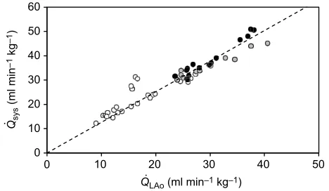
The signal from one of the systemic flow probes was temporarily lost during the measurement periods for some of the N21 animals. In these cases, missing blood flows were estimated using the linear
regression through the origin,Q_sys¼1:2595Q_LAo(see Results,
Fig. 1). AsQ_sys¼Q_RAoþQ_LAo, the equation was rearranged so
thatQ_RAo¼0:2595Q_LAo. Thus, only one of the systemic flow
probes was needed to estimate the flow through the other probe, and
therefore Q_sys and all parameters derived from it (as described
above).Q_RAowas estimated for two of the six N21 turtles at 12 h
after feeding, but only one at 24 h after feeding because the signal
returned in the other flow probe.Q_LAo was estimated in one N21
turtle at 12 and 24 h after feeding.
All studies were carried out according to the approved animal care protocol of the University of North Texas Institutional Animal Care and Use Committee no. 1403-04.
Statistical analysis
Postprandial organ masses were compared between N21 and H10
animals with separate two-tailed unpaired Student’st-tests. Organ
masses were also calculated in terms of percentage animal mass
{organ mass (g)×[animal mass (g)]−1×100}. These proportional
data were then arcsine square transformed before performingt-tests,
which were also used for absolute organ masses, to compare between the two turtle groups (Crossley et al., 2003; Eme et al., 2011; Tate et al., 2016). Cardiovascular responses to feeding in N21 and H10 turtles were assessed using a one-way repeated-measures (RM) ANOVA, with incubation oxygen (i.e. N21 or H10) as an independent variable and postprandial time as the repeated factor, for each parameter (Statistica v13.0, StatSoft, Tulsa, OK). To assess
these effects on and between Q_RAo and Q_LAo, a two-way RM
ANOVA was performed, with incubation oxygen and vessel (RAo or LAo) as independent variables, and postprandial time as the
repeated factor. Each ANOVA was followed by a Fisher’s Least
Significant Difference (LSD)post hoctest, withP<0.05 indicating a
significant difference. Data are presented as means±s.e.m.
RESULTS
Effects of embryonic hypoxia on postprandial organ masses Postprandial organ masses of 5-year-old turtles were unaffected by hypoxic embryonic development, both before and after normalizing to body mass (Table 1).
Systemic cardiac output in N21 turtles
There was a strong positive linear relationship between left aortic
output (Q_LAo) and total systemic output (Q_sys) in fasted and fed
5-year-old N21 turtles, with feeding increasing bothQ_LAoandQ_sys
above fasted values (Q_sys¼1:2595Q_LAo,R2=0.89022; Fig. 1).
_
Qsys=Q_LAovalues were not plotted for three of the six turtles at 12 h
or two of the turtles at 24 h owing to loss of signals from one of the
two systemic flow probes preventing measurement ofQ_LAoorQ_sys
(see Materials and methods).
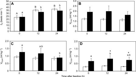
Effects of embryonic hypoxia on fasted and postprandial heart rate and stroke volume
Pre-feedingfHwas 23±3 and 24±4 beats min−1for the N21 and H10
animals, respectively (Fig. 2A). Feeding elicited an ∼1.5-fold
increase infH(P<0.0001) that lasted up to 24 h after the meal in N21
and H10 turtles, but hypoxic incubation had no effect on fH
0 10 20 30 40 50 60
0 10 20 30 40 50
Q
. sys
(ml min
–1
kg
–1
)
[image:3.612.323.557.58.194.2]Q.LAo (ml min–1 kg–1)
Fig. 1. Linear relationship between the measured rate of blood flow to the systemic circuit (Q_sys) and rate of blood flow through the left aorta (Q_LAo) at 0, 12 and 24 h after feeding in 5-year-old control [21% O2(N21)] snapping turtles.0 h (white circles), 12 h (grey circles) and 24 h (black circles) after feeding.N=6. Dotted line fitted through origin with equation
_
Qsys¼1:2595Q_LAo,R2=0.89022. Each circle represents a measurement from
an individual turtle, with data from a total of six turtles contributing to this dataset.
Journal
of
Experimental
(P=0.6947). Prior to feeding, total ventricular stroke volume (VS,tot)
was 1.24±0.09 and 1.93±0.49 ml kg−1for N21 and H10 animals,
respectively (Fig. 2B). Neither hypoxic incubation (P=0.1510) nor
feeding (P=0.0838) had a significant impact onVS,tot, although H10
animals tended to have higher (42–66%) stroke volumes at each
time point compared with N21 animals. Significant differences
did become evident, however, when looking at VS,pul and VS,sys
separately (Fig. 2C,D). The N21 group of turtles maintained similar VS,pul, which was 0.94±0.07 ml kg−1prior to feeding, throughout
the postprandial period (Fig. 2C). However, VS,pul in the H10
group decreased significantly (by 30%; P=0.0096) from
1.26±0.32 to 0.88±0.16 ml kg−1at 24 h post-feeding. Embryonic
hypoxia had no effect on VS,pul (P=0.4056). Both hypoxic
incubation (P=0.0364) and feeding (P=0.0130) increased VS,sys
(Fig. 2D). H10 had slightly (2.3-fold) but not significantly
(P=0.0618) higher VS,sys before feeding compared to N21, and
had significantly higherVS,sys12 h (2.4-fold;P=0.0243) and 24 h
(2.4-fold; P=0.0355) after feeding. However, feeding had no
significant effect on VS,sys in N21 animals. In addition, H10
turtles exhibited a small (23.8%) but significant (P=0.0043)
increase in VS,sys, from a fasting VS,sys of 0.67±0.18 to
0.83±0.21 ml kg−1 at 12 h, whereas N21 turtles maintained fasted
VS,syslevels (0.29±0.04 ml kg−1) throughout the postprandial period.
Effects of embryonic hypoxia on fasted and postprandial _
QRAoandQ_LAo
Both hypoxic incubation (P<0.0001) and feeding (P<0.0001)
affected Q_RAo (Fig. 3A). Fasted H10 animals had significantly
(4.7-fold; P=0.0002) higher Q_RAo than N21 (7.73±0.65 versus
1.66±0.35 ml min−1kg−1), and this difference persisted during the
12 h (P<0.0001) and 24 h (P<0.0001) time points. Notably,
postprandialQ_RAobecame elevated (53–55%;P<0.0002) compared
to fasted values in H10 turtles only, but there was no postprandial response in N21 animals. However, feeding elicited an increase in
_
QLAo in both N21 and H10 turtles (P<0.0001), with peak flow
occurring at 12 h after the meal (2.11-fold and 1.90-fold increase from
fastedQ_LAo for N21 and H10, respectively; Fig. 3B). Interestingly,
_
QRAowas significantly lower thanQ_LAoin N21 animals at 0 h (66%;
P=0.0211), 12 h (78%;P<0.0001) and 24 h (74%;P<0.0001) after
[image:4.612.49.563.70.149.2]feeding, but was similar toQ_LAoin H10 animals.
Table 1. Effects of embryonic hypoxia on postprandial organ masses
Group Heart Liver Lungs Kidneys Stomach Small intestine Colon
Organ mass (g)
N21 5.5±0.6 160.3±23.0 17.8±2.3 9.5±0.7 28.0±4.0 41.7±5.7 21.5±2.7 H10 5.1±0.4 131.7±20.6 15.7±1.7 7.6±0.9 22.0±1.7 31.6±4.4 18.0±2.1
Relative organ mass (%)
N21 0.16±0.01 4.47±0.27 0.50±0.02 0.27±0.02 0.78±0.05 1.18±0.10 0.60±0.05 H10 0.16±0.02 3.96±0.40 0.48±0.032 0.24±0.03 0.68±0.03 0.95±0.03 0.55±0.02
Means±s.e.m. absolute and relative (% animal mass) organ masses of 5-year-old N21 (N=6) and H10 (N=5) snapping turtles 48 h after an∼5% body mass meal.
A
B B
a
b b
0 5 10 15 20 25 30 35 40 45 50
0 12 24
0 0.5 1.0 1.5 2.0 2.5 3.0
0 12 24
a a,b
b
0 0.5 1.0 1.5 2.0
0 12 24
Time after feeding (h) a
*
b
*
a,b0 0.5 1.0 1.5 2.0
0 12 24
C
B
D
A
VS,sys
(ml kg
–1
)
VS,pul
(ml kg
–1
)
VS,tot
(ml kg
–1
)
fH
(beats min
–1
)
Fig. 2. Effects of embryonic hypoxia on fasted and postprandial heart rate and stroke volume.Mean heart rate (fH; A), total cardiac stroke volume (VS,tot; B),
cardiac stroke volume to pulmonary circuit (VS,pul; C) and cardiac stroke volume to systemic circuit (VS,sys; D) before and after feeding in 5-year-old control
snapping turtles [21% O2(N21); white bars;N=6] and turtles exposed to hypoxia as embryos [10% O2(H10); black bars;N=5]. Following significant one-way
repeated-measures (RM) ANOVA, different uppercase letters (within N21) or lowercase letters (within H10) indicate significant differences (P<0.05) between feeding time points within an embryonic O2treatment based on Fisher’s Least Significant Difference (LSD)post hoctest. * indicate a significant difference
(P<0.05) between N21 and H10 animals at that feeding time point based on Fisher’s LSDpost hoctest. Data are presented as means±s.e.m.
Journal
of
Experimental
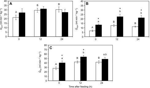
[image:4.612.83.531.417.672.2]Effects of embryonic hypoxia on fasted and postprandial cardiac output
Feeding increasedQ_pul (P=0.0448) in 5-year-old turtles (Fig. 4A).
Specifically, N21 animals exhibited a significant (41%;P=0.0119)
increase from the fasted Q_pul of 21.57±2.63 to 30.47
±2.97 ml min−1kg−1at 24 h after feeding. However, although H10
animals were similar at all time points to the N21 turtles (P=0.7221),
post hocanalysis revealed no significant impact of feeding onQ_pul
in the H10 group. Meanwhile, Q_sys increased following feeding
(P<0.0001), and differed between the experimental groups (P=0.0018;
Fig. 4B). FastedQ_syswas 13.27±0.97 ml min−1kg−1in H10 turtles,
which was ∼2-fold (P=0.0109) that of the N21 turtles (6.54
±0.90 ml min−1 kg−1). Feeding caused an increase in Q_sys, which
peaked at 12 h after the meal in both groups of animals (1.9-fold and
1.7-fold increase from fastingQ_sysfor N21 and H10, respectively), but
remained above fasted levels at 24 h post-feeding (1.7-fold and
1.6-fold increase from fastingQ_sysfor N21 and H10, respectively). Fasted
_
Qtot was also higher in H10 (43%;P=0.0211) compared with N21
*
a*
b
*
b0 5 10 15 20
0 12 24
‡ A
‡
B ‡ B
a
b
b
0 5 10 15 20
0 12 24 Time after feeding (h)
A
B
Q
. LAo
(ml min
–1
kg
–1
)
Q
. RAo
(ml min
–1
kg
–1
[image:5.612.84.536.57.186.2])
Fig. 3. Effects of embryonic hypoxia on fasted and postprandial mean systemic blood flows.Mean rate of blood flow through the branches of the brachiocephalic artery (Q_RAo; A) and through the left aorta (Q_LAo; B) before and after feeding in 5-year-old control snapping turtles [21% O2(N21); white bars;N=6]
and turtles exposed to hypoxia as embryos [10% O2(H10); black bars;N=5]. Following significant two-way repeated-measures (RM) ANOVA, different
uppercase letters (within N21) or lowercase letters (within H10) indicate significant differences (P<0.05) between feeding time points within an embryonic O2
treatment based on Fisher’s Least Significant Difference (LSD)post hoctest. * indicate a significant difference (P<0.05) between N21 and H10 animals at that feeding time point, and‡indicate a significant difference betweenQ_RAoandQ_LAovalues within the same experimental group at each time point, based on Fisher’s
LSDpost hoctest. Data are presented as means±s.e.m.
A
B
B
*
a
*
b
*
b0 5 10 15 20 25 30 35 40
0 12 24
A
B B
0 5 10 15 20 25 30 35 40
0 12 24
A
B B
*
a*
b
a,b
0 10 20 30 40 50 60 70 80
0 12 24
Time after feeding (h)
A
C
B
Q
. tot
(ml min
–1
kg
–1
)
Q
. sys
(ml min
–1
kg
–1
)
Q
. pul
(ml min
–1
kg
–1
[image:5.612.67.556.386.674.2])
Fig. 4. Effects of embryonic hypoxia on fasted and postprandial cardiac output.Mean rate of blood flow to the pulmonary circuit (Q_pul; A), to the systemic
circuit (Q_sys; B) and in total (Q_tot; C) before and after feeding in 5-year-old control snapping turtles [21% O2(N21); white bars;N=6] and turtles exposed to hypoxia
as embryos [10% O2(H10); black bars;N=5]. Following significant one-way repeated-measures (RM) ANOVA, different uppercase letters (within N21) or
lowercase letters (within H10) indicate significant differences (P<0.05) between feeding time points within an embryonic O2treatment based on Fisher’s
Least Significant Difference (LSD)post hoctest. * indicate a significant difference (P<0.05) between N21 and H10 animals at that feeding time point based on Fisher’s LSDpost hoctest. Data are presented as means±s.e.m.
Journal
of
Experimental
turtles (40.15±4.48 versus 28.11±3.01 ml min−1kg−1, respectively), a
difference that remained 12 h after feeding (P=0.0262; Fig. 4C).
Effects of embryonic hypoxia on fasted and postprandial cardiac shunting
There was no main effect of either feeding (P=0.7596) nor hypoxic
incubation (P=0.0967) onQ_shunt, which was positive throughout the
measurements, representing a net flow of blood to the pulmonary circulation, even during fasting conditions (Fig. 5A). By contrast,
compared with N21 turtles,Q_pul=Q_sys was significantly lower in
H10 turtles at 0 h (42% lower;P=0.0138) and 24 h (49% lower;
P=0.0268; Fig. 5B). Feeding caused a decrease inQ_pul=Q_sys (by
31%; P=0.0185) in N21 turtles at 12 h, with a trend toward a
reduction at 24 h (24% lower than at 0 h;P=0.0595).
DISCUSSION
Hypoxia has long been recognized as a physiological stressor that activates homeostatic mechanisms, acutely acting on cell signalling pathways as well as altering organ system function, modulating organismal phenotype. If experienced during ontogeny, reductions in oxygen availability can interact with developmental plasticity, eliciting effects that can persist into adult life. Our prior investigation of common snapping turtles demonstrated that embryonic hypoxia impacts metabolism and heart rate in juvenile animals 3 years after hatching (Wearing et al., 2016). In addition, despite likely differences in cardiac output to supply different oxygen demands, we found that arterial pressure in both fasted and fed juvenile turtles was unaffected by embryonic hypoxia. Although the findings of this study implied that convective transport capacity is changed by embryonic hypoxia, direct measurement of blood flows through the major cardiac outflow vessels are needed to fully investigate and confirm this possibility. Using feeding as a tool to
increase oxygen demand, we determined that Q_tot increased
significantly after feeding in N21 turtles, primarily owing to a postprandial tachycardia that was accompanied by a decrease in a
net left–right (L–R) shunt. Hypoxic incubation resulted in a
reduction in the L–R shunt in fasted H10 turtles relative to N21
animals, with maintenance of the magnitude of this shunt during the
postprandial period. The lower and constant net L–R shunt in H10
animals was the result of higherQ_sysbut similarQ_pulrelative to N21
turtles. Our findings indicate that hypoxic embryonic incubation produces an adult cardiovascular phenotype that is characterized by elevated blood delivery to the systemic circulatory circuit, which
may facilitate the greater postprandial metabolic response we previously reported in H10 snapping turtles (Wearing et al., 2016).
Fasted measurements
Heart rate (fH) of fasted 5-year-old N21 snapping turtles
(23±3 beats min−1; Fig. 2A) was similar to values previously
reported (Frische et al., 2000) but lower than when these turtles were
studied at the same temperature (30°C) at 3 years old
(∼33 beats min−1) in our previous study (Wearing et al., 2016).
Although not the primary focus of this investigation, the difference
in fasted fH of N21 turtles between the two studies may be a
consequence of body mass differences (1.32±0.20 kg at 3 years old versus 3.53±0.37 kg at 5 years old). However, given that both investigations were conducted on the same cohort of snapping turtles at two distinct ages, an age effect on heart rate cannot be ruled out.
UnlikefH, blood flow parameters were previously unstudied in
snapping turtles. In 5-year-old snapping turtles, fasted and
postprandial Q_pul values (∼21–31 ml min−1 kg−1; Fig. 4A) were
within the range for other turtle species at similar temperatures
(∼15–56 ml kg−1min−1; West et al., 1992; Wang and Hicks, 1996a;
Hicks and Wang, 1998; Galli et al., 2004). However,Q_sysof fasted
N21 snapping turtles (6.54±0.90 ml kg−1 min−1; Fig. 4B) was
considerably lower than values previously reported for other species
(∼17–73 ml kg−1min−1; Wang and Hicks, 1996a; Hicks and Wang,
1998; Galli et al., 2004). This was partially the result of a lowQ_RAo
(Fig. 3A), which also resulted in a reduced Q_tot and a net L–R
cardiac shunt (i.e.Q_shunt .0, andQ_pul=Q_sys.1) in N21 snapping
turtles (Fig. 5A,B). Over half a century ago, anaesthetized snapping
turtles (8–14 kg) were shown to demonstrate a large net L–R shunt,
through the use of a dye-dilution technique (Steggerda and Essex, 1957). The presence of this pulmonary shunt has been more
extensively described in red-eared slider turtles,Trachemys scripta
(White and Ross, 1966; Comeau and Hicks, 1994; Hicks et al., 1996; Crossley et al., 1998; Overgaard et al., 2002b; Krosniunas and Hicks, 2003; Galli et al., 2004), and is attributed to low vagal tone and/or high adrenergic stimulation (Hicks, 1994; Hicks and Comeau, 1994; Hicks and Wang, 1998; Wang et al., 2001b;
Overgaard et al., 2002b). Shunt ratio (Q_pul=Q_sys) is dependent on the
relationship of pulmonary to systemic vascular resistance in turtles
(Crossley et al., 1998). The high Q_pul=Q_sys of 3.58±0.62 in N21
snapping turtles (Fig. 5B) suggests that pulmonary resistance is
∼30% of systemic resistance in these animals. Although a detailed
0 5 10 15 20 25
0 12 24
Q
. shunt
(ml min
–1
kg
–1
)
A
B A,B
*
*
0 1 2 3 4 5 6
0 12 24
Q
. pul
/
Q
. sys
Time after feeding (h)
[image:6.612.90.538.546.683.2]A
B
Fig. 5. Effects of embryonic hypoxia on fasted and postprandial cardiac shunting.Mean net shunt (A) and shunt fraction (B) before and after feeding in 5-year-old control snapping turtles [21% O2(N21); white bars;N=6] and turtles exposed to hypoxia as embryos [10% O2(H10); black bars;N=5]. Following
significant one-way repeated-measures (RM) ANOVA, different uppercase letters indicate significant differences (P<0.05) between feeding time points in N21 turtles based on Fisher’s Least Significant Difference (LSD)post hoctest. * indicate a significant difference (P<0.05) between N21 and H10 animals at that feeding time point based on Fisher’s LSDpost hoctest. Data are presented as means±s.e.m.
Journal
of
Experimental
study of cardiovascular control is yet to be conducted in adult snapping turtles, our findings suggest that fasted snapping turtles maintain relatively low vagal tone and/or higher vascular adrenergic
tone to sustain the dominant L–R shunt.
Prior studies have also reported a net systemic blood flow pattern
[i.e. right–left (R–L) shunt] inT. scripta(Wang and Hicks, 1996a;
Hicks and Wang, 1998; Crossley et al., 2000; Krosniunas and Hicks, 2003; Galli et al., 2004; Keen et al., 2016). However, we are among the first to assess intracardiac shunting in the snapping turtle (Steggerda and Essex, 1957), and it is unclear whether the direction of shunting is similarly variable in this species. Krosniunas and
Hicks (2003) reported that a L–R shunt inT. scriptais dependent on
the activity level of the animal and ambient temperature. In our study, snapping turtles were maintained at 30°C and given the ability to submerge. It is possible that this may have contributed to
the resting L–R shunt, resulting in a net recirculation of oxygenated
blood to the lungs, with the potential cost of necessitating increased _
Q to maintain sufficient oxygen delivery to systemic tissues
(Krosniunas and Hicks, 2003). However, as previously discussed, the turtle lung is not a perfect gas exchanger, and recirculation of unsaturated blood immediately back to the lungs may increase
pulmonary venous partial pressure of oxygen (i.e. PO2), instead
resulting in increased systemic oxygen transport (Steggerda and Essex, 1957; Wang and Hicks, 1996b). In the case of the snapping turtle, determinations of the consequences of a resting net pulmonary shunt require further studies that simultaneously measure blood flow patterns and systemic blood oxygen content.
The long-term cardiovascular impact of developmental hypoxia
was evident in the marked differences inQ_sysandQ_totbetween H10
and N21 turtles. These were both significantly higher in H10 snapping turtles compared to the N21 animals during fasted and postprandial periods (Fig. 4B,C). Specifically, these increased
blood flows in H10 animals were the result of increased Q_RAo,
whereas Q_LAo was similar between the two groups (Fig. 3A,B).
Interestingly, N21 animals appeared to selectively perfuse the LAo branch of the systemic circulation, whereas H10 animals maintained similar flows through both systemic branches. Along with this, the
similar Q_pul in both animal groups also resulted in H10 turtles
maintaining a lower shunt fraction than the N21 animals (Fig. 5B).
To date, studies ofQ_ in surgically recovered, conscious turtles have
reported total systemic flow only (Wang and Hicks, 1996a; Hicks and Wang, 1998; Krosniunas and Hicks, 2003; Galli et al., 2004; Stecyk et al., 2004), so it is unclear whether this phenomenon is common to turtles, or whether it is unique to snapping turtles.
However,Q_RAohas been reported to be roughly double that ofQ_LAo
in anaesthetized T. scripta (Comeau and Hicks, 1994). If this
persists in recovered animals, snapping turtles may exhibit a different pattern of systemic blood flow distribution than the more extensively studied red-eared slider turtle. Anatomically, the RAo and LAo give rise to differing arterial trees. Notably, the LAo supplies blood to the stomach and viscera before it fuses with the descending aorta, whereas the RAo supplies blood to the anterior portion of the animal before also merging with the descending aorta (Bojanus, 1819). Based on the anatomy, differences in regulatory vasomotor tone may be responsible for differences in vascular resistances, and therefore blood flow, through the RAo and LAo of
N21 snapping turtles (Fig. 3A,B). By extension, the elevatedQ_RAo
of H10 turtles (Fig. 3A) may be attributed to reduced vascular resistance, due to phenotypic differences in vasomotor regulation and/or vessel morphology downstream of the RAo. A study of the aortic phenotypic response to chronic hypoxic exposure in hatchling
American alligators (Alligator mississippiensis) reported changes in
outflow vessel cross-sectional area (Owerkowicz et al., 2011). If hypoxic embryonic development also modulates outflow vessel morphology and physical properties, e.g. distensibility, in a way that
persists into adulthood, this may explain the disparity in Q_RAo
between 5-year-old N21 and H10 turtles. Importantly, embryonic angiogenesis and reductions in resistance in the RAo branch would aid in providing increased blood delivery and maintaining oxygen supply to the developing hypoxia-sensitive brain. To address this, effects of hypoxic development on vascular morphology and regulation of blood flow distribution need to be investigated in turtles of various ages.
Postprandial measurements
Changing heart rate is a prominent mechanism for adjusting cardiac output to meet increasing oxygen demands in terrestrial vertebrates (Burggren et al., 2011; Crossley et al., 2017a). The increase in oxygen demand associated with feeding in 5-year-old snapping
turtles was met by increasing fH ∼50% above fasted values
(Fig. 2A). Our previous study of juvenile snapping turtles found an
∼28% increase infHassociated with feeding (Wearing et al., 2016).
However, fastedfHwas 30% higher in 3-year-old turtles compared
with those at 5 years of age. Whereas fHincreased to meet the
aerobic demands of digestion,VS,totwas unaffected by feeding in
N21 snapping turtles (Fig. 2B–D). H10 snapping turtles exhibited
a similar postprandial increase in fH, whereas VS,tot remained
unchanged (Fig. 2A,B). However, whereas N21 animals maintained
constantVS,sysandVS,pul, H10 turtles decreasedVS,pulbut increased
VS,sys(Fig. 2C,D). The capacity to differentially adjustVS,sysand VS,pulhas been previously reported in turtles, with exercise inducing
a greater increase in VS,pul than in VS,sys (West et al., 1992;
Krosniunas and Hicks, 2003). The opposite was the case for H10 snapping turtles during the postprandial period (Fig. 2C,D), suggesting that relative stroke volumes of the heart depend on the aerobic requirements of the specific activity. This was also reflected
in the increase inQ_sysin H10 turtles, with no change inQ_pul(Fig. 4A,
B). Although the experimental groups differed in theirVSadjustments
in response to feeding,Q_totincreased in both groups, but N21 groups
maintained relatively low values (Fig. 4C). Owing to a resting net
L–R shunt, there may be less drive to further increase pulmonary
perfusion in snapping turtles, but more benefit in increasing blood delivery to the digestive tissues, particularly in H10 turtles.
The phenotypic consequences of developmental hypoxia were again evident in the postprandial flow profiles of the RAo in N21 and
the H10 turtles (Fig. 3A). Both groups increased Q_sys following
feeding (Fig. 4B). However, N21 turtles increased only Q_LAo
following feeding, whereas H10 animals increased both Q_LAo and
_
QRAo(Fig. 3A,B). Furthermore, the N21 group increasedQ_pul, albeit
to a lesser degree than theQ_sys, whereas H10 animals maintained
fasted Q_pul (Fig. 4A,B). This suggests that H10 animals have an
increased capacity to augment systemic flow, via the RAo, supporting the elevated postprandial metabolism previously reported for this phenotype (Wearing et al., 2016). Systemic vasodilation (i.e. decreased systemic resistance) is associated with digestion, resulting in shunting of blood to the systemic circulation, which may account for the postprandial systemic flow profiles of both experimental groups of turtles (Axelsson et al., 1991; Secor et al., 2000). In this case, a net shunt to the pulmonary circulation (i.e. a
L–R shunt) persists, albeit a smaller one. Maintaining a net L–R shunt
after feeding may be important for increasing blood oxygen saturation in snapping turtles if gas diffusion across the lung is limited (Steggerda and Essex, 1957; Wang and Hicks, 1996b). Importantly,
the persistence of the L–R shunt in both groups of snapping turtles
Journal
of
Experimental
differs from the response reported in alligators (Busk et al., 2000) and pythons (Overgaard et al., 1999; Secor et al., 2000), which both
exhibit a postprandial reduction of the fasting net R–L shunt (Wang
et al., 2001a,b). This would suggest that snapping turtles are more reliant on maximizing lung perfusion, therefore ensuring sufficient blood oxygen saturation before and after a meal.
Conclusions
Developmental hypoxia is established to affect both functional and structural maturation of the snapping turtle cardiovascular system (Eme et al., 2013, 2014; Tate et al., 2015). This is the second study to demonstrate that the effects of developmental phenotypic plasticity persist through programming cardiovascular physiology of common snapping turtles several years after hatching (Wearing et al., 2016). Both data sets support the hypothesis that developmental oxygen levels interact with the innate plasticity of developmental processes to alter the resulting phenotype of the juvenile and adult animal. Given that reductions in oxygen availability in the natural developmental environment have been noted for embryonic reptiles (Ackerman, 1977; Seymour and Ackerman, 1980; Booth, 1998, 2000; Grigg et al., 2010), this challenge may be a common determination of cardiovascular function of post-hatching animals. Our findings in the snapping turtle indicate that hypoxic embryonic incubation results in juvenile
and adult animals with increasedQ_ that persists during periods of
elevated oxygen demand, such as after feeding. If this capacity also persists during aerobic exercise, developmental hypoxia will further impact juvenile reptile and other vertebrate performance in natural settings. This highlights the importance of extended longitudinal studies of developmental hypoxia to comprehensively understand the effects of this embryonic challenge.
Acknowledgements
The authors thank the State of Minnesota Department of Natural Resources for special permit no. 18337 to collect turtle eggs.
Competing interests
The authors declare no competing or financial interests.
Author contributions
Conceptualization: O.H.W., D.A.C.; Methodology: O.H.W., D.A.C.; Formal analysis: O.H.W., D.A.C.; Investigation: O.H.W., J. Conner, D.N., J. Crossley, D.A.C.; Resources: D.A.C.; Writing - original draft: O.H.W.; Writing - review & editing: O.H.W., D.A.C.; Visualization: O.H.W.; Supervision: D.A.C.; Project administration: D.A.C.; Funding acquisition: D.A.C.
Funding
This work was supported by the National Science Foundation (NSF) Career Award IBN IOS-0845741 to D.A.C.
References
Ackerman, R. A.(1977). Respiratory gas exchange of sea turtle nests (Chelonia,
Caretta).Respir. Physiol.31, 19-38.
Andersen, J. B., Rourke, B. C., Caiozzo, V. J., Bennett, A. F. and Hicks, J. W. (2005). Postprandial cardiac hypertrophy in pythons.Nature434, 37-38.
Axelsson, M., Fritsche, R., Holmgren, S., Grove, D. J. and Nilsson, S.(1991).
Gut blood flow in the estuarine crocodile,Crocodylus porosus.Acta Physiol. Scand.142, 509-516.
Bojanus, L. H.(1819). Anatome Testudinis Europaeae, Vol. I. p. 74. Vilnae,
Lithuania: Impensis Auctoris, Typis Josephi Zawadzki, Typographi Universitatis.
Booth, D. T. (1998). Nest temperature and respiratory gases during natural
incubation in the broad-shelled river turtle, Chelodina expansa(Testudinata: Chelidae).Aust. J. Zool.46, 183-191.
Booth, D. T.(2000). The effect of hypoxia on oxygen consumption of embryonic
estuarine crocodiles (Crocodylus porosus).J. Herpetol.34, 478-481.
Burggren, W., Farrell, A. and Lillywhite, H.(2011). Vertebrate cardiovascular
systems. In Compr Physiol Suppl 30: Handbook of Physiology, Comparative Physiology(ed. W. H. Dantzler), pp. 215-308. New York: Oxford Press.
Busk, M., Overgaard, J., Hicks, J. W., Bennett, A. F. and Wang, T.(2000). Effects
of feeding on arterial blood gases in the American alligator Alligator mississippiensis.J. Exp. Biol.203, 3117-3124.
Comeau, S. G. and Hicks, J. W. (1994). Regulation of central vascular
blood flow in the turtle. Am. J. Physiol. Regul. Integr. Comp. Physiol. 36, R569-R578.
Crossley, D. A., II and Altimiras, J. (2005). Cardiovascular development in
embryos of the American alligatorAlligator mississippiensis: effects of chronic and acute hypoxia.J. Exp. Biol.208, 31-39.
Crossley, D. A., II, Altimiras, J. and Wang, T.(1998). Hypoxia elicits an increase in
pulmonary vascular resistance in anaesthetized turtles (Trachemys scripta).
J. Exp. Biol.201, 3367-3375.
Crossley, D. A., II, Wang, T. and Altimiras, J.(2000). Role of nitric oxide in the
systemic and pulmonary circulation of anesthetized turtles (Trachemys scripta).
J. Exp. Zool.286, 683-689.
Crossley, D. A., II, Hicks, J. W. and Altimiras, J.(2003). Ontogeny of baroreflex
control in the American alligatorAlligator mississippiensis.J. Exp. Biol.206, 2895-2902.
Crossley, D. A., II, Burggren, W. W., Reiber, C. L., Altimiras, J. and Rodnick,
K. J. (2017a). Mass transport: circulatory system with emphasis on
nonendothermic species.Compr. Physiol.7, 17-66.
Crossley, D. A., II, Ling, R., Nelson, D., Gillium, T., Conner, J., Hapgood, J.,
Elsey, R. M. and Eme, J.(2017b). Metabolic responses to chronic hypoxic
incubation in embryonic American alligators (Alligator mississippiensis).
Comp. Biochem. Physiol. A Mol. Integr. Physiol.203, 77-82.
Eme, J., Hicks, J. W. and Crossley, D. A.II. (2011). Chronic hypoxic incubation blunts a cardiovascular reflex loop in embryonic American alligator (Alligator mississippiensis).J. Comp. Physiol. B181, 981-990.
Eme, J., Rhen, T., Tate, K. B., Gruchalla, K., Kohl, Z. F., Slay, C. E. and Crossley, D. A.II. (2013). Plasticity of cardiovascular function in snapping turtle embryos (Chelydra serpentina): chronic hypoxia alters autonomic regulation and gene expression.Am. J. Physiol. Regul. Integr. Comp. Physiol.304, R966-R979. Eme, J., Rhen, T. and Crossley, D. A.II. (2014). Adjustments in cholinergic,
adrenergic and purinergic control of cardiovascular function in snapping turtle embryos (Chelydra serpentina) incubated in chronic hypoxia.J. Comp. Physiol. B
184, 891-902.
Enok, S., Simonsen, L. S., Pedersen, S. V., Wang, T. and Skovgaard, N.(2012).
Humoral regulation of heart rate during digestion in pythons (Python molurusand
Python regius).Am. J. Physiol. Regul. Integr. Comp. Physiol.302, R1176-R1183.
Enok, S., Simonsen, L. S. and Wang, T.(2013). The contribution of gastric
digestion and ingestion of amino acids on the postprandial rise in oxygen consumption, heart rate and growth of visceral organs in pythons. Comp. Biochem. Physiol. A Mol. Integr. Physiol.165, 46-53.
Enok, S., Leite, G. S. P. C., Leite, C. A. C., Gesser, H., Hedrick, M. S. and Wang, T.(2016). Improved cardiac filling facilitates the postprandial elevation of stroke volume inPython regius.J. Exp. Biol.219, 3009-3018.
Frische, S., Fago, A. and Altimiras, J.(2000). Respiratory responses to short term
hypoxia in the snapping turtle,Chelydra serpentina.Comp. Biochem. Physiol. A
126, 223-231.
Galli, G., Taylor, E. W. and Wang, T.(2004). The cardiovascular responses of the
freshwater turtleTrachemys scriptato warming and cooling.J. Exp. Biol.207, 1471-1478.
Galli, G. L. J., Crossley, J., Elsey, R. M., Dzialowski, E. M., Shiels, H. A. and Crossley, D. A.II. (2016). Developmental plasticity of mitochondrial function in American alligators,Alligator mississippiensis.Am. J. Physiol. Regul. Integr. Comp. Physiol.311, R1164-R1172.
Grigg, G. C., Thompson, M. B., Beard, L. A. and Harlow, P.(2010). Oxygen levels
in mound nests ofCrocodylus porosusandAlligator mississippiensisare high, and gas exchange occurs primarily by diffusion, not convection.Aust. Zool.35, 235-244.
Hicks, J. W.(1994). Adrenergic and cholinergic regulation of intracardiac shunting.
Physiol. Zool.67, 1325-1346.
Hicks, J. W. and Bennett, A. F.(2004). Eat and run: prioritization of oxygen delivery
during elevated metabolic states.Respir. Physiol. Neurobiol.144, 215-224.
Hicks, J. W. and Comeau, S. G.(1994). Vagal regulation of intracardiac shunting in
the turtlePseudemys scripta.J. Exp. Biol.186, 109-126.
Hicks, J. W. and Wang, T.(1998). Cardiovascular regulation during anoxia in the
turtle: anin vivostudy.Physiol. Zool.71, 1-14.
Hicks, J. W., Ishimatsu, A., Molloi, S., Erskin, A. and Heisler, N.(1996). The
mechanism of cardiac shunting in reptiles: a new synthesis.J. Exp. Biol.199, 1435-1446.
Hicks, J. W., Wang, T. and Bennett, A. F.(2000). Patterns of cardiovascular and
ventilator response to elevated metabolic states in the lizard Varanus exanthematicus.J. Exp. Biol.203, 2437-2445.
Kam, Y.-C.(1993). Physiological effects of hypoxia on metabolism and growth of
turtle embryos.Respir. Physiol.92, 127-138.
Keen, A. N., Shiels, H. A. and Crossley, D. A.II. (2016). Cardiovascular function, compliance, and connective tissue remodeling in the turtle,Trachemys scripta, following thermal acclimation.Am. J. Physiol. Regul. Integr. Comp. Physiol.311,
R133-R143.
Journal
of
Experimental
Krosniunas, E. H. and Hicks, J. W.(2003). Cardiac output and shunt during voluntary activity at different temperatures in the turtle, Trachemys scripta.
Physiol. Biochem. Zool.76, 679-694.
Marks, C., Eme, J., Elsey, R. M. and Crossley, D. A.II. (2013). Chronic hypoxic incubation blunts thermally dependent cholinergic tone on the cardiovascular system in embryonic American alligator (Alligator mississippiensis).J. Comp. Physiol. B183, 947-957.
Overgaard, J., Busk, M., Hicks, J. W., Jensen, F. B. and Wang, T.(1999).
Respiratory consequences of feeding in the snake Python molorus. Comp. Biochem. Physiol. A Mol. Integr. Physiol.124, 359-365.
Overgaard, J., Andersen, J. B. and Wang, T.(2002a). The effects of fasting
duration on the metabolic response to feeding inPython molurus: an evaluation of the energetic costs associated with gastrointestinal growth and upregulation.
Physiol. Biochem. Zool.75, 360-368.
Overgaard, J., Stecyk, J. A. W., Farrell, A. P. and Wang, T.(2002b). Adrenergic
control of the cardiovascular system in the turtleTrachemys scripta.J. Exp. Biol.
205, 3335-3345.
Owerkowicz, T., Elsey, R. M. and Hicks, J. W.(2009). Atmospheric oxygen
level affects growth trajectory, cardiopulmonary allometry and metabolic rate in the American alligator (Alligator mississippiensis). J. Exp. Biol. 212, 1237-1247.
Owerkowicz, T., Spikings, T. J., Elsey, R. M. and Hicks, J. W.(2011). Atmospheric
oxygen remodels cardiac outflow tract in the American alligator.FASEB. J.1 Suppl., 1045.10.
Secor, S. M. and Diamond, J.(1999). Maintenance of digestive performance in the
turtles Chelydra serpentina, Sternotherus odoratus, and Trachemys scripta.
Copeia1999, 75-84.
Secor, S. M. and White, S. E. (2010). Prioritizing blood flow: cardiovascular
performance in response to the competing demands of locomotion and digestion for the Burmese python,Python molurus.J. Exp. Biol.213, 78-88.
Secor, S. M., Hicks, J. W. and Bennett, A. F.(2000). Ventilatory and cardiovascular
responses of a python (Python molurus) to exercise and digestion.J. Exp. Biol.
203, 2447-2454.
Seymour, R. S. and Ackerman, R. A.(1980). Adaptations to underground nesting
in birds and reptiles.Am. Zool.20, 437-447.
Stecyk, J. A. W., Overgaard, J., Farrell, A. P. and Wang, T.(2004).α-Adrenergic
regulation of systemic peripheral resistance and blood flow distribution in the turtle
Trachemys scriptaduring anoxic submergence at 5°C and 21°C.J. Exp. Biol.207, 269-283.
Steggerda, F. R. and Essex, H. E.(1957). Circulation and blood pressure in the
great vessels and heart of the turtle (Chelydra serpentina).Am. J. Physiol.190, 320-326.
Tate, K. B., Kohl, Z. F., Eme, J., Rhen, T. and Crossley, D. A.II. (2015). Critical windows of cardiovascular susceptibility to developmental hypoxia in common snapping turtle (Chelydra serpentina) embryos. Physiol. Biochem. Zool. 88, 103-115.
Tate, K. B., Rhen, T., Eme, J., Kohl, Z. F., Crossley, J., Elsey, R. M. and Crossley, D. A.II. (2016). Periods of cardiovascular susceptibility to hypoxia in embryonic American alligators (Alligator mississippiensis).Am. J. Physiol. Regul. Integr. Comp. Physiol.310, R1267-R1278.
Wang, T. and Hicks, J. W.(1996a). Cardiorespiratory synchrony in turtles.J. Exp.
Biol.199, 1791-1800.
Wang, T. and Hicks, J. W.(1996b). The interaction of pulmonary ventilation and the
right-left shunt on arterial oxygen levels.J. Exp. Biol.199, 2121-2129.
Wang, T., Busk, M. and Overgaard, J.(2001a). The respiratory consequences of
feeding in amphibians and reptiles.Comp. Biochem. Physiol. A Mol. Integr. Physiol.128, 535-549.
Wang, T., Warburton, S., Abe, A. and Taylor, T.(2001b). Vagal control of heart rate
and cardiac shunts in reptiles: relation to metabolic state.Exp. Physiol.86, 777-784.
Wearing, O. H., Eme, J., Rhen, T. and Crossley, D. A.II. (2016). Phenotypic plasticity in the common snapping turtle (Chelydra serpentina): long-term physiological effects of chronic hypoxia during embryonic development.
Am. J. Regul. Integr. Comp. Physiol.310, R176-R184.
West, N. H., Butler, P. J. and Bevan, R. M.(1992). Pulmonary blood flow at rest and
during swimming in the green turtle,Chelonia mydas.Physiol. Zool.65, 287-310.
White, F. N. and Ross, G.(1966). Circulatory changes during experimental diving in
the turtle.Am. J. Physiol.211, 15-18.
Yntema, C. L.(1968). A series of stages in the embryonic development ofChelydra
serpentina.J. Morphol.125, 219-256.
Journal
of
Experimental