STRUCTURAL AND FUNCTIONAL STUDIES OF
PORINS FROM PATHOGENIC BACTERIA
Luana G. M. Ferrara
A Thesis Submitted for the Degree of PhD
at the
University of St Andrews
2018
Full metadata for this item is available in
St Andrews Research Repository
at:
http://research-repository.st-andrews.ac.uk/
Please use this identifier to cite or link to this item:
http://hdl.handle.net/10023/15639
Structural and functional studies of porins
from pathogenic bacteria
Luana G.M. Ferrara
This thesis is submitted in fulfillment for the degree of
Doctor of Philosophy
October 2017
I. Declarations
I, Luana G.M. Ferrara, hereby certify that this thesis, which is approximately 24,000 words in length, has been written by me, and that it is the record of work carried out by me, or principally by myself in collaboration with others as acknowledged, and that it has not been submitted in any previous application for a higher degree.
I was admitted as a research student in October 2013 and as a candidate for the degree of Doctor of Philosophy in October 2017; the higher study for which this is a record was carried out in the University of St Andrews between 2013 and 2017.
Date……….. Signature of candidate………...
I hereby certify that the candidate has fulfilled the conditions of the Resolution and Regulations appropriate for the degree of Doctor of Philosophy in the University of St Andrews and that the candidate is qualified to submit this thesis in application for that degree.
Date……… Signature of supervisor
05/03/2018
In submitting this thesis to the University of St Andrews I understand that I am giving permission for it to be made available for use in accordance with the regulations of the University Library for the time being in force, subject to any copyright vested in the work not being affected thereby. I also understand that the title and the abstract will be published, and that a copy of the work may be made and supplied to any bona fide library or research worker, that my thesis will be electronically accessible for personal or research use unless exempt by award of an embargo as requested below, and that the library has the right to migrate my thesis into new electronic forms as required to ensure continued access to the thesis. I have obtained any third-party copyright permissions that may be required in order to allow such access and migration, or have requested the appropriate embargo below.
The following is an agreed request by candidate and supervisor regarding the publication of this thesis:
Access to printed copy and electronic publication of the thesis through the University of St Andrews.
II. Abstract
Multi-drug resistant bacteria have become a real threat to public health worldwide. Gram-negative bacteria, in particular, have shown high level of antibiotic resistance due to the presence of an additional membrane, known as outer-membrane (OM), that acts as an extra barrier. Most antibiotics enter the cells via a particular class of outer-membrane proteins (OMPs) known as porins. Porins are β-barrel channels that allow the passive diffusion of hydrophobic compounds. The porins are known to select against molecules on the basis of size and charge. When exposed to antibiotics, bacteria can modify the OM permeability by altering their porins profile. Mutations affecting the size and conductivity of the pore channel, and modification of the level of porins expression are just few examples of how the bacteria can decrease the influx of antibiotics.
III. Acknowledgments
Firstly, I would like to express my sincere gratitude to my supervisor Prof. James H. Naismith for giving me the opportunity to join his team for the great journey that was my PhD, for his guidance and for his continuous support through all my research. A special thanks goes to Dr. Huanting Liu for his help and advice on gene cloning and gene mutation, but also for his wisdom and kind friendship. I would also like to thank Dr. Lucile Moynié for helping me with crystals fishing and data collection. I might not have been the perfect PhD student, but I am pretty sure I was her favorite. A big thanks goes also to Mr. Antoni Tortajada for his help and lovely chats. And surely, I can’t not express my gratitude to all Prof Naismith’s lab team for the useful and motivating discussions and for the inevitable (and very much appreciated) coffee breaks.
My PhD wouldn’t have been the same without my girls, Laura W, Laura G, Emilia, Audrey and Miriam. Our Friday nights pub, shopping afternoons and home-made dinners made these past four years much easier, and for this I would like to thank them.
A very special thanks goes to Joe, for his endless support and care (especially in the last few months). I have the feeling that writing up my dissertation was as painful for me as it was for him.
IV.Table of Contents
I. Declarations……….…...………..i
II. Abstract ... iii
III. Acknowledgment ... v
IV.Table of Contents ... vii
V. List of figure and tables ... xii
VI. Abbreviations ... xv
Chapter 1: Structure and function of outer-membrane proteins ... 2
1.1 Introduction ... 3
1.1.1 Gram-negative outer-membrane: an overview ... 3
1.1.2 Outer-membrane proteins ... 6
1.1.3 Passive transport through the OM: Porins ... 6
1.1.4 Active transport through OM: TonB-dependent transporters ... 13
1.1.5 OMPs with enzymatic activity ... 16
1.1.6 Biosynthesis of OMPs ... 18
1.1.7 Antibiotic resistance: role of the OM and OMPs ... 23
Chapter 2: Major outer-membrane protein (MOMP) from Campylobacter jejuni ... 28
2.1 Introduction ... 29
2.1.1 Campylobacter: an overview ... 29
2.1.2 Campylobacter jejuni infection ... 30
2.1.5 Aims ... 36
2.2 Material and Methods ... 38
2.2.1Campylobacter jejuni culture ... 38
2.2.2 MOMP 85H purification ... 39
2.2.3 MOMP 85H crystallization ... 40
2.2.4 Data collection, Structure determination and refinement ... 41
2.2.5 Single channel conductance measurements of MOMP ... 42
2.3 Results ... 44
2.3.1 Campylobacter jejuni 85H culture preparation ... 44
2.3.2 MOMP extraction and purification ... 44
2.3.3 MOMP crystallization and structure determination ... 47
2.3.4 Crystal structure of MOMP ... 49
2.3.5 The electrostatic potential of MOMP ... 52
2.3.6 Calcium binding sites in MOMP ... 54
2.3.7 Single channel conductance measurements ... 56
2.4 Discussion ... 60
2.4.1 MOMP crystal structure ... 60
2.5.2 MOMP pore activity analysis in single channel experiments ... 62
2.5.3 Structural role of the Ca2+ at the constriction zone ... 63
2.5.4 Functional role of the Ca2+ at the constriction zone (Collaboration with the University of Cagliari) ... 64
2.5.5 Comparison between recombinant and native MOMP ... 65
Chapter 3: Omp50 from Campylobacter jejuni ... 70
3.1 Introduction ... 71
3.1.1 Omp50 from Campylobacter jejuni ... 71
3.1.2 Aims ... 72
3.2 Material and methods ... 74
3.2.1 Omp50 purification from the native C. jejuni 85H strain ... 74
3.2.2 Omp50 cloning ... 74
3.2.3 Single base site-directed mutagenesis ... 75
3.2.4 Small-scale expression ... 77
3.2.5 Large-scale expression ... 77
3.2.6 Purification of Omp50 Wt and Omp50 Y348F ... 78
3.2.7 Omp50 inclusion bodies (IB) purification ... 80
3.2.8 Crystallization ... 81
3.2.9 Lipid cubic phase (LCP) ... 81
3.2.10 Single channel conductance analysis of Omp50 ... 82
3.2.11 Western blot with antibodies anti phosphorylated tyrosine ... 83
3.2.12 Radioactive-thin layer chromatography (TLC) ... 84
3.3.Results ... 86
3.3.1 Omp50 purification from the native C. jejuni 85H strain ... 86
3.3.2 Omp50 recombinant expression and purification ... 86
3.3.3 Omp50 crystallization ... 89
3.3.4 Lipid cubic phase (LCP) ... 90
3.3.5 Single channel conductance measurements of Omp50 ... 91
3.3.7 Radioactive-thin layer chromatography (TLC) ... 94
3.4 Discussion ... 96
3.4.1 Purification and crystallization of Omp50 ... 96
3.4.2 Homology predictions ... 96
3.4.4 Omp50 putative tyrosine kinase activity ... 98
3.5 Conclusion and future work ... 100
Chapter 4: Omp35 and Omp36 from Enterobacter aerogenes ... 102
4.1 Introduction ... 103
4.1.1 Enterobacter aerogenes infections and antibiotic resistance ... 103
4.1.2 Enterobacter aerogenes porins ... 104
4.1.3 Aims ... 105
4.2 Materials and methods ... 106
4.2.1 Cloning and expression ... 106
4.2.2. Omp35 and Omp36 purification ... 107
4.2.3 Omp35 and Omp36 crystallization ... 108
4.2.4 Omp35 and Omp36 data collection and structure determination ... 108
4.2.5 Liposome swelling assay ... 109
4.3 Results ... 111
4.3.1 Omp35 and Omp36 purification ... 111
4.3.2 Omp35 and Omp36 crystallization and structure determination ... 113
4.3.3 Crystal structure of Omp35 and Omp36 ... 115
4.4.2 Structural comparison of OmpC-like and OmpF-like porins in
Enterobacteriaceae ... 122
4.4.3 Liposome swelling assay ... 125
4.4 Conclusions ... 129
5. General conclusions and outlooks ... 131
6. Bibliography ... 136
7. Appendices ... 158
A. Buffer and media composition ... 158
B. Crystallization screenings ... 162
C. PDB codes ... 163
V. List of figure and tables
V.1 List of figures
Figure 1.1: LPS schematic representation. ... 5
Figure 1.2: Cartoon representation of OmpC from E. coli. ... 8
Figure 1.3: Greasy slide in LamB from E. coli.). ... 9
Figure 1.4: Basic ladder in OccD1 from Pseudomonas aeruginosa. ... 10
Figure 1.5: Cartoon representation of 14-β-stranded porins. ... 12
Figure 1.6: FhuA, TonB dependent transporter of E. coli. ... 14
Figure 1.7: Schematic representation of TBDT mediated transport. ... 15
Figure 1.8: SusCD complex from Bacteroides thetaiotaomicron. ... 17
Figure 1.9: OMPs enzymes. ... 18
Figure 1.10: Cartoon representation of the OMPs assembly. ... 21
Figure 1.11: Crystal structures of BamA and BamB. ... 22
Figure 1.12: Schematic representation of porins regulation under antibiotics pressure. ... 25
Figure 2.1: Mimicry mechanism in GBS associated to C. jejuni.. ... 32
Figure 2.2: C. jejuni antibiotic resistance. ... 34
Figure 2.3: Single channel experiments cuvette. Picture of the cuvette used for single channel experiments. ... 42
Figure 2.7: N-terminus of MOMP. ... 50
Figure 2.8: Electric potential of MOMP. ... 52
Figure 2.9: Calcium-binding sites in MOMP. ... 54
Figure2.10: Single channel recording of MOMP. ... 56
Figure 2.12: Single channel recording of MOMP. ... 57
Figure 2.13: α-helices N-terminus.. ... 60
Figure 2.14: “Most statistically relevant conformations for the chosen minima are depicted EX, CR1 and CR2, for both scenarios". ... 62
Figure 2.15: Superimposition of MOMP native (cyan) and MOMP recombinant (orange). ... 65
Figure 3.1: Schematic representation of primers used for site-directed mutagenesis.. ... 74
Figure 3.2 Omp50 purification: ... 85
Figure 3.3: Crystallisation of Omp50. ... 86
Figure 3.4: SDS gel of Omp50 proteolitic products. ... 87
Figure 0.5Figure 3.5: Single channel measurement. ... 88
Figure 3.6: WB against phosphorylated tyrosine.. ... 89
Figure 3.7: WB against phosphorylated tyrosine. ... 90
Figure 3.8: Radioactive TLC of Omp50 and Omp50 Y348F. ... 92
Figure 3.9: 3D homology model of Omp50. ... 94
Figure 4.1: Schematic representation of liposome swelling assay. ... 107
Figure 4.2: Omp35 and Omp36 purification.. ... 109
Figure 4.3: Omp35 and Omp36 crystals. ... 110
Figure 4.5: Crystal structures of Omp35 and Omp36. ... 114
Figure 4.6: Electric potential of Omp35 and Omp36. ... 115
Figure 4.7: Liposome swelling assay chart. ... 118
Figure 4.8. Superimposition of OmpF and OmpC orthologs.. ... 121
Figure 4.9: Liposome swelling assay of Enterobacteriacea orthologs. ... 123
Figure 4.10: Antibiotics molecular structures and dipole momento. ... 125
V.2 List of tables Table 2.1. Optimization of MOMP crystallization conditions. ... 41
Table 2.2. Data collection, refinement and validation statistics. ... 48
Table 2.3. Bond distances of the two calcium binding site in MOMP. ... 56
Table 2.4. α-helices N-terminus bond distances. Measurements were generated with PISAePDB. ... 62
Table 3.1. Phyre2 Omp50 prediction models ... 97
Table 4.1. Omp35 and Omp36 crystallization conditions. ... 108
Table 4.2. Data collection, refinement and validation statistics. ... 114
Table 4.3: Charge and molecular weight of the antibiotics used for the LSA. ... 120
VI. Abbreviations
A
ADP Adenosine diphosphate
AMP Adenosine monophosphate
ATP Adenosine triphosphate
B
BAM β-barrel assembly machinery
BME β-mercaptoethanol
β-OG n-Octyl-β-D-glucoside
C
CAMPs Cationic Antimicrobial Peptides
CCP4 Collaborative Computing Project Number 4
CD Circular Dichroism
C8E4 Tetraethylene glycol monooctyl ether
CMC Critical Micelle Concentration
C-terminus Carboxyl terminus
CV Column Volume
D
DDM n-Dodecyl-β-maltopyranoside
DNA Deoxyribonucleic acid
E
EDTA Ethylenediaminetetracetic acid
EM Electron microscopy
F
FQ Fluoroquinolones
FS Fisher Syndrome
G
GBS Guillian-Barré Syndrome
GF Gel Filtration
H
Hep Heptose
HR High Resolution
HPS Heat Shock Protein
I
IB Inclusion Body
IBS Irritable Bowel Syndrome
IM Inner-Membrane
IPTG Isopropyl β-D-thiogalactopyranoside
K
kDa kilo Dalton
Kdo 3-deoxy-D-manno-octulosonic acid
L
LB Luria Bertani
LDAO N,N-Dimethyldodecylamine N-oxide
LOS Lipooligosacharide
M
MDR Multi Drug Resistance
MIC Minimal Inhibitory Concentration
MOMP Major Outer-Membrane Protein
MR Molecular Replacement
MS Mass Spectrometry
MW Molecular Weight
N
Ni-NTA Nickel Nitrilotriacetic acid
N-terminus amino terminus
O
Occ Outer-membrane Carboxyl acid
OD Optical Density
OM Outer-membrane
OMP Outer-membrane Protein
OPOE n-Octyl-oligo-oxyethylene
P
PAGE Polyacrylamide Gel Electrophoresis
PCR Polymerase Chain Reaction
PDB Protein Data Bank
PE Periplasm
PEG Polyethylene glycol
PEI Polyethylenimine
PPi Pirophosphate
pS pico Siemens
PVDF Polyvinylidene Difluoride
R
RNA Ribonucleic acid
RMSD Root-Mean-Square-Deviation
RMSF Root-Mean-Square-Flactuation
RPM Revolution Per Minute
S
SDS Sodium Dodecyl Sulphate
SEC Size Exclusion Chromatography
Skp Seventeen Kilodalton Protein
T
T3SS Type 3 Secretory System
TAT Twin-Arginine Translocation
TB Terrific Broth
TCEP Tris(2-Carboxyethyl)phosphine
TE Tris-EDTA
TEV Tobacco Etch Virus protease
TLC Thin Liquid Chromatography
TLS Translation, Libration and Screw-rotation
U
UV Ultraviolet
Amino Acids
Alanine Ala A Leucine Leu L
Arginine Arg R Lysine Lys K
Asparagine Asn N Methionine Met M
Aspartic Acid Asp D Phenylalanine Phe F
Cysteine Cys C Proline Pro P
Glutamic acid Glu E Serine Ser S
Glutamine Gln Q Threonine Thr T
Glycine Gly G Tryptophan Trp W
Histidine His H Tyrosine Tyr Y
Isoleucine Ile I Valine Val V
Nucleic acids
Chapter 1: Structure and function of outer-membrane proteins
1.1 Introduction
1.1.1 Gram-negative outer-membrane: an overview
Bacteria, like all living cells, possess an envelope that allows them to protect the cytoplasmic contents from the surrounding environment. In addition to this membrane, Gram-positive and Gram-negative bacteria [1] have a peptidoglycan layer. Gram-negative bacteria possess a further additional ‘outer’ membrane that has a distinctive composition. This introduction will focus on the membrane structure and composition of the Gram-negative bacteria, with a particular interest in the outer-membrane.
Gram-negative bacteria are composed of two chemically distinct membranes, the inner membrane (IM) (separates cytoplasm from periplasm), and the outer-membrane (OM) (separates periplasm from extra-cellular space). The periplasm, is an aqueous compartment containing a layer of peptidoglycan. While the IM is composed of a bilayer of phospholipids (with ethanolamine and phosphatidyl glycerol being the most abundant phospholipids), the composition of the OM is more complex. The inner leaflet of the OM (facing the periplasm) shows a similar composition to the IM, whereas the outer leaflet is mainly composed of glycolipids, which form the lipopolysaccharide (LPS) [2].
Chapter 1: Structure and function of outer-membrane proteins
Chapter 1: Structure and function of outer-membrane proteins
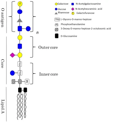
Figure 1.1: LPS schematic representation. The three main regions forming the LPS
Chapter 1: Structure and function of outer-membrane proteins
1.1.2 Outer-membrane proteins
The outer-membrane is embedded with proteins that display a characteristic β-barrel arrangement. To date, many outer-membrane protein (OMP) structures have been solved by X-ray crystallography and almost all of these share common features that can be summarized as follows: i) they are made of antiparallel β-strands connected to each other (the number of the β-β-strands differs amongst the OMPs, ranging from a minimum of 8 to a maximum of 26), (Wza was the first exception to this rule [9]) ii) both the N- and the C-terminus face the periplasmic space, iii) the β-strands are connected by longs loops (L) facing the extra-cellular space and short turns (T) facing the periplasmic space, iv) the loops represent the most variable part of the barrel, vi) in most (but not in all) the OMPs the L3 folds back inside the channel to form the so-called constriction zone or eyelet, vii) when trimerized, a non-polar core is formed along the threefold axis, viii) the external surface of the barrel (embedded in the lipid bilayer) consists mainly of hydrophobic, uncharged amino-acids[10],[11].
The OMPs play multiple roles that are essential for the viability of bacteria, they form channels for the uptake of nutrients, maintain the bilayer asymmetry, are involved in cellular adhesion, and can also act as enzyme (e.g. protease) [12],[13].
1.1.3 Passive transport through the OM: Porins
Chapter 1: Structure and function of outer-membrane proteins
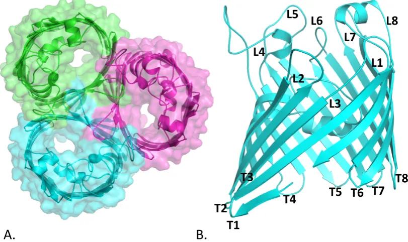
OMP, porins show a β-barrel structure with several long external loops and short periplasmic turns (Figure 1.2). Porins can adopt either a monomeric or (homo)trimeric conformation [15],[16]. The latter is stabilized due to the interaction within the L2 of one monomer and the β-strands of the neighbor monomer. In vitro, this interaction can be disturbed using chaotropic agents or by heat, leading to the partial or complete dissociation of the trimer [17]. β-barrel proteins benefit from a high level of stability due to numerous hydrogen bonds formed among the β-sheets [18]. Several studies, however, have identified “weakly stable” regions within the transmembrane domain of trimeric porins [19],[20],[21]. Interestingly, those regions are stabilized by the formation of protein-protein interfaces (oligomerization). In OmpF, a trimeric porin of E. coli, the amino acids R100, G19, G135, and N141 have been identified as weakly stable residues [21]. Site-directed mutagenesis of these residues leads to both a stable dimer and monomer of OmpF [21].
Chapter 1: Structure and function of outer-membrane proteins
Figure 1.2: Cartoon representation of OmpC from E. coli. (A) Extracellular view of
OmpC trimer (B) Side view of OmpC monomer. Extracellular loops are indicated with L and periplasmic turns with T. (PDB code: 2J1n)
Non-specific (also known as general) and specific porins are differentiated based on the level of specificity shown for their substrates. Normally 16-β-stranded, general porins allow permeation of substances with a MW lower than 600 Da with a slight selectivity for cations. Cation-selectivity in general porins – such as in OmpC [25] and OmpF [26] from E. coli, and in OmpK36 and OmpK35 from Keisbiella pneumonia [27] – has been largely demonstrated by single channel experiments. Selectivity is caused by an excess of negatively charged residues located inside the channel [28],[29],[30],[27]. The majority of Gram-negative bacteria possess at least one general porin, typically expressed in high copies number [31]. For instance, it has been estimated that OmpF from E. coli is normally present at 105 copies per cells [31].
L1 L2
L3 L4
L5 L6
L7 L8
T1 T2
T3
T4 T5 T6 T7
T8
Chapter 1: Structure and function of outer-membrane proteins
[image:31.595.182.417.390.663.2]The maltoporin LamB from E. coli [32] and the sucrose-specific porin SrcY from Salmonella typhimurium [33],[34] are well-characterized examples of specific (sugar) porins. A common feature of sugar transporting porins is an array of aromatic amino acids which run in lanes inside the inner wall of the channel, commonly known as “greasy slide” (Figure 1.3) [35]. Site-directed mutagenesis studies have shown the importance of the greasy slide on sugar translocation [36]. The aromatic residues help lining the sugars inside the channel by interacting with their hydroxide groups, lowering the energy barrier for permeation [37],[38]. Specific porins are expressed in low copy numbers and their expression depends upon certain limiting growth conditions.
Figure 1.3: Greasy slide in LamB from E. coli.LamB monomer is colored in green. The
F227 W358
W420 Y6
Chapter 1: Structure and function of outer-membrane proteins
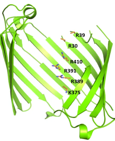
[image:32.595.187.405.420.688.2]Pseudomonas aeruginosa possess another class of substrate-specific porins. This is called the “Outer membrane Carboxylic acid Channel” (Occ)[39]. Interestingly, no general porins have been identified in P. aeruginosa, perhaps correlating with this pathogens inherent resistance to permeation of antibiotics [40],[41]. The Occ porins can be divided in two sub families OccD (specific for amino acids) and OccK (specific for cyclic negatively charged molecules) [42],[43]. In the Occ porins the substrates are guided through the pore due to a so-called “basic ladder”. This is a string of basic amino acids (Lys and Arg) that interact with the substrate’s carboxyl group [44] (Figure 1.4). To date, several structures of the Occ members have been solved, showing that although trimeric in solution, these porins crystallize as monomers of 18-β-strands [45].
Figure 1.4: Basic ladder in OccD1 from Pseudomonas aeruginosa. OccD1 monomer is
colored in green. Amino acids forming the so-called “basic ladder” are shown in sticks and colored in orange (PDB code: 4FOZ).
R39 R30
R410 R391
R389
Chapter 1: Structure and function of outer-membrane proteins
In the last decade, an increased number of porin structures have become available [10], uncovering the presence of porins with unusual characteristics [46]. For example, recently, the x-ray structure of the cyclodextrin metabolism A (CymA) porin from Klebsiella oxytoca has uncovered a diverse organization of the internal loops and thus a peculiar mechanism of uptake [47],[48]. CymA is a 14-β-sheets cyclodextrine-specific channel with shorts extracellular loops. Interestingly, none of them fold back to constrict the pore [48] (Figure 1.5 A-B). In CymA, in fact, the pore is kept closed due to an N-terminus inner plug that interacts with the wall of the channel. Such interaction is disturbed when the cyclodextrines approach, leading to the opening of the channel and subsequent translocation [48].
Chapter 1: Structure and function of outer-membrane proteins
Figure 1.5: Cartoon representation of 14-β-stranded porins. (A) Side view of CymA.
The barrel is colored in green and the inner plug in magenta. (B) Extracellular view of CymA. The substrate, cyclodextrine, is bound to the binding site. The molecule of cyclodestrine is displayed in stick and colored in magenta. (C) Side and (D) extracellular view of OmpG from E. coli (PDB code: 4D5B and 2F1C, respectively).
A.
B.
C.
D.
Chapter 1: Structure and function of outer-membrane proteins
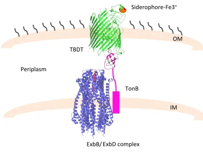
1.1.4 Active transport through OM: TonB-dependent transporters
[image:35.595.101.505.380.676.2]TonB-dependent transporters (TBDTs) are a class of OMPs which allow active translocation of essential substances such as iron (chelates to specific molecules called siderophore), cobalt and nickel. It also allows the translocation of substances such as vitamin B12 and carbohydrates [49]. To date, several structures of TBDTs have been reported, including FhuA [50], FepA [51], PiuA, PirA [52] and BtuB [53]. Those structures revealed that the TBTDs are 22-β stranded barrel with an inner “plug domain”, folded back inside the barrel (Figure 1.6).
Figure 1.6: FhuA, TonB dependent transporter of E. coli. (A) FhuA in complex to TonB
C-terminus. FhuA is colored in yellow, the inner plug in magenta and TonB in blue. (B)
A.
B.
Chapter 1: Structure and function of outer-membrane proteins
[image:36.595.92.494.394.695.2]As no source of energy is available at the level the OM, active translocation occurs by transducing the proton motive force (pmf) generated in the IM by a protein complex, namely TonB-ExbB-ExbD [49][54][55]. Specifically, as a result of the binding of the ligand, the TBDTs undertake conformational changes that lead the N-terminal of the plug domain, also known as TonB box, to reach and interact with the C-terminal domain of the TonB protein, at the level of the periplasmic space. Such interaction is necessary to transfer the energy for the ligand translocation (Figure 1.7) [56],[57]. Although the crystal structure of the TonB-ExbB-ExbD complex has been recently determined [58],[59], the transport mechanism has yet to be understood.
Figure 1.7: Schematic representation of TBDT mediated transport. Transport of the
siderophore-Fe3+occurs via specific TBTD receptor (green, PDB code: 2GRX). Upon ligand interaction the receptor interacts with the C-terminus of TonB (magenta, PDB code: 2GRX) giving the power stroke for translocation. The ExbB/ExbD complex is depicted in
TBDT
TonB
ExbD complex ExbB/
Siderophore-Fe3+
OM
Chapter 1: Structure and function of outer-membrane proteins
[image:37.595.181.420.390.696.2]Chapter 1: Structure and function of outer-membrane proteins
1.1.5 OMPs with enzymatic activity
To date, only few outer-membrane proteins have been characterized as enzymes [61],[62]. Those OMPs have adapted to modify the outer-membrane structure as a response to external stimuli and threats [61]. For instance, OmpT porin from E. coli, a well-characterized member of the omptine family, is involved in the catalysis of antimicrobial peptide [63]. The x-ray structure of OmpT showed a 10 β-sheets barrel with the catalytic residues (D83, D85, D210, H212) placed at the level of the extracellular loops (Figure 1.9 A) [64]. The structural organization of the catalytic pocket is stabilized due to the interaction with the LPS, so that the LPS is considered as a cofactor of OmpT [65]. In silico modeling analysis have suggested that a molecules of water, located in between the residues D83 and H212, it is directly involved in the hydrolysis of the peptide bound of two consecutives basic amino acids [66].
PagP and PagL from E. coli are another example of enzymatic OMPs. Both porins are monomeric and 8-β-stranded and are involved in the modification of the LPS [67],[68]. Specifically, PagP and PagL undertake the palmitoylation and the deacylation of the Lipid A, respectively[69],[70]. Such modification have been linked to an increase (palmytoylation) and a decrease (deacylation) of LipidA endotoxicity [71].
Chapter 1: Structure and function of outer-membrane proteins
glycerophospholipids in the outer leaflet of the outer-membrane [72]. Interestingly the monomeric form of this protein is inactive [73]. Activation of OMPLA occurs via dimerization and it is triggered by alteration of the LPS asymmetry [73],[74] (Figure 1.9 B).
Further, LpxR and LpxQ have too been identified as OMP enzymes. They are involved in the acyloxyacylation and oxidation of the Lipid A, respectively [75],[76]. No structural information is yet available for either of those enzymes.
Figure 1.9: OMPs enzymes. (A) Crystal structure of OmpT is colored in yellow (PDB
code: 1I78). The amino acids proposed as catalytic domain are shown in sticks and colored in blue. (B) Dimer of OMPLA (colored in pea-green) (PDB code: 1QD6). The calcium molecules are depicted as spheres and colored in purple.
D83
D85 H212
D210
Chapter 1: Structure and function of outer-membrane proteins
1.1.6 Biosynthesis of OMPs
The biosynthesis of OMPs starts, like any other protein, in the cytoplasm. Several proteins, often associated to form multi-protein complexes, are involved in the OMPs assembly [77],[78]. Chaperones, both in the cytoplasm and in the periplasm, keep the precursors of the OMPs (pre-OMPs) unfolded [79],[80], the SEC complex allows the pre-OMPs to pass through the IM and finally the β- barrel assembly machinery, also known as BAM, fold and insert the OMPs in the OM (Figure 1.10)[81].
Figure 1.10: Cartoon representation of the OMPs assembly. The pre-OMPs are
targeted to the IM via the SecAB complex, and reached the periplasm through the secYEG translocone. Here several chaperons bind the pre-OMPS and deliver them to the BAM complex. Figure adapted from [81]
BamA
BamB
BamD BamC BamE
Primary pathway SurA
Missfolding Degrada<on Secondary pathway
SecA
SecB Skp
DegP
SecYEG IM
Chapter 1: Structure and function of outer-membrane proteins
The majority of OMPs contains a signal peptide that allows the bacteria to recognize and target the protein to the correct translocon. The signal peptide is a three-part sequence, located at N-terminus of the pre-OMPs and it is usually 18-20 amino acids long [82]. The three motifs that comprised the signal peptide are identified as n-region (at the N-terminus of the sequence), h-region (composed of hydrophobic amino acids) and c-region (at C-terminus of the sequence, often contains an A-A consensus)[83].
A two proteins complex, SecAB, is responsible to deliver the pre-OMPs to the SEC translocon [84]. SecA is an ATP-powered protein, whose role is to carry the pre-OMPs to the translocon, while the chaperon SecB which bounds the pre-pre-OMPs in specific sequences, usually masked in the folded protein, keeps them unfolded [85].
Although the majority of the proteins cross the IM maintaining an unfolded conformation, fully folded proteins can also translocate through the IM, by using an alternative translocon, known as twin-arginine-translocase (TAT) [86]. In E. coli the TAT system consists of three inner membrane proteins, TatA, TatB and TatC. The signal peptide, that target the proteins to the TAT complex includes the consensus sequence Ser/Thr-Arg-Arg-(X)-Phe-Leu-Lys (with X being any polar amino acid)[86]. When the proteins reach the TAT complex, a H+ proton gradient
Chapter 1: Structure and function of outer-membrane proteins
The SecYEG translocon is a multi-protein complex composed of the three proteins SecY, SecE and SecG [88]. A homologue of the bacteria SEC translocon, the translocon Sec61αβγ, has been identified in Eukaryotes [89]. Although the mechanism of translocation through the SEC complex has not been fully understood, it is clear that SecA plays a major role [90]. During translocation, in fact, SecA partially insert itself through the translocon pore and through several cycle of binding and hydrolysis of ATP gives the energy necessary for the pre-OMPs to translocate [91][90].
In the SEC pathway, once the protein has crossed the IM, a signal peptidase cleaves the signal peptide and releases the OMPs in the periplasm. The pre-OMPs are recognizes by different chaperons (SurA, Skp, FkpA and Spy) whose role is to keep the pre-OMPs unfolded, avoiding their aggregation [92][93]. In case of misfolded proteins, a particular chaperone with protease activity, DegP, binds and hence degrades the OMPs [94]. Once the chaperons bind the pre-OMPs, they are delivered to the BAM complex.
Chapter 1: Structure and function of outer-membrane proteins
interaction. Deletion of the last three POTRAs (P3-5) is lethal for E. coli, but not for Neisseria gonorrhea [99], which implies that the importance of the single POTRAs on the bacterial viability is species-specific.
Figure 1.11: Crystal structures of BamA and BamB. (A) BamA structure is colored in
yellow and POTRAs domains in blue. (B) The lipoprotein BamB in complex with P3-5 POTRAs domains of BamA (PDB codes: 4K3B and4XGA, respectively).
BamA interacts to BamC and BamE through BamD. While deletion of BamB, BamC and BamE is tolerable, deletion of BamA and BamD severely impairs the OMPs assembly leading to the bacteria death. BamA and BamD the only essential
Chapter 1: Structure and function of outer-membrane proteins
Chapter 1: Structure and function of outer-membrane proteins
1.1.7 Antibiotic resistance: role of the OM and OMPs
Antibiotic resistance has now become a real concern for public health. Particular alarming is the increasing pace at which resistance pathogens are emerging, while the Pharma industries are struggling to find new classes of antibiotics [106]. Gram-negative bacteria show “intrinsic” resistance due to the presence of the OM that act as barrier against external agents. However, hydrophobic antibiotics, such as aminoglycoside and chloramphenicol can penetrate the bacterial cell by slowly diffuse through the OM [107]. As a resistance mechanism, bacteria can modify the OM composition and hence alter its permeability. For instance, the pathogenic bacteria, Salmonella enterica [108], has showed the ability to adjust the composition of the LPS, following the host colonization. This mechanism is due to the presence of a two components regulatory system PmrB-PmrA, that regulates the expression of genes involved in the modification of the LPS [109],[110]. Such
modifications include the incorporation of aminoarabinose and
Chapter 1: Structure and function of outer-membrane proteins
The number and the types of channels also determine the level of permeability of the OM, and therefore they play an important role in antibiotic resistance. It is well established that antibiotics, such as β-lactams, cefalosphorins and fluoroquinolones can cross the OM via general (non-specific) porins [113]. In pathogens, such as P. aeruginosa, the absence of general channels is associated with low susceptibility to antibiotics [40].
Mutations affecting the size and conductivity of the channel, modification of the level of porins expression, alongside with the complete loss of one or more general porins, are examples of how the bacteria are able to modify the OM permeability when exposed to antimicrobials agents (acquired resistance) (Figure 1.12) [114],[115].
Chapter 1: Structure and function of outer-membrane proteins
[image:47.595.152.453.309.590.2][117],[118],[119]. Those genes are activated by several external stimuli, such us oxidative stress, pH changes and antibiotics [120]. Once activated, they up-regulate the expressions of efflux pumps, such as TolC-AcrAB and small RNAs like micF and down-regulate the expression of porins such as OmpF [121],[122][123]. Point mutation, insertion element and deletion of those genes lead to a MDR phenotype and to an increased minimum inhibitory concentration (MIC) for different classes of antibiotics[116], [124],.
Figure 1.12: Schematic representation of porins regulation under antibiotics
pressure. Antibiotics exposure leads the bacteria to alter the permeability of the OM, by
modifying the expression level of its porins. Picture adapted from [115].
Porin loss Smaller size pore Pore muta1on An1bio1cs exposure
Chapter 1: Structure and function of outer-membrane proteins
Mutation of amino acids located in the loop 3 has also been associated to a decreasing antibiotic susceptibility [125]. G119D mutation in OmpF and similarly G112D mutation in Omp36 from Enterobacter aerogene and D116A mutation in OmpU from Vibro cholera have been found in clinic isolates resistant to β-lactams and cefalosphorins [22], [126]. In most organisms, different mechanisms synergistically function to protect the bacterial cell from the effect of antibiotics. In this phenomenon, known as MIC creep, alongside an altered OM the bacteria would increase the expression of efflux pumps (thus decrease the amount of antibiotic inside the cells) and increase the production of enzymes targeting the antibiotics (impeding their functions) [115].
Chapter 2: Major outer-membrane protein from Campylobacter jejuni
2.1 Introduction
2.1.1 Campylobacter: an overview
First isolated in 1913 from an aborted lamb, Campylobacters were originally wrongly classified in the genus Vibrio (Vibrio fetus), and they were subsequently identified as a new genus (Campylobacter) in 1973 [127]. To date 15 species and 6 subspecies (both commensal and pathogenic) have been annotated [128].
Campylobacter spp. are gram-negative bacteria belonging to the class of ε-proteobacteria [129]. Usually spiral or curve shaped, Campylobacter spp are motile by means of a single or bipolar flagellum [129]. They belong to the group of microaerophilic and thermophilic bacteria, meaning that a modified atmosphere with a low concentration of oxygen and a high concentration of CO2,as well as
high temperatures (37-44°C), are necessary for optimal growth [6], [131],[132]. Growth at lower temperatures has been proven to be slow [132].
Chapter 2: Major outer-membrane protein from Campylobacter jejuni
glucose-derivatives that are essential for lipopolysaccharide (LPS) and capsule biosynthesis and hence campylobacter virulence [134].
2.1.2 Campylobacter jejuni infection
Chapter 2: Major outer-membrane protein from Campylobacter jejuni
Chapter 2: Major outer-membrane protein from Campylobacter jejuni
Figure 2.1: Mimicry mechanism in GBS associated to C. jejuni. Schematic
Chapter 2: Major outer-membrane protein from Campylobacter jejuni
2.1.3 Campylobacter jejuni antibiotic resistance
Antibiotic resistance in C. jejuni has become a real concern in public health. As Campylobacter contamination among people is rare, the use of antibiotics in animal farming is considered to be the main cause of resistance [157]. Although the European Union has prohibited the addition of antibiotics in animal feed for non-therapeutic purpose (i.e. growth promotion of livestock), in other part of the world, the use of antibiotics as growth agents is still allowed [158]. C. jejuni strains isolated from contaminated poultry have shown resistance to different classes of antibiotics such as fluoroquinolone (FQ), macrolide [159], β-lactam and tetracycline [160][161][162] (Figure 2.2). A multi-drug resistance (MDR) phenotype has also been observed [163][164]. The macrolide erythromycin (Ery) is used as first choice treatment when campylobacteryosis is diagnosed. However, as campylobacteryosis is indistinguishable from other enteric infections, Ciprofloxacin – an FQ with a broad spectrum activity against Gram-negative bacteria – is often preferred [160].
Chapter 2: Major outer-membrane protein from Campylobacter jejuni
therefore translocation of antibiotics is decreased [125]. Unlike E. coli, C. jejuni has only one major outer membrane protein (MOMP), which has a molecular weight limit of 360 Da, thus molecules with higher molecular weight cannot permeate inside the cell [168]. Additionally, being cationic selective, MOMP represent a barrier for translocation of anionic antibiotics [168].
Figure 2.1: C. jejuni antibiotic resistance. Schematic representation of C. jejuni
mechanisms of antibiotic resistance.
> 360 MW
23 rRNA* gyrA* FQ
Macrolide CmeB CmeC
CmeA
Chapter 2: Major outer-membrane protein from Campylobacter jejuni
2.1.4 Major outer membrane protein (MOMP)
Chapter 2: Major outer-membrane protein from Campylobacter jejuni
Surface exposed antigens stimulate the immune system to produce antibodies [180]. In chicks, maternal antibodies have been proven to serve as protection against C. jejuni infections [181]. This inspired the idea to use C. jejuni surface proteins as a target to develop vaccines. However, in chickens and mice models, immunization using CmeC subunit, the lipoprotein CjaA and PEB1 antigen (all surface-exposed proteins), have failed to protect from C. jejuni colonization [182],[183],[184]. On the contrary, MOMP-derived vaccines have shown promise. MOMP contains antigenically variable regions at the level of the loops and conserved regions present in the β-strands [170]. Islam and co-workers have already shown the protective effect of a recombinant MOMP-vaccine tested in mice models[185]. This study can be used as a starting point to generate a vaccine for human use. Given that C. jejuni live attenuated vaccines could potentially trigger auto-immune diseases, the use of a MOMP-derived vaccines would represent a safer alternative.
2.1.5 Aims
Although MOMP from C. jejuni has been extensively studied over the last three decades, only recently its crystal structure has became available (the structure was solved by Dr. Gregor Wallat for Naismith’s lab). Being C. jejuni a fastidious organism to cultivate, Dr. Wallat successfully over-expressed and purified MOMP from E. coli.
Chapter 2: Major outer-membrane protein from Campylobacter jejuni
Chapter 2: Major outer-membrane protein from Campylobacter jejuni
2.2 Material and Methods
2.2.1Campylobacter jejuni culture
Campylobacter jejuni 85H strain [45] was grown according to Bolla et al. [171]. The C. jejuni strain was spread onto a blood agar plate and incubated for 24 hours at 42 °C. Bacteria were rinsed with 1 ml of 2YT medium and agitated for 15 minutes. Subsequently, 250 µl of the recovered bacteria were spread onto 4 Columbia agar plates supplemented with the appropriate amount of Campylobacter selective antibiotics supplement (Oxoid), and incubated for 48 hours at 42 °C. The plates were rinsed with 1 ml of 2YT medium and left rocking for 15 minutes. The recovered bacteria were subsequently inoculated onto 40 plates of Columbia agar (antibiotic-free) and incubated for 48 hours at 42 °C. Each plate was then rinsed using 5 ml of 10 mM Tris-EDTA (TE) buffer pH 7.4 and agitated for 15 minutes at room temperature. The bacterial suspension was recovered and the OD600 checked to be around 2. The bacteria were pelleted by
Chapter 2: Major outer-membrane protein from Campylobacter jejuni
2.2.2 MOMP 85H purification
Cells were re-suspended in 300 ml of lysing buffer (Appendix A.1) and lysed by using two passes at 30 Kpsi through a high-pressure cell disruption for micro volumes (Constant System Ltd). Unbroken cells were removed by spinning the cell lysate at 10,000xg for 30 minutes at 4 °C. Membranes were recovered by ultracentrifugation of the supernatant at 100,000 x g for 1 hour at 4 °C. The pellet was then homogenized in 10 mM Tris-HCl pH 7.4 and 0.1% (w/v) of sodium lauryl sarcosinate (Sigma) and left rocking for 30 minutes at 4 °C. The outer membrane was recovered by ultracentrifugation at 100,000xg for 1 hour at 4 °C. The supernatant containing the inner membrane proteins fraction was discarded and the pellet was homogenized with 20 mM sodium phosphate buffer pH 7.4 and 1% of n-octylpolyoxyethylene (Octyl-POE) (Bachem AG) and left rocking at 4 °C for 30 minutes. Solubilized outer membrane proteins were recovered by ultracentrifugation at 100,000xg for 1 hour at 4 °C. At this stage, the pellet was discarded and the supernatant was loaded onto a MonoQ HR ion exchange column (GE Healthcare) and equilibrated with 5 column volumes (CV) of buffer A (30 mM Na2HPO4, 10 mM NaCl and 0.6% Octyl-POE). The bound proteins were
Chapter 2: Major outer-membrane protein from Campylobacter jejuni
ether (C8E4) (Bachem). Fractions containing MOMP were combined and
concentrated to 10 mg ml-1.
2.2.3 MOMP 85H crystallization
Initial crystallization trials were carried out using several commercial screens (Appendix B). The screens were dispensed into a 96-well sitting drop plate (Intelli-Plate®, ARI)using a Gryphon robot, at a protein:reservoir ratio of 1:1 and
1:2. First hits appeared within a week. Crystals were manually optimized by the hanging-drop vapor diffusion technique.
Chapter 2: Major outer-membrane protein from Campylobacter jejuni
Table 2.1. Optimization of MOMP crystallization conditions.
2.2.4 Data collection, Structure determination and refinement
X-ray data from a crystal of MOMP purified from C. jejuni 85H were collected in house using a Rigaku Micromax™-007HF Cu anode with VariMax optics and a Rigaku Saturn 944+CCD detector and processed with Xia2 [186]. The structure was solved to a resolution of 2.9 Å by molecular replacement using the recombinant MOMP structure (PDB code: 5LDV) as a search model using the program MR Phaser [187] . The model was manually built in Coot [188] and refined with REFMAC5 [189]. Eventually, 15 TLS groups were used in the final refinement cycle. The webserver “CheckmyMetal” [190] was used to validate the presence of calcium molecules at the constriction zone of the protein.
pH 7
pH 7.5
pH 8
pH 8.5
30%
A1
A2
A3
A4
31%
B1
B2
B3
B4
32%
C1
C2
C3
C4
0.1 M Tris-HCl+
0.05 M CaCl
2+ 0.05 BaCl
2PE
G
400
(v/
Chapter 2: Major outer-membrane protein from Campylobacter jejuni
2.2.5 Single channel conductance measurements of MOMP
A planar lipid bilayer was formed using the solvent free lipid bilayer technique [191]. Briefly, the cuvettes used for experiments consist of two separate chambers, named cis and trans. A 25-µm thick Teflon film (Goodfellow) with an aperture of 40–100 µm diameter is located in between the chambers (Figure 2.3). The aperture in the Teflon film was made by using a high voltage cathode discharge (ElectrotechniProducts). The aperture was pre-painted with 1 µl of 1% hexadecane in hexane using a pipette tip. The chambers were filled with electrolyte solution consisting 1 M KCl, 10 mM MES pH 6.0 to a total solution volume of 2.5 ml. Then, 5% solution of diphytanoyl phosphatidylcholine (DPhPC, Avanti Polar Lipids) was added to both chambers. At this point, the solution was slowly pipetted up and down to allow the formation of the bilayer. MOMP was added to the cis side (which is the electrical ground) of the chamber at a final concentration of 2-3 ng ml-1 and the channel insertion was eased by quickly
mixing the solution contained in the chamber while applying a trans-membrane potential of –199 mV. Electrical recordings were made through a pair of Ag/AgCl2
electrodes (World Precision Instruments) attached to an Axon Instruments 200B amplifier (Axon Instruments Inc.). A low pass Bessel filter at 10 kHz was used to filter the data and saved with a sampling frequency of 50 kHz. Data analyses were performed using Clampfit 10.0 software (Axon Instruments Inc).
Chapter 2: Major outer-membrane protein from Campylobacter jejuni
Additionally, measurements were taken in the presence of 10 mM of the chelating agent ethylene glycol-bis (β-aminoethyl ether)-N,N,N',N'-tetraacetic acid (EGTA). The protein was incubated overnight with 10 mM EGTA.
Figure 2.2: Single channel experiments cuvette. Picture of the cuvette used for single
Chapter 2: Major outer-membrane protein from Campylobacter jejuni
2.3 Results
2.3.1 Campylobacter jejuni 85H culture preparation
Campylobacter jejuni bacteria were spread onto selective blood-agar plates and grown under microaerophilic condition at 42 °C. At an OD600 of 2, the bacteria
were harvested by centrifugation. Due to a particular pigment produced by Campylobacter, the cell paste displayed a pink coloration.
2.3.2 MOMP extraction and purification
Chapter 2: Major outer-membrane protein from Campylobacter jejuni
crystallography experiments. The eluted protein was then concentrated to 10 mg ml-1. SDS -PAGE was run to evaluate the purity of the protein (Figure 2.4 B). Gel
Chapter 2: Major outer-membrane protein from Campylobacter jejuni
Figure 2.3: MOMP purification. (A). Profile of the anion-exchange chromatography.
Two peaks are present: one on the left elutes at 22% buffer B, and the second on the right elutes at 70% buffer B. (B). SDS-PAGE of chosen fractions from the anion-exchange chromatography. M is the ladder, lanes 1-4 show two bands: one at 50 kDa (Omp50) and one at 45 kDa as confirmed by mass spectrometry. C. Profile of the SEC performed using a 16/600 200pg (GE) column. The elution volume of MOMP was compared to the elution volumes of molecular weight (MW) markers (Bio-rad) (used to calibrate the column) in order to analyse its oligomeric state. MOMP was found to be trimeric. (D) SDS-PAGE of chosen fractions from the SEC. M is the ladder, lane 1 is the sample before SEC and lanes 2 to 6 are fractions from the main peak.
M 1 2 3 4 5 6 7 8 9
A.
B.
0 100 200 300 400 500 600 700 800 900
0 30 60 90 120
Ab so rb an ce at 28 0 (mAU )
Elu@on volume (mL) Lane 5-9 Lane 1-4 97- 66- 45- 30- 20- 14.4- kDa 0 50 100 150 200 250 300
0 50 100 150
Ab so rb an ce at 28 0 (mAU )
Elu@on volume (mL) Lane 2-6
M 1 2 3 4 5 6
25- 37- 50- 75- 110- kDa
Chapter 2: Major outer-membrane protein from Campylobacter jejuni
2.3.3 MOMP crystallization and structure determination
The MOMP crystals appeared after three days. Crystals were found in condition B3 of the Mem Gold I crystallization screening (Molecular dimension). After optimization using the hanging drop vapor technique, the largest crystals were found in 0.05M Calcium chloride, 0.05 M barium chloride, 0.1 M Tris-HCl pH 8.5 and 30% PEG 400 (Figure 2.5). The crystals were analyzed with an in house X-ray source and found to diffract to 2.9 Å. A dataset was collected and processed using Xia2 [186] in the space group P212121. Analysis of the Matthew’s coefficient
(Matthew’s coefficient of 2.64) suggested that three molecules were present in the asymmetric unit (AS). The structure was solved by molecular replacement using MR Phaser from the CCP4 suite [187]. As search model was used the recombinantly expressed MOMP (PDB code: 5LDV). The quality of the structure was assessed using Molprobity. Data collection and refinement statistics are shown in table 2.2.
Chapter 2: Major outer-membrane protein from Campylobacter jejuni
Table 2.2.Data collection, refinement and validation statistics.
Space group P212121
Cell dimensions a,b,c (Å) 94.4, 99.4,172.2
Cell dimensions α,β,γ (°) 90, 90, 90
Resolution (Å) 47.75-2.9(3.06-2.9)
Rmerge 0.14 (0.86)
Completeness (%) 99.9 (99.8)
Multiplicity 7.3 (7.2)
I/σ(I) 12.9 (3.0)
CC 1/2 0.9 (0.7)
Refinement
Rfactor/Rfree (%) 23.9/27.1
N° of unique reflections 36693
N° of residues 405
Water 5
Rmsd
Bonds length (Å) 0.01
Bonds angles (Å) 1.9
MolProbity
Ramachandran outliers (%) 0
Ramachandran favored (%) 95.41
Clash score 1.7
Molprobity score 1.68
Chapter 2: Major outer-membrane protein from Campylobacter jejuni
2.3.4 Crystal structure of MOMP
MOMP is a homotrimer composed of 18-β-stranded monomers, with a characteristic elliptical shape. Each monomer consists of 405 amino acids. Common for porins, the β-strands (β) are linked together by long extracellular loops (L) and short periplasmic turns (T) (Figure 2.6 A). As predicted by the software SignalP 4.0 [192], the signal peptide of the native MOMP was cleaved between Ala22 and Thr23. Consequently, the Thr23 is the first residue in the tertiary structure and as such was renumbered to Thr1.
Chapter 2: Major outer-membrane protein from Campylobacter jejuni
Figure 2.5: Crystal structure of MOMP. (A) Cartoon of MOMP trimer view from the
extracellular space (left) and from the side (right) of the membrane. The three chain are colored in magenta, green and cyan. (B) Monomer of MOMP, with highlighted loops involved in the formation of the constriction zone and L2. L3 is colored in green, L4 in orange, L6 in cherry red and L2 in purple. Major and minor axis measure approximately 37 and 19 Å, respectively.
L3
L2
L4
L6
A.
B.
19
Å
37
Chapter 2: Major outer-membrane protein from Campylobacter jejuni
8 amino acids (Thr1 to Lys8) at the N-terminus form an α-helix that points away from the barrel wall, toward the adjacent monomer. In this way, the three α-helices (one for each monomer) form a triangle that faces the periplasmic space (Figure 2.7).
Figure 2.6: N-terminus of MOMP. (A) In grey is depicted the MOMP monomer, in
magenta the N-terminus. (B) Periplasmic view of the N-terminus complex (color scheme as in 2.5) in MOMP trimer.
Chapter 2: Major outer-membrane protein from Campylobacter jejuni
2.3.5 The electrostatic potential of MOMP
The electrostatic potential of MOMP was analyzed using CCP4mg program [193]. The eyelet, viewed from the extra-cellular (EX) (Figure 2.8 A) and periplasmic (PE) (Figure 2.8 B) side, exhibited a clear separation of positive and negative charges with Asp116 (L3), Asp 120 (L3) and Asp155 (L4) facing Arg17, Arg19 and Lys43 on the opposite side.
Chapter 2: Major outer-membrane protein from Campylobacter jejuni
Figure 2.7: Electric potential of MOMP. (A) View of the channel from the extra-cellular
space. In the black circle is a zoom in of the eyelet. The charged amino acids forming the eyelet are in sticks. (B) View of the channel from the periplasmic space. (C) View of the channel from the side. (D) View of the trimeric MOMP from the extra-cellular space.
C.
A.
EX
PE
Asp116 Asp120 Asp155
Arg17 Arg19 Lys43
B.
Chapter 2: Major outer-membrane protein from Campylobacter jejuni
2.3.6 Calcium binding sites in MOMP
Chapter 2: Major outer-membrane protein from Campylobacter jejuni
Figure 2.8: Calcium-binding sites in MOMP. (A) Extra-cellular view of MOMP trimer
and the calcium binding sites. Theβ-barrel is colored in cyan, the loop in sand and the calcium molecules in purple. Zoom in of the calcium-binding site inside the channel (B) and between monomers. (C) The amino acids involved in the binding are shown in sticks. Calcium is colored in purple and water in red. (D) F0-F2 and 2F0-F2 electron density map at 5σ and 2σ, respectively, of the amino acids involved in the calcium binding site at the
A.
B.
C.
Asp145 Gly72 Asn77
Asn180
Asp 116
Asp289 Asp155
Glu 288 Gln 152
Asp 120
Asp 116
Asp289 Asp155
Glu 288
Gln 152
Asp 120
Chapter 2: Major outer-membrane protein from Campylobacter jejuni
Table 2.3.Bond distances of the two calcium binding sites in MOMP.
2.3.7 Single channel conductance measurements
MOMP conductivity was assessed by single channel measurements in 10 mM Hepes pH 6 and 1 M KCl. MOMP was randomly inserted into the bilayer as either a monomer or a trimer. Consequently, we detected two different levels of conductance, a lower one of 0.7 ± 0.2 nS, and a higher one of 2.2 ± 0.2 nS. These corresponded to the monomeric and trimeric states, respectively. The electrical signature showed that at negative voltages, long upward spikes occurred for both
Interacting molecule Bond length (Å)
Calcium binding site at the constriction zone
Asp120
(Oxygen and side
chain)
Ca2+ 2.33/2.29
Gln152 Ca2+ 2.32
Asp155 Ca2+ 2.32
Glu288 Ca2+ 2.31
H2O Ca2+ 2.49
Calcium binding site in between monomers
Asp145
(Oxygen and side
chain)
Ca2+ 2.29/2.3
Asn180 Ca2+ 2.26
Gly72 Ca2+ 2.27
Chapter 2: Major outer-membrane protein from Campylobacter jejuni
[image:79.595.93.510.197.499.2]monomeric and trimeric MOMP. In contrast, at positive voltages, while the monomer trace was noisy (displaying long downward spikes), the trimer trace was silent (Figure 2.10).
Figure 2.9: Single channel recording of MOMP. Electric trace of (A) MOMP monomer
and (B) MOMP trimer at +100 mV (right) and at -100 mV (left).
To elucidate the effect of Ca2+ on the channel, conductance measurements were
also taken in presence of 10mM CaCl2. Interestingly, the presence of calcium
eliminates the long spikes previously observed at negative voltage. The calcium makes the channel stable (at both positive and negative voltage) (Figure 2.11 A).
A.
Chapter 2: Major outer-membrane protein from Campylobacter jejuni
buffer at a final concentration of 10 mM). Not surprisingly, the electrical trace was silent at positive voltage but noisy at negative voltage, as with the native protein (Figure 2.11 B).
In a control experiment, we slowly replaced the buffer containing EGTA with a buffer containing 10 mM CaCl2. As expected the calcium reverted the effect of the
chelating agent, resulting in an entirely silent channel.
Figure 2.10: Single channel recording of MOMP. Electric trace of MOMP trimer with
(A) and without (B) at +100 mV (right) and at -100 mV (left).
A.
[image:80.595.88.513.304.660.2]Chapter 2: Major outer-membrane protein from Campylobacter jejuni
particular, conductance measurements were taken in the presence of 10mM of either MgCl2 or ZnCl2. Neither the magnesium nor the zinc was able to stabilize
[image:81.595.92.503.219.619.2]the channel. At negative voltage the channel showed the same long spikes detected in absence of calcium (Figure 2.12).
Figure 2.11: Single channel recording of MOMP. Electric trace of MOMP trimer with 10
mM MgCl2 (A) and 10 mM of ZnCl2 (B) at +100 mV (right) and at -100 mV (left).
A.
Chapter 2: Major outer-membrane protein from Campylobacter jejuni
2.4 Discussion
2.4.1 MOMP crystal structure
In this study, we present the first X-ray structure of the natively expressed MOMP from Campylobacter jejuni strain 85 H. Although previous analysis of two-dimensional and three-two-dimensional crystals of MOMP provided a clear indication of the trimeric arrangement of the protein, similar to that observed for OmpC and OmpF in E. coli, they failed to give complete structural data [194]. The crystal packing of the structure we obtained confirmed this trimeric arrangement. Each monomer-monomer interface buries over 1200 Å2 of surface area, while the
trimer buries almost 10,000 Å2 of surface area. Overall, the structural features
observed in MOMP are common to all the porins characterized so far. A barrel consisting of 18-anti-parallel β-strands joined together by long extracellular loops and short periplasmic turns. The L3 folds back inside the barrel to form the eyelet of the pore, a region characterized of negatively and positively charged residues facing each other that create a strong electric field. L2 stabilizes the trimer as suggested by the several hydrogen bonds formed with the β-strands of the adjacent monomers.
Chapter 2: Major outer-membrane protein from Campylobacter jejuni
[image:83.595.103.510.367.692.2]not involved in the trimerizazion since both proteins exist as monomer. Interestingly, the sucrose-specific porin (ScrY) from Salmonella typhimurium contains a coiled-coil structure at the N-terminal domain [196],[197]. Unlike PagP and OprB, ScrY oligomerises form a homo-trimer. Spectroscopic and biophysical analysis suggested that the ScrY N-terminus α-helical coiled-coil complex might be involved in the oligomer stabilization; however, no structural data of this domain are available [196]. Although the role of the N-terminal α–helix in MOMP has yet to be investigated, PDBePISA analysis suggests it might contribute to the stability of the trimer (Table 2.4).
Figure 2.12: α-helices N-terminus. N-terminus of PagP (A) and OprB (B). β-barrels are