Received 24 May 2019 Accepted 30 January 2020
Edited by C. Massera, Universita` di Parma, Italy
Keywords:crystal structure; nickel complexes; tren; tripodal ligand; hydrogen bonding.
CCDC reference:1911584
Supporting information:this article has supporting information at journals.iucr.org/e
Crystal structure of a nickel compound comprising
two nickel(II) complexes with different ligand
environments: [Ni(tren)(H
2O)
2][Ni(H
2O)
6](SO
4)
2Karilys Gonza´lez Nievesa* and Dalice M. Pin˜ero Cruzb
a
Department of Natural Sciences, University of Puerto Rico, Carolina Campus, 2100 Avenida Sur, Carolina, PR 00987, Puerto Rico, andbDepartment of Chemistry, University of Puerto Rico, Rio Piedras Campus, Ponce de Leon Avenue, San Juan, PR 00931, Puerto Rico. *Correspondence e-mail: [email protected]
The title compound, diaqua[tris(2-aminoethyl)amine]nickel(II) hexaaqua-nickel(II) bis(sulfate), [Ni(C6H18N4)(H2O)2][Ni(H2O)6](SO4)2 or
[Ni(tren)-(H2O)2][Ni(H2O)6](SO4)2, consists of two octahedral nickel complexes within
the same unit cell. These metal complexes are formed from the reaction of [Ni(H2O)6](SO4) and the ligand tris(2-aminoethyl)amine (tren). The crystals of
the title compound are purple, different from those of the starting complex [Ni(H2O)6](SO4), which are turquoise. The reaction was performed both in a 1:1
and 1:2 metal–ligand molar ratio, always yielding the co-precipitation of the two types of crystals. The asymmetric unit of the title compound, which crystallizes in the space groupPnma, consists of two half NiIIcomplexes and a sulfate counter-anion. The mononuclear cationic complex [Ni(tren)(H2O)2]
2+
comprises an Ni ion, the tren ligand and two water molecules, while the mononuclear complex [Ni(H2O)6]
2+
consists of another Ni ion surrounded by six coordinated water molecules. The [Ni(tren)(H2O)2] and [Ni(H2O)6] subunits are connected to the
SO4 2
counter-anions through hydrogen bonding, thus consolidating the crystal structure.
1. Chemical context
Tris(2-aminoethyl)amine (tren) has been used extensively as an ancillary tripodal ligand for capping transition metals to form mononuclear and polynuclear complexes. The tren ligand has the capacity to chelate metal ions through its central tertiary amine and through its three terminal amine groups in a spider-like conformation, leaving one or two positions available for additional ligand coordination (Marzotto et al., 1993; Albertinet al., 1975; Blackman, 2005; Brineset al., 2007). Metal complexes with a variety of ligands in which also tren is coordinating to the metal center have been proposed for applications in catalysis (Ruffinet al., 2017), sensors, and as precursors of bioinorganic reactions (Sakaiet al., 1996). For instance, Ni(tren) complexes have been proposed for applications in biological systems (Salam & Aoki, 2001) or as a model to study enantioselective synthesis or asymmetric catalysis (Raoet al., 2009), and as coordination polymers in magnetism, electrical conductivity and ion exchange (Park et al., 2001; Tanase et al., 1996). [Ni(tren)(H2O)2] was reported previously (Chenet al., 2001;
Pedersenet al., 2014); however, to our knowledge, this is the first report of it co-crystallizing with the hexaaquo nickel complex [Ni(H2O)6](SO4).
2. Structural commentary
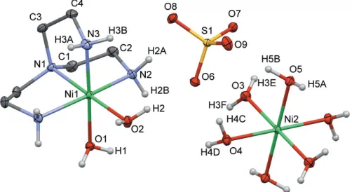
Fig. 1 shows the molecular structure of the title compound, which crystallizes in the space group Pnma. Its asymmetric unit comprises two half NiIIcomplexes and a sulfate counter-anion. Each Ni complex shows a different ligand environment: (i) the mononuclear cationic complex [Ni(tren)(H2O)2]
2+
includes Ni1, the tren ligand and two water molecules; (ii) the mononuclear complex [Ni(H2O)6]
2+
consists of Ni2
surrounded by six coordinated water molecules.
Ni1 exhibits an octahedral geometry of the type N4O2, with
the central N1 atom of the tren ligand occupying one of the axial positions and atoms N2, N3 and N2ioccupying three of the equatorial positions [symmetry code: (i)x,y+12,z]. The
remaining two positions, one axial (O2) and one equatorial (O1), are occupied by two oxygen atoms from the two water molecules. The bond lengths are similar for the Ni1—N bonds that are trans to oxygen atoms; for instance, Ni1—N1ax is
2.064 (2) A˚ and Ni1—N3eq is 2.069 (2) A˚ ; a longer bond
distance is observed between Ni1—N2eq, 2.122 (2), which is
transby symmetry to another nitrogen atom, N2i. The nickel– oxygen bond length is shorter for Ni1—O2axat 2.094 (2) A˚ , in
comparison to Ni1—O1eq, which is 2.140 (2) A˚ . The N3 and
C3 atoms of the tren ligand lie on a mirror plane perpendicular to [010]. This results in a symmetry-induced disorder of the N3/C4/C3 fragment. The octahedral geometry around the Ni1 ion is reflected by the angles N1—Ni1—O2 = 178.42 (8),
N2—Ni1—N2i= 164.74 (9), and N3—Ni1—O1 = 177.27 (8).
The Ni2 ion of the mononuclear complex [Ni(H2O)6] 2+
also shows an octahedral geometry. In the asymmetric unit, the atom Ni2 sits on an inversion center on a screw axis along the b-axis direction. The Ni2—Owaterbond lengths with O3, O4
and O5 range between 2.051 (1) and 2.074 (1) A˚ , respectively, with angles of 180due to symmetry.
3. Supramolecular features
The crystal structure of the title compound is consolidated through intermolecular hydrogen bonding between the water molecules from the [Ni(tren)(H2O)2] complex, the sulfate
oxygen atoms and the water molecules from the [Ni(H2O)6]
complex (Fig. 2 and Table 1). In particular, the two water molecules of [Ni(tren)(H2O)2] form O1—H1 O8
i
and O2— H2 O6 hydrogen bonds of 2.05 (2) and 1.96 (2) A˚ respec-tively, involving two neighboring SO4
2
anions [symmetry code: (i)x+1
2,y,z+ 3
2). The [Ni(H2O)6] complex is hydrogen
bonded to adjacent SO4 2
anions through O3—H3E O9ii, O3—H3F O7i, O4—H4C O6, O4—H4D O8i, O5— H5B O7, O5—H5A O7iii contacts [symmetry codes: (ii)
x + 1
2, y + 1, z 1
2; (iii) x + 1
2, y + 1, z + 1 2]. These
hydrogen-bond distances range from 1.905 (15) to
2.047 (18) A˚ . Additional weak hydrogen bonds are formed between the hydrogen atoms from the primary amine groups of the tren ligand and the sulfate oxygen atoms.
research communications
Acta Cryst.(2020). E76, 314–317 Gonzalez Nieves and Pin˜ero Cruz [Ni(C
[image:2.610.60.277.79.188.2]6H18N4)(H2O)2][Ni(H2O)6](SO4)2
315
Figure 2The hydrogen-bonding network (cyan dotted lines) in the title compound. Symmetry codes: (i)x+1
2,y,z+ 3
2; (ii)x+ 1
2,y+ 1,z 1
2; (iii)x+ 1 2,
[image:2.610.315.566.88.209.2]y+ 1,z+1 2. Table 1
Hydrogen-bond geometry (A˚ ,).
D—H A D—H H A D A D—H A
O1—H1 O8i 0.78 (2) 2.05 (2) 2.8212 (16) 172 (2)
O2—H2 O6 0.81 (2) 1.96 (2) 2.7342 (15) 162 (3) O3—H3E O9ii 0.81 (2) 1.94 (2) 2.731 (2) 167 (2)
O3—H3F O7i 0.85 (2) 2.05 (2) 2.8403 (18) 155 (2) O4—H4C O6 0.83 (2) 1.91 (2) 2.7249 (18) 171 (2) O4—H4D O8i 0.83 (2) 1.95 (2) 2.7810 (18) 179 (2)
O5—H5A O7iii 0.88 2.02 2.8125 (19) 150
O5—H5B O7 0.88 1.95 2.7826 (17) 160
Symmetry codes: (i) xþ1 2;y;zþ
3
2; (ii) xþ 1
2;yþ1;z 1 2; (iii)
xþ1
[image:2.610.46.297.540.677.2]2;yþ1;zþ 1 2.
Figure 1
View of the molecular structure of the title compound with displacement ellipsoids drawn at the 20% probability level and labeling scheme for the symmetry-independent atoms. The CH2 hydrogen atoms have been omitted for clarity. The symmetry operations generating the equivalent atoms are 1 x, 1 y, 2zand x,1
2y,z for [Ni(H2O)6] 2+ and
[image:2.610.313.563.558.707.2]4. Database survey
A search for tris(2-aminoethyl)aminenickel complexes in the Cambridge Structural Database (CSD version 5.38, updated February 2019; Groom et al., 2016) yielded 222 hits. Among these results, 124 hits contained the ligand tris(2-aminoeth-yl)amine capping the nickel ion, along with other types of ligands on the remaining coordination sites. Only two hits contain the diaqua[tris(2-aminoethyl)amine]nickel(II) complex, [Ni(tren)(H2O)2] (LUMVIY; Chen et al., 2001;
TIYQAT; Tanaseet al., 1996). More precisely, the asymmetric unit in LUMVIY comprises the [Ni(tren)(H2O)2]
2+
cation with two independent halves of a 1,5-naphthalenedisulfonate (1,5nds) ligand as counter-anion. A common feature of this structure with the title compound is the hydrogen bond network formed between the water molecules on the Ni(tren) motif with the counter anions. However, in the title compound, also the hydrogen atoms on the primary amine groups form hydrogen bonds with the sulfate anions, albeit quite weak. In TIYQAT, sulfate anions act as counter-ions for the [Ni(tren)(H2O)2]
2+
complex, and uncoordinated water molecules are included in the crystal lattice. The angle between the Ni center and the two oxygen atoms from the coordinated water molecules are 86.52 (5) (O7—Ni1—O8)
and 86.9 (4) (O5—Ni1—O6) for LUMVIY and TIYQAT,
respectively. The corresponding angle O2—Ni—O1 in the tittle compound has a value of 88.70 (8), which is in good
agreement with the reported values. The title compound is the first example of a crystal structure of [Ni(tren)(H2O)2]2+
co-crystallizing with the [Ni(H2O)6]2+complex.
5. Synthesis and crystallization
The synthesis of the title compound is summarized in the reaction scheme shown in Fig. 3. NiSO46H2O and
tris(2-aminoethyl)amine (tren) were used without further purifica-tion. A methanolic solution of NiSO46H2O (0.0265 g,
0.1 mmol) was added slowly to a tren (0.0146 g, 0.1 mmol) solution (4 mL MeOH) at room temperature. The resulting solution was stirred for two h and it changed color from light green to purple. The solution was then filtered through celite and evaporated under reduced pressure. Single crystals of the title compound were obtained by vapor diffusion of methanol into 2-propanol. In the crystallization process, two types of crystal were formed: the starting reagent hexahydrate nickel (II) complex (turquoise crystals) and the nickel(II) tren complex (purple crystals, Fig. 4). The reaction was performed both in a 1:1 and 1:2 metal–ligand molar ratio, always yielding
the title compound. IR data: 3265 (m), 3171 (m), 2937 (w), 2891 (w), 1607 (m), 1472 (w) 1338 (w), 1054 (s), 984 (m), 885 (m), 750 (w), 685 (w).
6. Refinement
Crystal data, data collection and structure refinement details are summarized in Table 2. H atoms were included in geometrically calculated positions for the alkyl and amine groups using a riding model: C—H = 0.97 A˚ and N—H = 0.89 A˚ withUiso(H) =1.2Ueq(C, N). The hydrogen atoms of the
water molecules were located from the difference-Fourier map; they were refined freely in the case of O1 and O2, with a DFIX of 0.85 (2) A˚ andUiso(H) =1.5Ueq(O) in the case of O3
and O4, and riding with O—H = 0.88 A˚ and Uiso(H)
[image:3.610.314.564.88.385.2]=1.5Ueq(O) in the case of O5.
Figure 3
Reaction scheme for the synthesis of [Ni(tren)(H2O)2][Ni(H2O)6](SO4)2.
Figure 4
[image:3.610.311.566.630.717.2]Crystallization of [Ni(tren)(H2O)2][Ni(H2O)6](SO4)2and [Ni(H2O)6]SO4 in the same reaction vial.
Table 2
Experimental details.
Crystal data
Chemical formula [Ni(C6H18N4)(H2O)2
][Ni(-H2O)6](SO4)2
Mr 599.91
Crystal system, space group Orthorhombic,Pnma
Temperature (K) 293
a,b,c(A˚ ) 11.8937 (1), 21.3933 (2), 8.4468 (1)
V(A˚3) 2149.25 (4)
Z 4
Radiation type CuK
(mm1) 4.76
Crystal size (mm) 0.280.210.09
Data collection
Diffractometer Rigaku Oxford Diffraction
Super-Nova, Single source at offset/far, HyPix3000
Absorption correction Multi-scan (CrysAlis PRO; Rigaku OD, 2015)
Tmin,Tmax 0.353, 0.661
No. of measured, independent and observed [I> 2(I)] reflections
17858, 2044, 1996
Rint 0.023
(sin/)max(A˚
1) 0.605
Refinement
R[F2> 2(F2)],wR(F2),S 0.023, 0.063, 1.12
No. of reflections 2044
No. of parameters 173
No. of restraints 8
H-atom treatment All H-atom parameters refined max,min(e A˚
3
) 0.37,0.35
Computer programs:CrysAlis PRO(Rigaku OD, 2015),olex2.solve(Bourhiset al., 2015),
[image:3.610.45.294.661.728.2]research communications
Acta Cryst.(2020). E76, 314–317 Gonzalez Nieves and Pin˜ero Cruz [Ni(C
6H18N4)(H2O)2][Ni(H2O)6](SO4)2
317
The N3 and C3 atoms of the tren ligand lie on a mirror planeperpendicular to [010]. This results in a symmetry-induced disorder of the N3/C4/C3 fragment.
Acknowledgements
We are grateful to the Department of Natural Science at UPR Carolina Campus (Department of Education, grant No. PO31S130068; however, those contents do not necessarily represent the policy of the Department of Education, and you should not assume endorsement by the Federal Government) and the University of Puerto Rico’s Molecular Sciences Research Center for the use of the Rigaku XTLab SuperNova diffractometer. Special thanks to Dr Indranil Chakraborty for consultation on the final refinement of the structure.
Funding information
This material is based upon work supported by the National Science Foundation under grant No. 1626103. This study was supported by an Institutional Development Award (IDeA) INBRE grant No. P20GM103475 from the National Institute of General Medical Sciences (NIGMS), a component of the National Institutes of Health (NIH), and the Bioinformatics Research Core of the INBRE. Its contents are solely the responsibility of the authors and do not necessarily represent the official view of NIGMS or NIH.
References
Albertin, G., Bordignon, E. & Orio, A. A. (1975).Inorg. Chem.14,
1411–1413.
Blackman, A. G. (2005).Polyhedron,24, 1–39.
Bourhis, L. J., Dolomanov, O. V., Gildea, R. J., Howard, J. A. K. &
Puschmann, H. (2015).Acta Cryst.A71, 59–75.
Brines, L. M., Shearer, J., Fender, J. K., Schweitzer, D., Shoner, S. C.,
Barnhart, D., Kaminsky, W., Lovell, S. & Kovacs, J. A. (2007).Inorg.
Chem.46, 9267–9277.
Chen, C., Cai, J., Feng, X. & Chen, X. (2001).J. Chem. Crystallogr.31,
271–280.
Dolomanov, O. V., Bourhis, L. J., Gildea, R. J., Howard, J. A. K. &
Puschmann, H. (2009).J. Appl. Cryst.42, 339–341.
Groom, C. R., Bruno, I. J., Lightfoot, M. P. & Ward, S. C. (2016).Acta
Cryst.B72, 171–179.
Marzotto, A., Clemente, D. A., Ciccarese, A. & Valle, G. (1993).J.
Crystallogr. Spectrosc. Res.23, 119–131.
Park, H. W., Sung, S. M., Min, K. S., Bang, H. & Suh, M. P. (2001).Eur.
J. Inorg. Chem.pp. 2857–2863.
Pedersen, K. S., Bendix, J. & Cle´rac, R. (2014).Chem. Commun.50,
4396–4415.
Rao, S. A., Pal, A., Ghosh, R. & Das, S. K. (2009).Inorg. Chem.48,
10476.
Rigaku OD (2015). CrysAlis PRO. Rigaku Oxford Diffraction,
Yarnton, England.
Ruffin, H., Boussambe, G. N. M., Roisnel, T., Dorcet, V., Boitrel, B. &
Le Gac, S. (2017).J. Am. Chem. Soc.139, 13847–13857.
Sakai, K., Yamada, Y. & Tsubomura, T. (1996). Inorg. Chem. 35,
3163–3172.
Salam, A. Md. & Aoki, K. (2001).Inorg. Chim. Acta,314, 71–82.
Sheldrick, G. M. (2015).Acta Cryst.C71, 3–8.
Tanase, T., Doi, M., Nouchi, R., Kato, M., Sato, Y., Ishida, K.,
Kobayashi, K., Sakurai, T., Yamamoto, Y. & Yano, S. (1996).Inorg.
sup-1 Acta Cryst. (2020). E76, 314-317
supporting information
Acta Cryst. (2020). E76, 314-317 [https://doi.org/10.1107/S2056989020001358]
Crystal structure of a nickel compound comprising two nickel(II) complexes
with different ligand environments: [Ni(tren)(H
2O)
2][Ni(H
2O)
6](SO
4)
2Karilys Gonz
á
lez Nieves and Dalice M. Pi
ñ
ero Cruz
Computing details
Data collection: CrysAlis PRO (Rigaku OD, 2015); cell refinement: CrysAlis PRO (Rigaku OD, 2015); data reduction:
CrysAlis PRO (Rigaku OD, 2015); program(s) used to solve structure: olex2.solve (Bourhis et al., 2015); program(s) used
to refine structure: SHELXL2016 (Sheldrick, 20156); molecular graphics: OLEX2 (Dolomanov et al., 2009); software
used to prepare material for publication: OLEX2 (Dolomanov et al., 2009).
Diaqua[tris(2-aminoethyl)amine]nickel(II) hexaaquanickel(II) bis(sulfate)
Crystal data
[Ni(C6H18N4)(H2O)2][Ni(H2O)6](SO4)2 Mr = 599.91
Orthorhombic, Pnma
a = 11.8937 (1) Å
b = 21.3933 (2) Å
c = 8.4468 (1) Å
V = 2149.25 (4) Å3 Z = 4
F(000) = 1256
Dx = 1.854 Mg m−3
Cu Kα radiation, λ = 1.54184 Å Cell parameters from 14387 reflections
θ = 3.7–68.8°
µ = 4.76 mm−1 T = 293 K Block, clear violet 0.28 × 0.21 × 0.09 mm
Data collection
Rigaku Oxford Diffraction SuperNova, Single source at offset/far, HyPix3000
diffractometer
ω scans
Absorption correction: multi-scan (CrysAlis PRO; Rigaku OD, 2015)
Tmin = 0.353, Tmax = 0.661
17858 measured reflections
2044 independent reflections 1996 reflections with I > 2σ(I)
Rint = 0.023
θmax = 68.9°, θmin = 4.1°
h = −14→14
k = −25→25
l = −10→10
Refinement
Refinement on F2
Least-squares matrix: full
R[F2 > 2σ(F2)] = 0.023 wR(F2) = 0.063 S = 1.12 2044 reflections 173 parameters 8 restraints
Primary atom site location: dual
Secondary atom site location: difference Fourier map
Hydrogen site location: mixed All H-atom parameters refined
w = 1/[σ2(F
o2) + (0.0312P)2 + 1.2986P]
where P = (Fo2 + 2Fc2)/3
(Δ/σ)max < 0.001
Δρmax = 0.37 e Å−3
Δρmin = −0.35 e Å−3
Extinction correction: SHELXL2016 (Sheldrick, 2015),
Fc*=kFc[1+0.001xFc2λ3/sin(2θ)]-1/4
supporting information
sup-2 Acta Cryst. (2020). E76, 314-317
Special details
Geometry. All esds (except the esd in the dihedral angle between two l.s. planes) are estimated using the full covariance matrix. The cell esds are taken into account individually in the estimation of esds in distances, angles and torsion angles; correlations between esds in cell parameters are only used when they are defined by crystal symmetry. An approximate (isotropic) treatment of cell esds is used for estimating esds involving l.s. planes.
Fractional atomic coordinates and isotropic or equivalent isotropic displacement parameters (Å2)
x y z Uiso*/Ueq Occ. (<1)
Ni1 0.31517 (3) 0.250000 0.58049 (4) 0.01987 (12)
O1 0.49430 (15) 0.250000 0.5573 (2) 0.0285 (4)
H1 0.523 (2) 0.2795 (11) 0.592 (3) 0.042 (7)*
O2 0.33527 (17) 0.250000 0.8268 (2) 0.0315 (4)
H2 0.304 (2) 0.2795 (11) 0.867 (3) 0.056 (8)*
N1 0.29062 (16) 0.250000 0.3386 (2) 0.0237 (4)
N2 0.32729 (13) 0.34830 (7) 0.55183 (18) 0.0291 (3)
H2A 0.275056 0.367313 0.609911 0.035*
H2B 0.394537 0.361563 0.583583 0.035*
N3 0.14137 (18) 0.250000 0.5913 (3) 0.0330 (5)
H3A 0.116429 0.211740 0.613150 0.040* 0.5
H3B 0.118127 0.275722 0.667351 0.040* 0.5
C1 0.34686 (17) 0.30780 (9) 0.2826 (2) 0.0336 (4)
H1A 0.327506 0.315269 0.172689 0.040*
H1B 0.427761 0.302752 0.289509 0.040*
C2 0.31074 (17) 0.36314 (9) 0.3821 (2) 0.0362 (4)
H2C 0.354856 0.399608 0.353854 0.043*
H2D 0.232177 0.372476 0.362353 0.043*
C3 0.1684 (2) 0.250000 0.3008 (3) 0.0344 (6)
H3C 0.145952 0.208172 0.269741 0.041* 0.5
H3D 0.154994 0.277594 0.211689 0.041* 0.5
C4 0.0975 (3) 0.27067 (19) 0.4375 (5) 0.0384 (10) 0.5
H4A 0.092804 0.315934 0.437065 0.046* 0.5
H4B 0.022018 0.254369 0.424014 0.046* 0.5
Ni2 0.500000 0.500000 1.000000 0.02058 (12)
O3 0.46413 (12) 0.51163 (6) 0.76229 (15) 0.0337 (3)
H3E 0.4398 (19) 0.5433 (9) 0.723 (2) 0.051*
H3F 0.5103 (18) 0.4984 (11) 0.693 (2) 0.051*
O4 0.47512 (11) 0.40611 (6) 0.96546 (16) 0.0291 (3)
H4C 0.4079 (14) 0.3972 (9) 0.956 (3) 0.044*
H4D 0.5051 (17) 0.3935 (9) 0.883 (2) 0.044*
O5 0.33269 (10) 0.51618 (6) 1.05567 (15) 0.0309 (3)
H5A 0.320545 0.510448 1.156884 0.046*
H5B 0.288625 0.490680 1.003402 0.046*
S1 0.14894 (3) 0.39419 (2) 0.92733 (4) 0.02016 (12)
O6 0.26060 (10) 0.36508 (6) 0.92041 (16) 0.0339 (3)
O7 0.15935 (10) 0.46130 (5) 0.88353 (15) 0.0297 (3)
O8 0.07525 (11) 0.36247 (6) 0.81077 (16) 0.0341 (3)
sup-3 Acta Cryst. (2020). E76, 314-317
Atomic displacement parameters (Å2)
U11 U22 U33 U12 U13 U23
Ni1 0.0238 (2) 0.0199 (2) 0.0160 (2) 0.000 −0.00128 (15) 0.000
O1 0.0262 (9) 0.0235 (9) 0.0357 (10) 0.000 −0.0058 (8) 0.000
O2 0.0465 (11) 0.0277 (10) 0.0203 (9) 0.000 −0.0002 (8) 0.000
N1 0.0257 (10) 0.0280 (10) 0.0172 (9) 0.000 −0.0027 (8) 0.000
N2 0.0326 (8) 0.0234 (7) 0.0313 (8) 0.0027 (6) −0.0018 (6) −0.0024 (6)
N3 0.0275 (11) 0.0381 (12) 0.0335 (12) 0.000 0.0076 (9) 0.000
C1 0.0396 (10) 0.0397 (11) 0.0214 (9) −0.0057 (8) 0.0016 (8) 0.0098 (8)
C2 0.0439 (10) 0.0254 (9) 0.0392 (11) −0.0015 (8) −0.0059 (9) 0.0115 (8)
C3 0.0316 (13) 0.0423 (15) 0.0292 (13) 0.000 −0.0114 (11) 0.000
C4 0.0252 (16) 0.046 (2) 0.044 (2) 0.0077 (14) −0.0074 (15) −0.0045 (16)
Ni2 0.0226 (2) 0.0206 (2) 0.0185 (2) −0.00029 (15) −0.00085 (15) −0.00050 (15)
O3 0.0442 (8) 0.0364 (7) 0.0206 (6) 0.0102 (6) −0.0007 (6) 0.0031 (5)
O4 0.0297 (6) 0.0264 (6) 0.0312 (7) −0.0037 (5) 0.0030 (6) −0.0036 (5)
O5 0.0258 (6) 0.0392 (7) 0.0279 (6) −0.0029 (5) 0.0000 (5) −0.0073 (6)
S1 0.0224 (2) 0.0187 (2) 0.0193 (2) −0.00018 (14) −0.00065 (14) 0.00073 (14)
O6 0.0255 (6) 0.0294 (7) 0.0468 (8) 0.0032 (5) −0.0021 (6) −0.0059 (6)
O7 0.0369 (7) 0.0207 (6) 0.0316 (7) −0.0039 (5) −0.0083 (5) 0.0049 (5)
O8 0.0378 (7) 0.0280 (6) 0.0364 (7) −0.0079 (5) −0.0130 (6) 0.0017 (5)
O9 0.0594 (9) 0.0344 (7) 0.0284 (7) 0.0040 (7) 0.0168 (6) 0.0024 (6)
Geometric parameters (Å, º)
Ni1—O1 2.1395 (18) C2—H2D 0.9700
Ni1—O2 2.0940 (19) C3—H3C 0.9700
Ni1—N1 2.0640 (19) C3—H3Ci 0.9700
Ni1—N2 2.1217 (15) C3—H3D 0.9700
Ni1—N2i 2.1217 (15) C3—H3Di 0.9700
Ni1—N3 2.069 (2) C3—C4 1.496 (4)
O1—H1 0.78 (2) C4—H4A 0.9700
O1—H1i 0.78 (2) C4—H4B 0.9700
O2—H2 0.81 (2) Ni2—O3ii 2.0678 (13)
O2—H2i 0.81 (2) Ni2—O3 2.0678 (13)
N1—C1 1.483 (2) Ni2—O4ii 2.0511 (13)
N1—C1i 1.483 (2) Ni2—O4 2.0511 (13)
N1—C3 1.488 (3) Ni2—O5 2.0739 (12)
N2—H2A 0.8900 Ni2—O5ii 2.0739 (12)
N2—H2B 0.8900 O3—H3E 0.808 (15)
N2—C2 1.481 (2) O3—H3F 0.851 (15)
N3—H3Ai 0.8900 O4—H4C 0.826 (15)
N3—H3A 0.8900 O4—H4D 0.830 (15)
N3—H3B 0.8900 O5—H5A 0.8756
N3—H3Bi 0.8900 O5—H5B 0.8759
N3—C4 1.468 (4) S1—O6 1.4679 (13)
C1—H1A 0.9700 S1—O7 1.4878 (12)
supporting information
sup-4 Acta Cryst. (2020). E76, 314-317
C1—C2 1.514 (3) S1—O9 1.4537 (13)
C2—H2C 0.9700
O2—Ni1—O1 88.70 (8) C1—C2—H2D 109.8
O2—Ni1—N2 96.06 (4) H2C—C2—H2D 108.2
O2—Ni1—N2i 96.06 (4) N1—C3—H3Ci 109.06 (3)
N1—Ni1—O1 92.87 (8) N1—C3—H3C 109.1
N1—Ni1—O2 178.42 (8) N1—C3—H3Di 109.07 (10)
N1—Ni1—N2i 84.07 (4) N1—C3—H3D 109.1
N1—Ni1—N2 84.07 (4) N1—C3—C4 112.6 (2)
N1—Ni1—N3 84.39 (9) H3C—C3—H3Ci 134.6
N2i—Ni1—O1 85.52 (4) H3Ci—C3—H3Di 107.8
N2—Ni1—O1 85.52 (4) H3C—C3—H3D 107.8
N2i—Ni1—N2 164.74 (9) H3C—C3—H3Di 35.2
N3—Ni1—O1 177.27 (8) H3D—C3—H3Ci 35.2
N3—Ni1—O2 94.03 (9) H3D—C3—H3Di 75.0
N3—Ni1—N2i 94.18 (4) C4—C3—H3C 109.1
N3—Ni1—N2 94.18 (4) C4—C3—H3Ci 77.37 (16)
Ni1—O1—H1i 113.6 (18) C4—C3—H3Di 133.34 (17)
Ni1—O1—H1 113.6 (18) C4—C3—H3D 109.1
H1—O1—H1i 109 (3) N3—C4—H3Ai 34.21 (10)
Ni1—O2—H2 111.3 (19) N3—C4—C3 113.2 (3)
Ni1—O2—H2i 111.3 (19) N3—C4—H4A 108.9
H2—O2—H2i 103 (4) N3—C4—H4B 108.9
C1i—N1—Ni1 104.58 (11) C3—C4—H3Ai 136.9 (3)
C1—N1—Ni1 104.58 (11) C3—C4—H4A 108.9
C1—N1—C1i 113.0 (2) C3—C4—H4B 108.9
C1—N1—C3 111.85 (12) H4A—C4—H3Ai 76.7
C1i—N1—C3 111.85 (12) H4A—C4—H4B 107.8
C3—N1—Ni1 110.50 (15) H4B—C4—H3Ai 109.6
Ni1—N2—H2A 110.0 O3ii—Ni2—O3 180.0
Ni1—N2—H2B 110.0 O3ii—Ni2—O5 89.88 (5)
H2A—N2—H2B 108.4 O3—Ni2—O5 90.12 (5)
C2—N2—Ni1 108.29 (11) O3ii—Ni2—O5ii 90.12 (5)
C2—N2—H2A 110.0 O3—Ni2—O5ii 89.88 (5)
C2—N2—H2B 110.0 O4—Ni2—O3ii 92.87 (5)
Ni1—N3—H3A 110.0 O4ii—Ni2—O3 92.87 (5)
Ni1—N3—H3Ai 110.008 (12) O4—Ni2—O3 87.13 (5)
Ni1—N3—H3Bi 110.01 (5) O4ii—Ni2—O3ii 87.13 (5)
Ni1—N3—H3B 110.0 O4ii—Ni2—O4 180.0
H3A—N3—H3Ai 133.8 O4ii—Ni2—O5ii 93.28 (5)
H3A—N3—H3B 108.4 O4ii—Ni2—O5 86.72 (5)
H3A—N3—H3Bi 34.7 O4—Ni2—O5ii 86.72 (5)
H3Ai—N3—H3Bi 108.4 O4—Ni2—O5 93.28 (5)
H3B—N3—H3Ai 34.7 O5—Ni2—O5ii 180.00 (7)
H3B—N3—H3Bi 76.4 Ni2—O3—H3E 124.9 (15)
C4—N3—Ni1 108.42 (19) Ni2—O3—H3F 119.7 (15)
sup-5 Acta Cryst. (2020). E76, 314-317
C4—N3—H3Ai 77.77 (16) Ni2—O4—H4C 112.3 (14)
C4—N3—H3B 110.0 Ni2—O4—H4D 112.1 (14)
C4—N3—H3Bi 135.62 (17) H4C—O4—H4D 105 (2)
N1—C1—H1A 109.6 Ni2—O5—H5A 110.9
N1—C1—H1B 109.6 Ni2—O5—H5B 110.8
N1—C1—C2 110.31 (15) H5A—O5—H5B 107.8
H1A—C1—H1B 108.1 O6—S1—O7 108.92 (8)
C2—C1—H1A 109.6 O6—S1—O8 108.32 (8)
C2—C1—H1B 109.6 O8—S1—O7 109.01 (7)
N2—C2—C1 109.38 (14) O9—S1—O6 110.55 (9)
N2—C2—H2C 109.8 O9—S1—O7 110.37 (8)
N2—C2—H2D 109.8 O9—S1—O8 109.63 (8)
C1—C2—H2C 109.8
Ni1—N1—C1—C2 −48.90 (17) N1—C3—C4—N3 −35.8 (3)
Ni1—N1—C3—C4 18.68 (18) C1i—N1—C1—C2 −162.01 (12)
Ni1—N2—C2—C1 −27.22 (18) C1—N1—C3—C4 −97.4 (2)
Ni1—N3—C4—C3 34.2 (3) C1i—N1—C3—C4 134.7 (2)
N1—C1—C2—N2 52.2 (2) C3—N1—C1—C2 70.7 (2)
Symmetry codes: (i) x, −y+1/2, z; (ii) −x+1, −y+1, −z+2.
Hydrogen-bond geometry (Å, º)
D—H···A D—H H···A D···A D—H···A
O1—H1···O8iii 0.78 (2) 2.05 (2) 2.8212 (16) 172 (2)
O2—H2···O6 0.81 (2) 1.96 (2) 2.7342 (15) 162 (3)
O3—H3E···O9iv 0.81 (2) 1.94 (2) 2.731 (2) 167 (2)
O3—H3F···O7iii 0.85 (2) 2.05 (2) 2.8403 (18) 155 (2)
O4—H4C···O6 0.83 (2) 1.91 (2) 2.7249 (18) 171 (2)
O4—H4D···O8iii 0.83 (2) 1.95 (2) 2.7810 (18) 179 (2)
O5—H5A···O7v 0.88 2.02 2.8125 (19) 150
O5—H5B···O7 0.88 1.95 2.7826 (17) 160
![Figure 4Crystallization of [Ni(tren)(H2O)2][Ni(H2O)6](SO4)2 and [Ni(H2O)6]SO4in the same reaction vial.](https://thumb-us.123doks.com/thumbv2/123dok_us/375552.534907/3.610.314.564.88.385/figure-crystallization-tren-so-ni-so-reaction-vial.webp)
