(
R
)-(
)-Quinuclidin-3-ol
Yoann Rousselin,a* Alexandre Clavelband Isabelle Bonnaventureb
a
Institut de Chimie Moleculaire de l’Universite de Bourgogne - ICMUB, UMR CNRS 6302, Universite de Bourgogne, 9, Av. Alain Savary, 21078 Dijon Cedex, France, andbCordenPharma – Synkem, 47 rue de Longvic, 21301 Chenove, France Correspondence e-mail: [email protected]
Received 6 September 2013; accepted 1 October 2013
Key indicators: single-crystal X-ray study;T= 100 K; mean(C–C) = 0.003 A˚; Rfactor = 0.023;wRfactor = 0.064; data-to-parameter ratio = 14.6.
The structure of the title compound [alternatively called (R )-()-1-azabicyclo[2.2.2]octan-3-ol], C7H13NO, at 100 K has hexagonal (P61) symmetry. The structure shows a twist along the C—N pseudo-threefold axis. In the crystal, molecules are linkedviaO—H N hydrogen bonds, forming infinite chains along thec-axis direction. The crystal studied was twinned by merohedry (twin law: 010, 100, 001; population: 0.925:0.075)
Related literature
The title compound is a key building block for the syntheses of muscarinic receptor ligands, including solifenacin (Naitoet al., 2005), revatropate (Alabaster, 1997) and talsaclidine (Leusch et al., 2000). For properties of the title compound, see: Bosaket al. (2005); Carroll et al. (1991); Frackenpohl & Hoffmann (2000); Day & Motherwell (2006); Malone & Armstrong (2006); Siczek & Lis (2008); Sterling et al.(1988). For puck-ering parameters, see: Cremer & Pople (1975); For absolute configuration, see: Flack (1983); The twin law was determined usingTwinRotMatimplemented inPLATON(Spek, 2009).
Experimental
Crystal data
C7H13NO
Mr= 127.18
a= 6.2076 (3) A˚
c= 29.8731 (13) A˚
Z= 6
CuK1radiation = 0.67 mm1
T= 100 K
0.580.440.32 mm
Data collection
Bruker D8 VENTURE diffractometer
Absorption correction: numerical (SADABS; Bruker, 2012)
Tmin= 0.58,Tmax= 0.74
15447 measured reflections 1240 independent reflections 1240 reflections withI> 2(I)
Rint= 0.026
Refinement
R[F2> 2(F2)] = 0.023
wR(F2) = 0.064
S= 1.15 1240 reflections 85 parameters 1 restraint
H-atom parameters constrained
max= 0.23 e A˚3
min=0.12 e A˚3
Absolute structure: Parsons & Flack (2004).
Absolute structure parameter: 0.01 (4)
Table 1
Hydrogen-bond geometry (A˚ ,).
D—H A D—H H A D A D—H A
O1—H1 N1i 0.84 2.00 2.8366 (19) 176
Symmetry code: (i)y1;xþy;z1 6.
Data collection:APEX2(Bruker, 2012); cell refinement:SAINT
(Bruker, 2012); data reduction:SAINT; program(s) used to solve structure:SHELXS97(Sheldrick, 2008); program(s) used to refine structure:SHELXL97(Sheldrick, 2008); molecular graphics:OLEX2
(Dolomanov et al., 2009); software used to prepare material for publication:OLEX2.
We thank Ms Marie-Jose Penouilh for the NMR and ESI mass spectra.
Supplementary data and figures for this paper are available from the IUCr electronic archives (Reference: BG2517).
References
Alabaster, V. A. (1997).Life Sci.60, 1053–1060.
Bosak, A., Primozic, I., Orsulic, M., Tomic, S. & Simeon-Rudolf, V. (2005).
Croat. Chem. Acta,78, 121–128.
Bruker (2012).APEX2,SAINTandSADABS. Bruker AXS Inc., Madison, Wisconsin, USA.
Carroll, F. I., Abraham, P., Gaetano, K., Mascarella, S. W., Wohl, R. A., Lind, J. & Petzoldt, K. (1991).J. Chem. Soc. Perkin Trans. 1, pp. 3017–3026. Cremer, D. & Pople, J. A. (1975).J. Am. Chem. Soc.97, 1354–1358. Day, M. G. & Motherwell, W. D. S. (2006).Cryst. Growth Des.6, 1985–1990. Dolomanov, O. V., Bourhis, L. J., Gildea, R. J., Howard, J. A. K. & Puschmann,
H. (2009).J. Appl. Cryst.42, 339–341. Flack, H. D. (1983).Acta Cryst.A39, 876–881.
Frackenpohl, J. & Hoffmann, H. M. R. (2000).J. Org. Chem.65, 3982–3996. Leusch, A., Tro¨ger, W., Greischel, A. & Roth, W. (2000).Xenobiotica,30, 797–
813.
Malone, K. Y. & Armstrong, E. P. (2006).Pharmacotherapy,26, 1694–1702. Naito, R., Yonetoku, Y., Okamoto, Y., Toyoshima, A., Ikeda, K. & Takeuchi,
M. (2005).J. Med. Chem.48, 6597–6606. Parsons, S. & Flack, H. (2004).Acta Cryst.A60, s61. Sheldrick, G. M. (2008).Acta Cryst.A64, 112–122. Siczek, M. & Lis, T. (2008).Acta Cryst.E64, o842. Spek, A. L. (2009).Acta Cryst.D65, 148–155.
Sterling, G. H., Doukas, P. H., Sheldon, R. J. & O?Neill, J. J. (1988).Biochem. Pharmacol.37, 379–384.
Acta Crystallographica Section E
Structure Reports Online
supporting information
Acta Cryst. (2013). E69, o1672 [doi:10.1107/S1600536813026998]
(
R
)-(−)-Quinuclidin-3-ol
Yoann Rousselin, Alexandre Clavel and Isabelle Bonnaventure
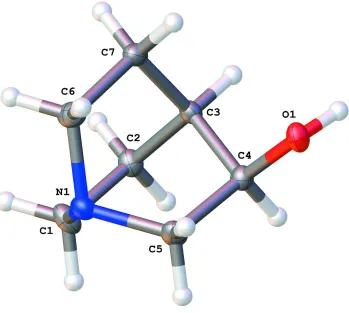
S1. Comment
(R)-(-)-quinuclidin-3-ol (Figure 1) is a key building block for the syntheses of muscarinic receptor ligands, including
solifenacin (M3 receptor antagonist),(Naito et al., 2005) revatropate (M3 receptor antagonist),(Alabaster, 1997) and
talsaclidine (M1 receptor agonist),(Leusch et al., 2000).
The asymetric unit of the crystal (Figure 1) consists of one single (R)-(-)-quinuclidin-3-ol molecule.
The quinuclidinol moiety has pseudo-threefold symmetry about the N1-C3 axis with C6-N1-C1, C1-N1-C5 and
C5-N1-N6 angles of 108.86 (15)°, 108.73 (14)° and 108.72 (14)° respectively, and N1-C1-C2-C3, N1-C6-C7-C3 and
N1-C5-C4-C3 torsion angles of -0.9 (3)°, 0.4 (2)° and 0.2 (2)° respectively.
The three piperidine rings formed by (N1, C1, C2, C3, C4, C5), (N1, C5, C4, C3, C7, C6) and (N1, C6, C7, C3, C2, C1)
adopt a boat conformation with total puckering amplitutdes QT of 0.8218 (0) (with Θ = 91.84 (0)° and φ = -0.2 (0)°), QT
of 0.8141 (1) (with Θ = 91.55 (0)° and φ = 0.19 (0)°) and QT of 0.8123 (0) (with Θ = 90.95 (0)° and φ = -0.17 (0)°),
respectively (Cremer & Pople, (1975)).
There is a hydrogen bond (Table 1) which links molecules into infinite chains along the c axis (Figure 2).
S2. Refinement
All H atoms, on carbon atom or oxygen atom, were placed at calculated positions using a riding model with C-H = 1 Å
(methine), 0.99 Å (methylene) or O-H = 0.84 Å with Uiso(H) = 1.2Ueq(CH), Uiso(H) = 1.2Ueq(CH2) or Uiso(H) =
1.5Ueq(OH).
TWIN/BASF refinement type was used to determine absolute configuration from anomalous scattering using the Flack
method. (Flack, 1983). The structure display a merohedral twinning and the twin law was found by using TwinRotMat
implemented in Platon (Spek, 2009). The use of twin law (0 1 0 1 0 0 0 0 -1) with a population of 0.925/0.075 reduced
Figure 1
A view of (R)-(-)-quinuclidin-3-ol with atom labelling scheme. The thermal displacement ellipsoids are drawn at 50%
probability level.
Figure 2
A view of molecular packing showing chains running along the c direction. The hydrogen bonds are shown as dashed
lines. The thermal displacement ellipsoids are drawn at 50% probability level.
(R)-(-)-Quinuclidin-3-ol
Crystal data
C7H13NO
Mr = 127.18
Hexagonal, P61
[image:3.610.127.483.438.583.2]c = 29.8731 (13) Å
V = 996.91 (11) Å3
Z = 6
F(000) = 420
Dx = 1.271 Mg m−3
Melting point: 492(2) K
Cu Kα1 radiation, λ = 1.54178 Å
µ = 0.67 mm−1
T = 100 K
Prism, clear light colourless 0.58 × 0.44 × 0.32 mm
Data collection
Bruker D8 VENTURE diffractometer
Radiation source: sealed X-ray tube, high brilliance microfocus sealed tube, Cu Graphite monochromator
Detector resolution: 1024 x 1024 pixels mm-1
φ and ω scans
Absorption correction: numerical (SADABS; Bruker, 2012)
Tmin = 0.58, Tmax = 0.74
15447 measured reflections 1240 independent reflections 1240 reflections with I > 2σ(I)
Rint = 0.026
θmax = 69.2°, θmin = 4.4°
h = −7→7
k = −7→7
l = −34→35
Refinement
Refinement on F2
Least-squares matrix: full
R[F2 > 2σ(F2)] = 0.023
wR(F2) = 0.064
S = 1.15
1240 reflections 85 parameters 1 restraint 0 constraints
Primary atom site location: structure-invariant direct methods
Hydrogen site location: inferred from neighbouring sites
H-atom parameters constrained
w = 1/[σ2(F
o2) + (0.0394P)2 + 0.1183P]
where P = (Fo2 + 2Fc2)/3
(Δ/σ)max < 0.001
Δρmax = 0.23 e Å−3
Δρmin = −0.12 e Å−3
Extinction correction: SHELXL97 (Sheldrick,
2008), Fc*=kFc[1+0.001xFc2λ3/sin(2θ)]-1/4
Extinction coefficient: 0.0156 (15)
Absolute structure: Parsons & Flack (2004). Absolute structure parameter: 0.01 (4)
Special details
Geometry. All e.s.d.'s (except the e.s.d. in the dihedral angle between two l.s. planes) are estimated using the full covariance matrix. The cell e.s.d.'s are taken into account individually in the estimation of e.s.d.'s in distances, angles and torsion angles; correlations between e.s.d.'s in cell parameters are only used when they are defined by crystal symmetry. An approximate (isotropic) treatment of cell e.s.d.'s is used for estimating e.s.d.'s involving l.s. planes.
Refinement. Refined as a 2-component twin.
Fractional atomic coordinates and isotropic or equivalent isotropic displacement parameters (Å2)
x y z Uiso*/Ueq
O1 −0.1928 (2) 0.4528 (2) 0.48963 (4) 0.0175 (3)
H1 −0.2253 0.4264 0.4622 0.026*
N1 0.3057 (3) 0.6832 (3) 0.56404 (5) 0.0157 (4)
C2 0.5095 (3) 0.8018 (3) 0.48879 (6) 0.0166 (4)
H2A 0.5325 0.9500 0.4722 0.020*
H2B 0.6378 0.7625 0.4786 0.020*
C4 0.0565 (3) 0.6495 (3) 0.49520 (5) 0.0149 (4)
H4 0.0812 0.7999 0.4785 0.018*
C1 0.5354 (4) 0.8550 (4) 0.53973 (6) 0.0208 (4)
H1B 0.5744 1.0283 0.5450 0.025*
C7 0.2139 (3) 0.3542 (3) 0.50634 (5) 0.0159 (4)
H7A 0.3369 0.3067 0.4966 0.019*
H7B 0.0450 0.2111 0.5013 0.019*
C5 0.0986 (4) 0.7091 (4) 0.54597 (5) 0.0188 (4)
H5A 0.1340 0.8814 0.5510 0.023*
H5B −0.0557 0.5956 0.5624 0.023*
C3 0.2487 (3) 0.5801 (3) 0.47977 (6) 0.0141 (4)
H3 0.2279 0.5406 0.4471 0.017*
C6 0.2509 (4) 0.4253 (4) 0.55649 (6) 0.0204 (4)
H6A 0.0984 0.3095 0.5732 0.024*
H6B 0.3897 0.4068 0.5684 0.024*
Atomic displacement parameters (Å2)
U11 U22 U33 U12 U13 U23
O1 0.0134 (6) 0.0222 (6) 0.0150 (6) 0.0076 (5) −0.0012 (4) −0.0004 (5)
N1 0.0176 (7) 0.0155 (8) 0.0128 (7) 0.0074 (6) −0.0005 (6) −0.0014 (5)
C2 0.0149 (8) 0.0186 (9) 0.0145 (8) 0.0069 (7) 0.0009 (7) 0.0010 (6)
C4 0.0150 (8) 0.0161 (8) 0.0138 (8) 0.0079 (7) −0.0002 (6) 0.0006 (7)
C1 0.0176 (9) 0.0215 (9) 0.0164 (9) 0.0045 (8) −0.0020 (6) −0.0034 (7)
C7 0.0150 (8) 0.0151 (8) 0.0177 (8) 0.0077 (7) −0.0012 (6) −0.0028 (7)
C5 0.0207 (9) 0.0237 (9) 0.0153 (8) 0.0135 (8) −0.0006 (7) −0.0042 (7)
C3 0.0135 (8) 0.0158 (8) 0.0124 (8) 0.0068 (7) −0.0011 (6) −0.0024 (7)
C6 0.0292 (9) 0.0186 (9) 0.0159 (9) 0.0139 (8) −0.0003 (8) 0.0013 (7)
Geometric parameters (Å, º)
O1—H1 0.8400 C1—H1A 0.9900
O1—C4 1.423 (2) C1—H1B 0.9900
N1—C1 1.475 (2) C7—H7A 0.9900
N1—C5 1.475 (2) C7—H7B 0.9900
N1—C6 1.479 (2) C7—C3 1.530 (2)
C2—H2A 0.9900 C7—C6 1.546 (2)
C2—H2B 0.9900 C5—H5A 0.9900
C2—C1 1.548 (2) C5—H5B 0.9900
C2—C3 1.536 (2) C3—H3 1.0000
C4—H4 1.0000 C6—H6A 0.9900
C4—C5 1.552 (2) C6—H6B 0.9900
C4—C3 1.528 (2)
C4—O1—H1 109.5 C3—C7—H7A 110.1
C1—N1—C6 108.86 (15) C3—C7—H7B 110.1
C5—N1—C1 108.73 (14) C3—C7—C6 107.96 (13)
C5—N1—C6 108.72 (14) C6—C7—H7A 110.1
H2A—C2—H2B 108.4 C6—C7—H7B 110.1
C1—C2—H2A 110.0 N1—C5—C4 112.56 (14)
C3—C2—H2A 110.0 N1—C5—H5B 109.1
C3—C2—H2B 110.0 C4—C5—H5A 109.1
C3—C2—C1 108.29 (15) C4—C5—H5B 109.1
O1—C4—H4 109.6 H5A—C5—H5B 107.8
O1—C4—C5 107.46 (13) C2—C3—H3 109.9
O1—C4—C3 112.96 (14) C4—C3—C2 108.41 (14)
C5—C4—H4 109.6 C4—C3—C7 109.25 (14)
C3—C4—H4 109.6 C4—C3—H3 109.9
C3—C4—C5 107.45 (13) C7—C3—C2 109.45 (14)
N1—C1—C2 111.72 (14) C7—C3—H3 109.9
N1—C1—H1A 109.3 N1—C6—C7 112.19 (14)
N1—C1—H1B 109.3 N1—C6—H6A 109.2
C2—C1—H1A 109.3 N1—C6—H6B 109.2
C2—C1—H1B 109.3 C7—C6—H6A 109.2
H1A—C1—H1B 107.9 C7—C6—H6B 109.2
H7A—C7—H7B 108.4 H6A—C6—H6B 107.9
O1—C4—C5—N1 122.04 (17) C5—C4—C3—C2 −59.75 (17)
O1—C4—C3—C2 −178.09 (13) C5—C4—C3—C7 59.46 (17)
O1—C4—C3—C7 −58.88 (17) C3—C2—C1—N1 −0.9 (2)
C1—N1—C5—C4 59.38 (18) C3—C4—C5—N1 0.20 (19)
C1—N1—C6—C7 −59.78 (19) C3—C7—C6—N1 0.4 (2)
C1—C2—C3—C4 60.54 (19) C6—N1—C1—C2 59.72 (19)
C1—C2—C3—C7 −58.55 (18) C6—N1—C5—C4 −59.01 (19)
C5—N1—C1—C2 −58.6 (2) C6—C7—C3—C2 58.56 (18)
C5—N1—C6—C7 58.53 (19) C6—C7—C3—C4 −60.01 (17)
Hydrogen-bond geometry (Å, º)
D—H···A D—H H···A D···A D—H···A
O1—H1···N1i 0.84 2.00 2.8366 (19) 176