4-Diphenylphosphanyl-1,5-naphthyridine
Ya-Ming Wu
Department of Applied Chemistry, Nanjing College of Chemical Technology, No. 625 Geguan Road, Dachang, Nanjing 210048, People’s Republic of China Correspondence e-mail: [email protected]
Received 4 September 2012; accepted 20 September 2012
Key indicators: single-crystal X-ray study;T= 293 K; mean(C–C) = 0.004 A˚;
Rfactor = 0.047;wRfactor = 0.151; data-to-parameter ratio = 14.5.
The asymmetric unit of the title compound, C20H15N2P,
contains two independent molecules with similar structures. The 1,5-naphthyridine ring system is nearly planar, with maximum deviations of 0.010 (3) and 0.012 (3) A˚ ; its mean plane is oriented with respect to the two phenyl rings at 79.69 (12) and 84.00 (10)in one molecule, and at 74.25 (12) and 82.05 (11)in the other. The two phenyl rings are twisted with respect to each other with a dihedral angle of 75.96 (14) in one molecule and 86.30 (13) in the other.
Related literature
For applications of the title compound, see: Badawnehet al. (2001); Hawes et al. (1977); Goswami & Mukherjee (1997); Goswami et al. (2001, 2005). For the synthesis of the title compound, see: Chenet al.(2012).
Experimental
Crystal data
C20H15N2P Mr= 314.31
Triclinic,P1 a= 10.1103 (7) A˚ b= 11.7020 (8) A˚ c= 15.7060 (11) A˚
= 71.54 (3)
= 75.05 (3)
= 71.37 (3)
V= 1644.6 (4) A˚3
Z= 4
MoKradiation
= 0.17 mm 1
T= 293 K
0.300.200.20 mm
Data collection
Enraf–Nonius CAD-4 diffractometer
Absorption correction: scan (Northet al., 1968) Tmin= 0.952,Tmax= 0.967 6393 measured reflections
6024 independent reflections 4473 reflections withI> 2(I) Rint= 0.019
3 standard reflections every 200 reflections
intensity decay: 1%
Refinement
R[F2> 2(F2)] = 0.047
wR(F2) = 0.151 S= 1.00 6024 reflections
416 parameters
H-atom parameters constrained
max= 0.24 e A˚ 3
min= 0.18 e A˚ 3
Data collection: CAD-4 EXPRESS (Enraf–Nonius, 1994); cell refinement:CAD-4 EXPRESS; data reduction:XCAD4(Harms & Wocadlo, 1995); program(s) used to solve structure: SHELXS97 (Sheldrick, 2008); program(s) used to refine structure:SHELXL97 (Sheldrick, 2008); molecular graphics:SHELXTL(Sheldrick, 2008); software used to prepare material for publication:SHELXTL.
The author thanks the Center of Test and Analysis, Nanjing University, for data collection.
Supplementary data and figures for this paper are available from the IUCr electronic archives (Reference: XU5619).
References
Badawneh, M., Ferrarini, P. L., Calderone, V., Manera, C., Martinotti, E., Mori, C., Saccomanni, G. & Testai, L. (2001).Eur. J. Med. Chem.36, 925–934. Chen, C., Wang, K.-Y., Jiang, P., Song, G.-L. & Zhu, H.-J. (2012).Inorg. Chem.
Commun.17, 116–119.
Enraf–Nonius (1994).CAD-4 EXPRESS. Enraf–Nonius, Delft, The Nether-lands.
Goswami, S., Ghosh, K. & Mukherjee, R. (2001).Tetrahedron,57, 4987–4993. Goswami, S. & Mukherjee, R. (1997).Tetrahedron Lett.38, 1619–1621. Goswami, S., Mukherjee, R., Mukherjee, S., Jana, S., Maity, A. C. & Adak, A.
K. (2005).Molecules,10, 929–934.
Harms, K. & Wocadlo, S. (1995).XCAD4. University of Marburg, Germany. Hawes, E. M., Gorecki, D. K. J. & Gedir, G. G. (1977).J. Med. Chem.20, 838–
841.
North, A. C. T., Phillips, D. C. & Mathews, F. S. (1968).Acta Cryst.A24, 351– 359.
Sheldrick, G. M. (2008).Acta Cryst.A64, 112–122.
Acta Crystallographica Section E
Structure Reports
Online
supporting information
Acta Cryst. (2012). E68, o3011 [https://doi.org/10.1107/S1600536812039992]
4-Diphenylphosphanyl-1,5-naphthyridine
Ya-Ming Wu
S1. Comment
The title compound, (I), is an important intermediate in medicine (Badawneh et al., 2001; Hawes et al., 1977).
Naphthyridines are also used as a key molecule in molecular recognition chemistry (Goswami & Mukherjee, 1997;
Goswami et al., 2005, 2001).
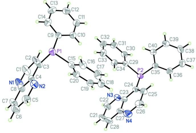
The molecular structure of (I) is shown in Fig. 1. The asymmetric unit of the title compound, C20H15N2P, contains two
independent molecules with the similar structure. The 1,5-naphthrydine ring system is nearly planar with the maximum
deviation of 0.010 (3) and 0.012 (3) Å, respectively; its mean plane is oriented with respect to the two phenyl rings at
79.69 (12) and 84.00 (10)° in one molecule and 74.25 (12) and 82.05 (11)° in the other. The two phenyl rings are twisted
to each other with a dihedral angle of 75.96 (14)° in one molecule and 86.30 (13)° in the other.
The crystal packing of the molecules in the crystal is influenced by van der Waals forces.
S2. Experimental
The title compound was synthesized according to the published procedure (Chen et al., 2012). Crystals suitable for X-ray
analysis were obtained by dissolving it(0.5 g) in tetrahydrofuran (20 ml) and evaporating the solvent slowly at room
temperature for about 5 d.
S3. Refinement
H atoms were positioned geometrically, with C—H = 0.93 Å for aromatic, and constrained to ride on their parent atoms,
Figure 1
The molecular structure of (I), with the atom-numbering scheme. Displacement ellipsoids are drawn at the 50%
[image:3.610.136.482.363.583.2]probability level.
Figure 2
A packing diagram of (I).
4-Diphenylphosphanyl-1,5-naphthyridine
Crystal data
C20H15N2P Mr = 314.31 Triclinic, P1 Hall symbol: -P 1
a = 10.1103 (7) Å
b = 11.7020 (8) Å
c = 15.7060 (11) Å
β = 75.05 (3)°
γ = 71.37 (3)°
V = 1644.6 (4) Å3
Z = 4
F(000) = 656
Dx = 1.269 Mg m−3
Mo Kα radiation, λ = 0.71073 Å
Cell parameters from 25 reflections
θ = 10–14°
µ = 0.17 mm−1 T = 293 K Block, yellow
0.30 × 0.20 × 0.20 mm
Data collection
Enraf–Nonius CAD-4 diffractometer
Radiation source: fine-focus sealed tube Graphite monochromator
ω/2θ scans
Absorption correction: ψ scan (North et al., 1968)
Tmin = 0.952, Tmax = 0.967 6393 measured reflections
6024 independent reflections 4473 reflections with I > 2σ(I)
Rint = 0.019
θmax = 25.4°, θmin = 1.4°
h = 0→12
k = −13→14
l = −18→18
3 standard reflections every 200 reflections intensity decay: 1%
Refinement
Refinement on F2
Least-squares matrix: full
R[F2 > 2σ(F2)] = 0.047 wR(F2) = 0.151 S = 1.00 6024 reflections 416 parameters 0 restraints
Primary atom site location: structure-invariant direct methods
Secondary atom site location: difference Fourier map
Hydrogen site location: inferred from neighbouring sites
H-atom parameters constrained
w = 1/[σ2(F
o2) + (0.1P)2 + 0.1P]
where P = (Fo2 + 2Fc2)/3
(Δ/σ)max < 0.001
Δρmax = 0.24 e Å−3
Δρmin = −0.18 e Å−3
Extinction correction: SHELXL97 (Sheldrick, 2008), Fc*=kFc[1+0.001xFc2λ3/sin(2θ)]-1/4
Extinction coefficient: 0.056 (4)
Special details
Geometry. All e.s.d.'s (except the e.s.d. in the dihedral angle between two l.s. planes) are estimated using the full covariance matrix. The cell e.s.d.'s are taken into account individually in the estimation of e.s.d.'s in distances, angles and torsion angles; correlations between e.s.d.'s in cell parameters are only used when they are defined by crystal symmetry. An approximate (isotropic) treatment of cell e.s.d.'s is used for estimating e.s.d.'s involving l.s. planes.
Refinement. Refinement of F2 against ALL reflections. The weighted R-factor wR and goodness of fit S are based on F2,
conventional R-factors R are based on F, with F set to zero for negative F2. The threshold expression of F2 > σ(F2) is used
only for calculating R-factors(gt) etc. and is not relevant to the choice of reflections for refinement. R-factors based on F2
are statistically about twice as large as those based on F, and R- factors based on ALL data will be even larger.
Fractional atomic coordinates and isotropic or equivalent isotropic displacement parameters (Å2)
x y z Uiso*/Ueq
C30 0.6058 (3) 0.8561 (2) 0.52547 (16) 0.0610 (6) H30A 0.6070 0.8830 0.5749 0.073* C31 0.4878 (3) 0.8223 (3) 0.5231 (2) 0.0713 (7) H31A 0.4111 0.8260 0.5708 0.086* C32 0.4836 (3) 0.7835 (3) 0.4509 (2) 0.0721 (7) H32A 0.4041 0.7613 0.4490 0.087* C33 0.5972 (3) 0.7775 (3) 0.3813 (2) 0.0725 (7) H33A 0.5944 0.7509 0.3320 0.087* C34 0.7168 (3) 0.8104 (2) 0.38273 (16) 0.0581 (6) H34A 0.7932 0.8056 0.3348 0.070* C35 0.9758 (2) 0.7539 (2) 0.53228 (14) 0.0457 (5) C36 1.0835 (3) 0.7605 (2) 0.56829 (17) 0.0596 (6) H36A 1.1033 0.8369 0.5566 0.072* C37 1.1621 (3) 0.6555 (3) 0.62125 (19) 0.0711 (7) H37A 1.2359 0.6611 0.6437 0.085* C38 1.1314 (3) 0.5431 (3) 0.64080 (19) 0.0710 (7) H38A 1.1833 0.4725 0.6773 0.085* C39 1.0240 (3) 0.5347 (2) 0.6064 (2) 0.0745 (8) H39A 1.0029 0.4584 0.6200 0.089* C40 0.9475 (3) 0.6386 (2) 0.55198 (17) 0.0614 (6) H40A 0.8761 0.6318 0.5281 0.074*
Atomic displacement parameters (Å2)
U11 U22 U33 U12 U13 U23
P2 0.0514 (3) 0.0457 (3) 0.0399 (3) −0.0145 (3) −0.0011 (2) −0.0099 (2) N3 0.0741 (14) 0.0552 (12) 0.0528 (12) −0.0134 (11) −0.0118 (10) −0.0054 (10) N4 0.0797 (16) 0.0955 (18) 0.0497 (13) −0.0305 (14) 0.0115 (11) −0.0212 (13) C21 0.131 (3) 0.076 (2) 0.0599 (18) −0.040 (2) −0.0297 (19) 0.0105 (15) C22 0.100 (2) 0.0591 (16) 0.0706 (18) −0.0137 (15) −0.0275 (16) −0.0041 (14) C23 0.0656 (15) 0.0512 (13) 0.0450 (12) −0.0242 (11) −0.0039 (11) −0.0102 (10) C24 0.0541 (13) 0.0511 (12) 0.0437 (12) −0.0201 (10) 0.0004 (10) −0.0113 (10) C25 0.0613 (15) 0.0650 (15) 0.0517 (14) −0.0169 (12) 0.0016 (11) −0.0161 (12) C26 0.0690 (17) 0.091 (2) 0.0613 (17) −0.0173 (15) 0.0115 (14) −0.0318 (16) C27 0.0821 (19) 0.0720 (16) 0.0451 (13) −0.0389 (15) −0.0019 (12) −0.0106 (12) C28 0.113 (3) 0.089 (2) 0.0459 (15) −0.046 (2) −0.0079 (16) 0.0005 (15) C29 0.0485 (12) 0.0437 (11) 0.0423 (11) −0.0096 (9) −0.0046 (9) −0.0066 (9) C30 0.0565 (14) 0.0710 (16) 0.0505 (13) −0.0174 (12) 0.0016 (11) −0.0166 (12) C31 0.0525 (15) 0.0784 (18) 0.0716 (17) −0.0200 (13) 0.0041 (13) −0.0130 (15) C32 0.0509 (15) 0.0708 (17) 0.093 (2) −0.0185 (13) −0.0168 (14) −0.0125 (16) C33 0.0710 (18) 0.0775 (18) 0.0796 (19) −0.0197 (14) −0.0203 (15) −0.0284 (15) C34 0.0558 (14) 0.0643 (15) 0.0550 (14) −0.0151 (12) −0.0057 (11) −0.0199 (12) C35 0.0473 (12) 0.0491 (12) 0.0387 (11) −0.0152 (10) −0.0005 (9) −0.0112 (9) C36 0.0610 (15) 0.0573 (14) 0.0650 (15) −0.0188 (12) −0.0118 (12) −0.0178 (12) C37 0.0668 (16) 0.0730 (18) 0.0808 (19) −0.0118 (14) −0.0299 (14) −0.0225 (15) C38 0.0730 (18) 0.0622 (16) 0.0714 (17) −0.0063 (13) −0.0283 (14) −0.0071 (13) C39 0.0841 (19) 0.0503 (14) 0.088 (2) −0.0216 (14) −0.0300 (16) 0.0002 (13) C40 0.0645 (15) 0.0556 (14) 0.0657 (15) −0.0242 (12) −0.0207 (12) −0.0015 (12)
Geometric parameters (Å, º)
C10—H10A 0.9300 C30—H30A 0.9300 C11—C12 1.359 (4) C31—C32 1.362 (4) C11—H11A 0.9300 C31—H31A 0.9300 C12—C13 1.365 (5) C32—C33 1.368 (4) C12—H12A 0.9300 C32—H32A 0.9300 C13—C14 1.363 (4) C33—C34 1.389 (4) C13—H13A 0.9300 C33—H33A 0.9300 C14—H14A 0.9300 C34—H34A 0.9300 C15—C16 1.392 (3) C35—C36 1.382 (3) C15—C20 1.394 (3) C35—C40 1.390 (3) C16—C17 1.375 (4) C36—C37 1.379 (3) C16—H16A 0.9300 C36—H36A 0.9300 C17—C18 1.380 (4) C37—C38 1.371 (4) C17—H17A 0.9300 C37—H37A 0.9300 C18—C19 1.381 (4) C38—C39 1.373 (4) C18—H18A 0.9300 C38—H38A 0.9300 C19—C20 1.377 (3) C39—C40 1.375 (3) C19—H19A 0.9300 C39—H39A 0.9300 C20—H20A 0.9300 C40—H40A 0.9300
C7—C8—C4 117.3 (2) C27—C28—H28A 120.3 C10—C9—C14 116.6 (2) C34—C29—C30 117.7 (2) C10—C9—P1 124.08 (18) C34—C29—P2 124.86 (17) C14—C9—P1 119.2 (2) C30—C29—P2 117.40 (17) C11—C10—C9 121.4 (2) C31—C30—C29 121.4 (2) C11—C10—H10A 119.3 C31—C30—H30A 119.3 C9—C10—H10A 119.3 C29—C30—H30A 119.3 C12—C11—C10 120.4 (3) C32—C31—C30 120.2 (2) C12—C11—H11A 119.8 C32—C31—H31A 119.9 C10—C11—H11A 119.8 C30—C31—H31A 119.9 C11—C12—C13 119.7 (3) C31—C32—C33 119.4 (2) C11—C12—H12A 120.1 C31—C32—H32A 120.3 C13—C12—H12A 120.1 C33—C32—H32A 120.3 C14—C13—C12 119.8 (3) C32—C33—C34 121.2 (3) C14—C13—H13A 120.1 C32—C33—H33A 119.4 C12—C13—H13A 120.1 C34—C33—H33A 119.4 C13—C14—C9 122.1 (3) C29—C34—C33 120.1 (2) C13—C14—H14A 119.0 C29—C34—H34A 119.9 C9—C14—H14A 119.0 C33—C34—H34A 119.9 C16—C15—C20 118.2 (2) C36—C35—C40 118.1 (2) C16—C15—P1 118.02 (17) C36—C35—P2 118.21 (17) C20—C15—P1 123.79 (17) C40—C35—P2 123.60 (17) C17—C16—C15 121.0 (2) C37—C36—C35 121.1 (2) C17—C16—H16A 119.5 C37—C36—H36A 119.5 C15—C16—H16A 119.5 C35—C36—H36A 119.5 C16—C17—C18 120.1 (2) C38—C37—C36 119.9 (2) C16—C17—H17A 120.0 C38—C37—H37A 120.0 C18—C17—H17A 120.0 C36—C37—H37A 120.0 C17—C18—C19 119.9 (2) C37—C38—C39 120.0 (2) C17—C18—H18A 120.1 C37—C38—H38A 120.0 C19—C18—H18A 120.1 C39—C38—H38A 120.0 C20—C19—C18 120.1 (2) C38—C39—C40 120.2 (2) C20—C19—H19A 120.0 C38—C39—H39A 119.9 C18—C19—H19A 120.0 C40—C39—H39A 119.9 C19—C20—C15 120.8 (2) C39—C40—C35 120.7 (2) C19—C20—H20A 119.6 C39—C40—H40A 119.6 C15—C20—H20A 119.6 C35—C40—H40A 119.6