RESEARCH ARTICLE
Should I stay or should I go? The settlement-inducing protein
complex guides barnacle settlement decisions
Manto Kotsiri1, Maria Protopapa1, Sofoklis Mouratidis1, Michael Zachariadis1,2, Demetrios Vassilakos1, Ioannis Kleidas1, Martina Samiotaki3and Skarlatos G. Dedos1,*
ABSTRACT
Reproduction in barnacles relies on chemical cues that guide their gregarious settlement. These cues have been pinned down to several sources of settlement pheromones, one of which is a protein termed settlement-inducing protein complex (SIPC), a large glycoprotein acting as a pheromone to induce larval settlement and as an adhesive in surface exploration by the cyprids. Settlement assays in laboratory conditions withAmphibalanus(=Balanus)amphitritecyprids in the presence of SIPC showed that cyprids exhibit settlement preference behaviour at lower concentrations of SIPC [half maximal effective concentration (EC50)=3.73 nmol l−1] and settlement avoidance behaviour at higher concentrations (EC50=101 nmol l−1). By using truncated fragments of SIPC in settlement assays, we identify that domains at the N-terminus of SIPC transduce settlement preference cues that mask the settlement avoidance cues transduced by domains at its C-terminus. Removing the N-terminal 600 amino acids from SIPC resulted in truncated fragments that transduced only settlement avoidance cues to the cyprids. From the sexual reproduction point of view, this bimodal response of barnacles to SIPC suggests that barnacles will settle gregariously when conspecific cues are sparse but will not settle if conspecific cues inform of overcrowding that will increase reproductive competition and diminish their reproductive chances.
KEY WORDS: Barnacles,Amphibalanus amphitrite, Settlement-inducing protein complex, Gregarious settlement, Predation, Biofouling
INTRODUCTION
The reproductive behaviour of barnacles has always fascinated scientists, among them Charles Darwin (Darwin, 1854), partly because their sessile trait makes them adopt reproductive strategies that rely on proximity to conspecifics (Barnes and Barnes, 1977; Crisp and Meadows, 1962; Crisp, 1961), but also because these animals are model organisms for marine biofouling (Aldred and Clare, 2008; Rittschof and Cohen, 2004; Rittschof et al., 1984, 1992). The life cycle of these animals consists of the nauplius (larval) stage, the cyprid stage, in which the barnacle actively seeks
a preferred substrate for settlement, after which it metamorphoses into a juvenile barnacle, and then the adult stage, in which it is capable of sexual reproduction (Høeg and Møller, 2006). Early observations on the surface exploration behaviour of barnacles (Yule and Walker, 1985; Yule and Crisp, 1983; Crisp and Meadows, 1962; Crisp, 1961) led to the discovery of the proteinaceous nature of the signals that these animals use to inform their conspecifics of their presence and thus increase their reproductive chances (Matsumura et al., 1998b; Dreanno et al., 2006c). Several earlier reports (see Rittschof and Cohen, 2004; Rittschof, 1990) and further research have shown that this reproductive strategy comes with a cost, because the same chemical cue acts as a potent feeding stimulant for a barnacle
predator, the whelkAcanthinucella spirata(Zimmer et al., 2016).
Intense research efforts culminated in the discovery of settlement-inducing protein complex (SIPC), a glycoprotein that induces
conspecific larval settlement inAmphibalanus amphitrite(Darwin
1854) (Matsumura et al., 1998b; Dreanno et al., 2006a,b,c). Originally shown to comprise 3 subunits of 98, 88 and 76 kDa (Matsumura et al., 1998b), cloning of the cDNA that encodes for
SIPC fromA. amphitriterevealed that SIPC is a single protein of
1547 amino acids with a calculated molecular mass of 171.7 kDa (Dreanno et al., 2006c). It is firmly established that SIPC is an N-linked glycan-containing protein and that glycans, such as mannose, can induce larval settlement behaviour (Pagett et al., 2012; Dreanno et al., 2006b; Thiyagarajan et al., 2002). Research on
another barnacle species,Balanus glandula, has identified another
orthologous protein termed MULTIFUNCin (Ferrier et al., 2016), which shares 78% nucleotide sequence identity with SIPC and is expressed as three subunits of 98, 88 and 76 kDa in addition to the 199 kDa of the full-length protein (Ferrier et al., 2016). Positive-settlement-directed purification of MULTIFUNCin and its subunits indicated that only the 199 kDa and the 98 kDa proteins were inducing settlement of the cyprids of this species (Ferrier et al., 2016). These authors reported failure to express MULTIFUNCin in
Escherichia coliandPichia pastorisheterologous systems (Ferrier
et al., 2016). Another study, however, reported the expression of a
recombinant SIPC fromA. amphitritein a baculovirus expression
system (Zhang et al., 2016). This recombinant SIPC from
A. amphitritewas lacking its N-terminal signal peptide sequence,
had an apparent molecular mass of around 200 kDa and did not induce cyprid settlement (Zhang et al., 2016). This inability of
heterologously expressedA. amphitriteSIPC to induce settlement
of the cyprids is in disagreement with results from another
study, which showed that native SIPC, purified fromA. amphitrite
homogenates, induces cyprid settlement with a half maximal
effective concentration (EC50) of 102 nmol l−1(Pagett et al., 2012).
Throughout the extensive literature pertaining to the
identification of SIPC or other orthologous proteins (Dreanno et al., 2007; Ferrier et al., 2016; Matsumura et al., 1998b), emphasis
Received 23 May 2018; Accepted 1 October 2018
1Department of Biology, National and Kapodistrian University of Athens,
Athens 157 84, Greece.2Institute of Biosciences and Applications, National Center
for Scientific Research‘Demokritos’, Agia Paraskevi, Athens 15310, Greece.
3Biomedical Sciences Research Center‘Alexander Fleming’, Fleming 34,
16672 Vari, Greece.
*Author for correspondence ([email protected])
M.K., 0000-0001-6949-8490; M.P., 9030-1469; S.M., 0002-0388-3369; D.V., 0003-2695-0795; I.K., 0003-3232-0167; S.G.D., 0000-0002-0432-338X
Journal
of
Experimental
is given to positive-settlement-directed purification, i.e. to identifying and reporting only subunits that exhibit a settlement-inducing response; therefore, it is critical to note that such positive-settlement assay designs may not allow for the identification of any
negative settlement cues. Therefore, comparing the results fromB.
glandulaMULTIFUNCin (Ferrier et al., 2016) and theA. amphitrite
SIPC (Pagett et al., 2012), there is ambiguity as to which of the SIPC subunits or the full-length protein exert the settlement behaviour. Is it the N-terminal 98 kDa subunit (Pagett et al., 2012; Ferrier et al., 2016; Matsumura et al., 1998b; Dreanno et al., 2006c) that exerts the settlement cues? Are the post-translational modifications (PTMs) of SIPC, which are critical for its activity as a pheromone cue (Aldred and Clare, 2008; Matsumura et al., 1998b; Zhang et al., 2016; Dreanno et al., 2007, 2006a), solely responsible for its biological
activity or is the reported EC50of 102 nmol l−1(Pagett et al., 2012)
of native SIPC a composite effect brought about by the simultaneous presence of all three subunits (Matsumura et al., 1998b)?
Attempting to answer these confounding questions, in this study we engineered, expressed and purified a recombinant form of SIPC
fromA. amphitriteas well as a systematic series of SIPC truncated
fragments to identify whether SIPC, its putative subunits (Matsumura et al., 1998b; Dreanno et al., 2006a) or its PTMs (Pagett et al., 2012) are responsible for its biological activity. We engineered a recombinant form of SIPC and its truncated fragments to contain an N-terminal myc-tag and a C-terminal 6×His-tag to be able to detect and purify the full-length protein or its fragments from heterologous expression systems. By developing a systematic series of truncated SIPC fragments that were overexpressed and purified from heterologous expression systems, we were aiming to identify,
in settlement bioassays ofA. amphitrite cyprids (Rittschof et al.,
1984, 1992), the minimal domain(s) that confer the species-specific biological activity of SIPC, be it protein domain(s) or domain-specific glycosylations (Pagett et al., 2012; Dreanno et al., 2007).
We show here that SIPC is a single glycoprotein, with a molecular mass substantially larger than its calculated molecular mass, that can transduce both settlement preference and settlement avoidance
cues and thus guide a cyprid’s decision to settle on a substrate or not.
We also show that domains in the N-terminal 600 amino acids of SIPC, possibly through their extensive glycosylations, act as settlement preference cues. When these 600 amino acids are sequentially removed in recombinant truncated fragments of SIPC, the remaining domains of SIPC dose-dependently transduce only settlement avoidance cues. Although puzzling at a first impression, the settlement avoidance cues conferred by high concentrations of the full-length recombinant SIPC may serve to reduce reproductive competition (Crisp, 1961) by conspecifics or may actually be a covert predator avoidance strategy (Zimmer et al., 2016).
MATERIALS AND METHODS Animals
Adult A. amphitrite (Cirripedia, Balanidae) were collected from
boat docks at Port Mikrolimano and Floisvos, Athens, Greece. Animals were cleaned of epibionts with a small hard brush and meticulously identified as individuals of the species (Bernard and Lane, 1962; Costlow, 1956). Adult barnacles were kept in separate
aerated glass tanks (20 l) containing 200-μm-filtered natural
seawater at 27°C and a 12 h:12 h light:dark photoperiod. Tanks
were fed 24-h-hatchedArtemiasp. (class: Branchiopoda) nauplii
and Tetraselmis suecica (class: Chlorodendrophyceae) and
Skeletonema costatum (class: Bacillariophyceae) algae each day,
and seawater was changed on alternate days. Upon stress to induce oviposition (24 h exposure to air or immersing in fresh water for
5 h), adults were returned to seawater for larval release. Hatched nauplii were attracted to a point light source, collected and placed
into a beaker containing 2 or 3 l of 0.7μm GF/F (Whatman)-filtered
natural seawater at a density of approximately 1–2 nauplii ml−1with
gentle aeration. Nauplii were maintained at 27°C at a 12 h:12 h
light:dark photoperiod on a diet of Chaetoceros gracilis (class:
Bacillariophyceae) provided at a density of 2×10⁵ cells ml−1
(Matsumura et al., 1998a; Thiyagarajan et al., 2002). Cultured in these conditions, nauplii metamorphosed to cyprids in 5 days. Aliquots of these cyprids were collected with a wide-mouthed Pasteur pipette and aged at 4°C (Phang et al., 2009) for 1 day prior to use in settlement assays. Only batches of cyprids that were active and had numerous oil cells, representing energy reserves (Lucas et al., 1979), were used in settlement assays (Rittschof et al., 1984, 1992).
Construction of recombinant SIPC and truncated SIPC fragments
We obtained a plasmid containing the full-length cDNA of the
A. amphitrite SIPC gene cloned in pCR4-TOPO (Dreanno et al.,
2006c) (Thermo Fisher Scientific). The full-length cDNA was
sequenced and then transferred as anEcoRI (NEB) fragment into
pENTR1α (Thermo Fisher Scientific). The sequencing results
revealed differences between the obtained clone and the original cDNA deposited in GenBank (acc. no.: AAR33079.1; see
Tables S1–S3). Differences were also observed between this
clone and the nucleotide and amino acid sequence of SIPC deposited by Zhang et al. (2016) (GenBank acc. no.: AMR58954.1;
see Tables S1–S3), which notably lacks the signal peptide sequence
entirely (Zhang et al., 2016). Sequence alignment of the cDNA we obtained and the two other cDNA sequences deposited in GenBank (Zhang et al., 2016; Dreanno et al., 2006c) showed that there are
differences between the three protein sequences (Tables S1–S3).
Bioinformatic analysis (see below) of the deduced amino acid sequence, encoded by the cDNA we obtained, revealed that the amino acid differences that we observed were not affecting any putative PTM site as judged by its primary structure.
We proceeded to correct the three amino acid differences (Tables S2 and S3) that we observed in the signal peptide sequence using a forward primer ( primer 1; Table S4) and a reverse primer ( primer 2; Table S4) designed to remove the
3′-untranslated region (UTR) and thus obtain the open reading
frame (ORF) of the gene. The PCR reaction conditions were: 95°C for 3 min as an initial step followed by 35 cycles of 95°C for 30 s,
46°C for 30 s and 68°C for 5 min usingPfxpolymerase (Thermo
Fisher Scientific) according to the manufacturer’s instructions. The
resulting PCR product of ∼4730 bp was cloned as an EcoRI
fragment in pENTR1α(Thermo Fisher Scientific) to generate clone
pENTR1α–SIPC–ORF.
To introduce a myc-tag (N–EQKLISEEDL–C) sequence
immediately after the cleavage site of the signal peptide, we used
primers 3 and 4 (see Table S4). Clone pENTR1α–SIPC–ORF was
used as template and PCR conditions were: 95°C for 3 min as an initial step followed by 35 cycles of 95°C for 30 s, 46°C for 30 s
and 68°C for 5 min using Pfx polymerase according to the
manufacturer’s instructions. Primers 3 and 4 were added at a final
concentration of 125 nmol l−1and primers 1 and 2 were added at a
final concentration of 300 nmol l−1 to the PCR reaction. The
resulting PCR product of ∼4760 bp was cloned as an EcoRI
fragment in pENTR1αto create pENTR1α–SIPC–Myc–ORF.
Next, we engineered a C-terminal His-tag on recombinant SIPC
by using a reverse primer ( primer 5; Table S4) that introduced a
Journal
of
Experimental
6×His tag just before the stop codon, and a forward primer ( primer 6; Table S4). The PCR conditions were identical to those described above but with shorter extension at 68°C for 1 min. The resulting
PCR product of 950 bp was digested withXbaI (NEB) and cloned
into pENTR1α–SIPC–Myc–ORF to create pENTR1α–SIPC–Myc–
ORF–6×His.
To this clone, which coded for an N-terminal myc-tagged and
C-terminal His-tagged SIPC, we introduced aSphI restriction site
just after the myc-tag using primers 7 and 8 (Table S4) and primers 1 and 5. The PCR conditions were: 95°C for 3 min as an initial step followed by 35 cycles of 95°C for 30 s, 46°C for 30 s and 68°C for
5 min using Pfx polymerase according to the manufacturer’s
instructions. Primers 7 and 8 were added at a final concentration
of 125 nmol l−1 and primers 1 and 5 were added at a final
concentration of 300 nmol l−1 to the PCR reaction. The resulting
PCR product of∼4760 bp was digested withEcoRI–XhoI (NEB)
and the resulting fragment of ∼2825 bp replaced a fragment of
similar size in the previous clone to create pENTR1α–SIPC–Myc–
SphI–ORF-6×His.
To generate the Venus variant of EGFP (Nagai et al., 2002), the pEGFP-N1 plasmid (Clontech) was used as template and the ORF of EGFP was amplified by PCR using primers 9 and 10 (Table S4)
and cloned as anEcoRI fragment in pENTR1α. This clone was used
as a template in PCR reactions that progressively mutated the ORF
of EGFP to its Venus variant using primers 11–22 (Table S4). The
PCR conditions were: 95°C for 3 min as an initial step followed by 35 cycles of 95°C for 30 s, 46°C for 30 s and 68°C for 1 min using
Pfx polymerase according to the manufacturer’s instructions.
Primers for mutagenesis ( primers 11–22) were added at a final
concentration of 125 nmol l−1and primers 9 and 10 were added at a
final concentration of 300 nmol l−1to the PCR reaction. Mutations
introduced to EGFP to change it to its Venus variant were: F47L, T66G, V69L, Q70M, S73A, M154T, V164A, S176G and T204Y (Nagai et al., 2002).
Upon completion of the introduced mutations, the resulting ORF
of Venus was subcloned as aSphI (NEB) fragment into pENTR1α–
SIPC–Myc–SphI–ORF–6×His to create pENTR1α–SIPC–Myc–
SphI–Venus–ORF–6×His.
To express the neurosecretory-vesicle-localised PTPRN2
(Caromile et al., 2010), first-strand cDNA was synthesised from
5μg total RNA from rat cerebellum (Takara) with 200 U
Superscript®III reverse transcriptase (Thermo Fisher Scientific) in
20μl reaction volume using an oligo(dT)20primer (Thermo Fisher
Scientific) plus random hexamers (Thermo Fisher Scientific) (50 ng
per 5μg total RNA) according to the manufacturer’s instructions.
The resulting cDNA was diluted with nuclease-free water (Thermo Fisher Scientific) before use in PCR. Using the NCBI reference sequence NM_031600.1 (Wasmeier and Hutton, 1996) as a guide,
first a 3.02 kb PCR product, which contained the ORF ofPTPRN2,
was amplified from the cDNA using primers 23 and 24 (Table S4). The PCR conditions were: 95°C for 3 min as an initial step followed by 35 cycles of 95°C for 30 s, 46°C for 30 s and 68°C for 4 min
usingPfxpolymerase according to the manufacturer’s instructions,
and the PCR product was digested withEcoRI–AgeI (NEB). Next,
the ORF of mTurquoise from mTurquoise2-ER (Goedhart et al., 2012) was amplified by PCR using primers 25 and 26 (Table S4). The PCR conditions were: 95°C for 3 min as an initial step followed by 35 cycles of 95°C for 30 s, 46°C for 30 s and 68°C for
1 min using Pfx polymerase according to the manufacturer’s
instructions. The PCR product was cloned as an EcoRI–XhoI
fragment in pENTR1α and then the resulting clone was digested
withEcoRI–AgeI and the 3.02 kbp PCR product of thePTPRN2
ORF was cloned in frame to generate pENTR1α–PTPRN2–
mTurquoise.
To replace the signal peptide sequence of SIPC, we used primer
27 (Table S4) and primer 5 and pENTR1α–SIPC–Myc–SphI–
ORF–6×His as template. The PCR conditions were: 95°C for 3 min
as an initial step followed by 35 cycles of 95°C for 30 s, 46°C for
30 s and 68°C for 5 min using Pfx polymerase according
to the manufacturer’s instructions. The resulting PCR product of
∼4760 bp was digested with EcoRI–XhoI and a fragment of
∼2825 bp replaced a fragment of similar size in pENTR1α–SIPC–
Myc–SphI–ORF–6×His to create pENTR1α–SpSIPC–Myc–SphI–
ORF–6×His. To this clone, the ORF of Venus was inserted in frame
at theSphI site to create pENTR1α–SpSIPC–Myc–SphI–Venus–
ORF–6×His.
To generate truncated fragments of SIPC, we used a structural model-based approach (see below) in conjunction with the bioinformatic analysis of the primary structure of SIPC (see
below). Using clone pENTR1α–SIPC–Myc–SphI-ORF–6×His as
template, we generated first 2 truncated SIPC fragments (Tr.2 and Tr.7) using primer 28 and 29 (Table S4), respectively, as forward primers and primer 5 as reverse primer. The PCR conditions were: 95°C for 3 min as an initial step followed by 35 cycles of 95°C for 30 s, 46°C for 30 s and 68°C for 4 min for Tr.2 and 2 min for Tr.7,
usingPfxpolymerase according to the manufacturer’s instructions.
The PCR products were cloned as SphI–XhoI fragments in
pENTR1α–SIPC–Myc–SphI–ORF–6×His to generate truncated
SIPC fragments Tr.2 and Tr.7. Subsequently, and based on the results we obtained from these truncated fragments, we generated truncated fragments Tr.1 and Tr.3 using primers 30 and 31 (Table S4) in an identical approach as above. Next, we generated truncated fragments Tr.4, Tr.5 and Tr.6 using primers 32, 33 and 34 (Table S4) in an identical approach as above by simply adjusting the PCR extension time at 68°C to 3 min. Finally, we generated truncated fragments Tr.8 and Tr.9 using primers 35 and 36 (Table S4) as forward primers and primer 37 (Table S4) as reverse primer in an identical approach as above by simply adjusting the PCR extension time at 68°C to 2 min. These last two PCR
products were digested with SphI–SpeI (NEB) and cloned into
pENTR1α–SIPC–Myc–SphI–ORF–6×His to generate truncated
fragments Tr.8 and Tr.9.
For expression in HEK293 cells, all constructs described above were transferred by recombination to the mammalian expression vector pcDNA3.2/V5-DEST (Thermo Fisher Scientific) using the Gateway LR Clonase II enzyme mix (Thermo Fisher Scientific)
according to the manufacturer’s instructions.
For baculovirus-mediated expression of SIPC in insect Sf9 cells,
constructs (1) pENTR1α–SIPC–Myc–SphI–ORF–6×His (hereafter
termed SIPC), (2) pENTR1α–SpSIPC–Myc–SphI–ORF–6×His
(hereafter termed SpSIPC), (3) pENTR1α–SIPC–Myc–SphI–
Venus–ORF–6×His (hereafter termed VSIPC) and (4) pENTR1α–
SpSIPC–Myc–SphI–Venus–ORF–6×His (hereafter termed
SpVSIPC) were recombined with baculovirus linear DNA using
the BaculoDirect™ C-Term Expression Kit (Thermo Fisher
Scientific) and the Gateway LR Clonase II enzyme mix (Thermo
Fisher Scientific) according to the manufacturer’s instructions.
All constructs were sequenced using the described primers and
additional primers 38–46 (Table S4) before transfer to expression
vectors. Notably, we observed no additional change or mutation in
the nucleotide sequence ofSIPCduring the construct engineering
process, an indication that theSIPC cDNA does not exhibit any
inherent instability during its propagation in E. coli that would
justify the presence of the observed nucleotide changes.
Journal
of
Experimental
Expression of recombinant SIPC in mammalian and baculovirus expression systems
HEK293 cells were seeded in culture flasks and transfected with expression vectors coding for SIPC, VSIPC and the various
truncated SIPC fragments when in density of 1×10⁶cells ml−1with
10μg of DNA per 1×10⁶cells ml−1of culture medium, using Escort
IV transfection reagent (Merck) at a 1:1 ratio of μg DNA:μl of
transfection reagent, according to the manufacturer’s instructions.
Medium and cells were separately harvested at 72 h
post-transfection. Medium was initially centrifuged at 90gfor 10 min
at 4°C to remove cells and then at 10,000gfor 10 min to remove
cellular debris, and then carefully aspirated and stored at −80°C
until use. Cells were removed by scraping in PBS and centrifuged
twice in the presence of PBS at 90gfor 10 min at 4°C to remove any
remaining medium, then resuspended in lysis buffer (50 mmol l−1
Tris pH 7.6, 150 mmol l−1 NaCl, 2 mmol l−1 EDTA, 1% Triton
X-100) and a protease inhibitor cocktail tablet/10 ml lysis buffer
(Roche Diagnostics) and stored at−80°C until use.
Recombinant baculoviruses expressing the various forms of full-length recombinant SIPC were propagated in Sf9 cells (Thermo Fisher Scientific) and, after 3 re-infections of Sf9 cells, the viral titre was determined using the end-point dilution assay. For large-scale
protein expression, Sf9 cells were cultured in Sf-900™ III SFM
medium and infected at a multiplicity of infection of 10. For each
batch, a total of 108 cells were infected. Medium and cells were
harvested at 72 h post-infection, and samples were processed
separately as described above and stored at−80°C until use.
Protein concentration in samples was determined with the RC
DC™Protein Assay kit II (Bio-Rad) using bovine serum albumin
(BSA) as standard. Protein samples were pre-incubated with
250 mmol l−1 dithiothreitol (DTT) before resolution in
one-dimensional (1D) SDS-PAGE gels of 5 or 8%
acrylamide/bis-acrylamide at 10μg protein/lane for cell samples and 20μl/lane for
culture medium samples.
For western blots, gels were wet transferred onto a nitrocellulose
membrane (Whatman) in transfer buffer [25 mmol l−1 Tris,
192 mmol l−1glycine, 20% (v/v) methanol] at 100 V constant at
4°C for 90 min. Nitrocellulose membranes were blocked by
incubation with 5% BSA in TBST (25 mmol l−1 Tris-HCl, pH
7.5, 150 mmol l−1 NaCl, 0.1% Tween 20) for 1 h at room
temperature. Membranes were then incubated for 1 h at room temperature with a Myc-Tag (9B11) mAb (Cell Signaling Technology) at 1:1000 dilution in TBST with 5% BSA, washed with TBST (3×5 min) and incubated with an HRP-conjugated anti-mouse antibody (1:1000, Cell Signaling Technology) for 1 h at room temperature. Immunoreactive bands were detected using the
20× LumiGLO® chemiluminescence kit (Cell Signaling
Technology) in an Alpha Innotech FluorChem 8800 imaging system.
Fluorescence confocal microscopy
HEK293 cells were seeded on poly-D-lysine (Merck)-coated
coverslips in 6-well plates at a density of 0.5×10⁶ cells/well and
transiently transfected with expression vectors for VSIPC and
PTPRN2–mTurquoise as described above. At 72 h
post-transfection, cells were washed 3 times with PBS ( pH 7.4), fixed in 1 ml of 4% PFA for 15 min at room temperature followed by 3
washes with PBS, and sealed with 10μl Duolink®In SituMounting
Medium with DAPI (Merck). Cells were imaged on a multiphoton confocal microscope Leica TCS SP8 MP equipped with an Argon laser (excitation lines at 458, 476, 488, 496 and 514 nm) and an IR MaiTai DeepSee Ti:Sapphire laser (Spectra-Physics) for
multiphoton applications. Images were acquired with the spectral detector of the microscope using appropriate emission wavelength
ranges: PTPRN2–mTurquoise was excited at 458 nm with the
Argon laser and emission was recorded between 465 and 500 nm, VSIPC was excited at 514 nm with the Argon laser and emission
recorded at 520–600 nm. DAPI was excited at 780 nm with the
MaiTai DeepSee laser and emission captured at 400–450 nm.
Images were acquired with the LAS X software (Leica Microsystems CMS GmbH). For imaging, suitable doubly
transfected cells were selected, and stacks of 20–30 optical
sections were recorded for each cell. Images are presented without any post-processing.
Purification of recombinant SIPC from insect cell culture medium
The 6×His-tagged recombinant SIPC (SpSIPC) recovered from the insect cell culture medium at 72 h post-infection was purified using Ni Sepharose 6 Fast Flow (GE Healthcare Life Science) and the following protocol: Ni beads prepared to 50% slurry with
binding buffer (20 mmol l−1sodium phosphate, 0.5 mol l−1NaCl,
20 mmol l−1imidazole, pH 7.4) were added at 1.5 ml per 30 ml of
culture medium and incubated at 4°C on a rotating shaker for 1 h. Beads were then washed (2×1 ml) with binding buffer and bound protein(s) were eluted from the beads with 4×0.5 ml elution buffer
(20 mmol l−1 sodium phosphate, 0.5 mol l−1 NaCl, 500 mmol l−1
imidazole, pH 7.4). Samples of the eluted fractions were analysed
by western blots and then fractions from several batches (n=6) were
pooled and dialysed against a HEPES buffer (20 mmol l−1HEPES,
200 mmol l−1NaCl, pH 7.4) using a Spectra/Por™cellulose ester
dialysis membrane (molecular mass cut-off 100 kDa; Thermo
Fisher Scientific). Samples were concentrated to 100μl using the
Amicon Ultra-15 centrifugal filter units (Merck) at 4000g for
15 min at 4°C. A fraction of these samples was reserved for liquid chromatography tandem-mass spectrometry (see below) and the remaining was dialysed against sterile artificial seawater (ASW)
(25‰) overnight in 4°C using Slide-A-Lyzer™dialysis cassettes
(molecular mass cut-off 100 kDa; Thermo Fisher Scientific).
Protein concentration was again determined using theRC DC™
Protein Assay kit II (Bio-Rad) and BSA as standard, and samples saved for use in settlement assays.
Purification of recombinant SIPC from mammalian cell culture medium
The 6×His-tagged recombinant SIPC (rSIPC) and its truncated fragments recovered from HEK293 cell culture medium were purified using Ni Sepharose 6 Fast Flow (GE Healthcare Life Science) and the following protocol: Ni beads prepared to 50%
slurry with binding buffer (20 mmol l−1 sodium phosphate,
0.5 mol l−1 NaCl, 20 mmol l−1imidazole, pH 7.4) were added at
1.5 ml per 30 ml of culture medium and incubated at 4°C on a rotating shaker for 1 h. Beads were then washed (2×1 ml) with binding buffer and bound protein(s) were eluted from the beads
with 4×0.5 ml elution buffer (20 mmol l−1 sodium phosphate,
0.5 mol l−1NaCl, 500 mmol l−1imidazole, pH 7.4). Samples of the
eluted fractions were analysed by western blot and then fractions
from several batches (n=4–7) were pooled and dialysed against
ddH2O for 2 h, then sterile sea water (36‰) for 2 h and finally
sterile ASW (25‰) overnight in 4°C using Spectra/Por™cellulose
ester dialysis membrane (molecular mass cut-off 100 kDa; Thermo Fisher Scientific). For truncated fragments of SIPC smaller than 100 kDa, a dialysis membrane with 14 kDa cut-off
was used (Merck). Protein concentration was determined using the
Journal
of
Experimental
RC DC™Protein Assay kit II (Bio-Rad) and BSA as standard, and samples saved for use in settlement assays.
Analysis of SIPC expression atA. amphitrite developmental stages
To identify whether the recombinant SIPC that we engineered has similar mobility in SDS-PAGE gels as the endogenous SIPC of
A. amphitrite, we generated a rabbit polyclonal antibody against a
peptide (N–IHKELKGGTERGGE–C) corresponding to amino
acids 1186–1199 of SIPC (GenBank acc. no.: AAR33079.1).
With a cysteine attached to its N-terminus, the 15 amino acid peptide was conjugated to keyhole limpet hecocyanin (KLH) and the conjugate was used to immunise rabbits. The antibody was produced and purified by GenScript (NJ, USA).
Monoclonal antibodies against the same peptide were produced according to a modified method of Köhler and Milstein (1975). Five BALB/c mice of 5 weeks of age were immunised intraperitoneally
(i.p.) with 25μg of the KLH-conjugated peptide ( peptide synthesis
and conjugation by GenScript). All immunisation and animal handling were in accordance with animal care guidelines as specified in EU Directive 2010/63/EU. After 5 cycles of immunisation, mice were euthanised and spleenocytes were
collected and fused with the P3X63Ag8.653 cell line (ATCC®
CRL1580™) according to the Köhler and Milstein (1975) fusion
protocol. Positive clones and antibody specificity were determined through immunoblotting and immunosorbent assays and, among the several positive clones, one was further propagated and used.
Nauplii, cyprids and freshly settled adults were cultured as described above. Nauplii were collected with a wide-mouthed
Pasteur pipette, whereas early attached cyprids of A. amphitrite
(24 h old) and metamorphosed juveniles (48 h old) were gently
scraped off the dishes. Animals were anaesthetised in 0.5 mol l−1
MgCl2and this solution was quickly replaced with 1μl/animal lysis
buffer (50 mmol l−1Tris, pH 7.6, 150 mmol l−1NaCl, 2 mmol l−1
EDTA, 1% Triton X-100) and a protease inhibitor cocktail tablet/
10 ml lysis buffer (Roche Diagnostics) and stored at−80°C until
use. After homogenisation with a small pestle, samples were sonicated 3 times (30 s with 2.5 min rest on ice) using an MSE Soniprep 150 ultrasonic disintegrator (Sanyo). Samples were
centrifuged at 15,000gfor 10 min at 4°C and the supernatant was
incubated with 250 mmol l−1 DTT and resolved in a 5%
acrylamide/bis-acrylamide SDS-PAGE gel.
Gels were wet transferred onto a nitrocellulose membrane in
transfer buffer [25 mmol l−1Tris, 192 mmol l−1glycine, 20% (v/v)
methanol] at 100 V constant at 4°C for 90 min. Nitrocellulose membranes were blocked by incubation with 5% BSA in TBST
(25 mmol l−1Tris-HCl, pH 7.5, 150 mmol l−1NaCl, 0.1% Tween
20) for 1 h at room temperature. Membranes were then incubated for 1 h at room temperature with the purified polyclonal anti-SIPC rabbit antibody at 1:1000 dilution in TBST with 5% BSA or with the mouse anti-SIPC monoclonal antibody at 1:100 dilution of the cloned cell line culture supernatant in TBST with 5% BSA. Membranes were then washed with TBST (3×5 min) and incubated with an HRP-conjugated anti-rabbit or HRP-conjugated anti-mouse antibody (1:1000, Jackson Laboratories) for 1 h at room temperature. Immunoreactive bands were detected using the 20×
LumiGLO®chemiluminescence kit (Cell Signaling Technology) in
an Alpha Innotech FluorChem 8800 imaging system.
Settlement bioassays
Assays were conducted by adding 12 cyprids into individual wells of a 24-well polystyrene sterile microplate (Orange Scientific)
containing 2 ml of ASW (25‰) and various concentrations of
recombinant SIPC. These experiments were repeated 3 times with
5 replicates for each concentration of recombinant SIPC (n=180
cyprids for each concentration of recombinant SIPC). An identical procedure was used for settlement assays in the presence of BSA (molecular biology grade from NEB).
Plates were covered and sealed with Parafilm® to avoid
evaporation, incubated at 25°C away from any light source, and examined after 24, 48, 72 and 96 h. Each animal was inspected under a stereomicroscope and its condition recorded. On each day, cyprids that did not move, had extended thoracopods and did not respond after a light touch with a Pasteur pipette were regarded as dead (Lau and Qian, 2000; Rittschof et al., 1992; Hellio et al., 2004). Both permanently attached and metamorphosed individuals were counted as settled or juveniles (Hellio et al., 2005; Rittschof
et al., 2003). The remainder were counted as ‘no settlement’.
Results were expressed as percentage of settlement, mortality and no settlement. For SIPC and its truncated fragments expressed in HEK293 cells, a similar approach was used but with fewer replicates
(n=48–96 cyprids) and fewer concentrations of each protein per
assay due to the substantially decreased yields of this expression system relative to the baculovirus expression system.
Adhesive properties of recombinant SIPC and truncated SIPC fragments
Adhesion assays of recombinant SIPC or its truncated fragments
(Tr.1 to Tr.9) were conducted by adding 300μl of each protein
sample (50 nmol l−1) into individual wells of a 24-well polystyrene
sterile microplate (Orange Scientific). Plates were covered and
sealed with Parafilm®to avoid evaporation and incubated at 25°C
away from any light source for 24 h. Then, samples were aspirated and wells were blocked by incubation with 5% BSA in TBST
(25 mmol l−1Tris-HCl, pH 7.5, 150 mmol l−1NaCl, 0.1% Tween
20) for 1 h at room temperature. Wells were then incubated for 1 h at room temperature with a Myc-Tag (9B11) mAb (Cell Signaling Technology) at 1:1000 dilution in TBST with 5% BSA, washed with TBST (3×5 min) and incubated with an HRP-conjugated anti-mouse antibody (1:1000, Cell Signaling Technology) for 1 h at room temperature. Wells were then washed with TBST (3×5 min)
and immunostaining was detected using the 20× LumiGLO®
chemiluminescence kit (Cell Signaling Technology) in a Bio-Rad ChemiDoc XRS+ Gel Photo Documentation system. Acquired high-resolution images were analysed with ImageJ 1.48v (National Institutes of Health) and data were exported and statistically analysed with GraphPad Prism v.6. These experiments were repeated 3 times with 2 replicates for each sample.
In experiments where the adhesive properties of native SIPC was assessed, settlement bioassays were conducted by adding 10 cyprids into individual wells of a 24-well polystyrene sterile microplate
(Orange Scientific) containing 2 ml of ASW (25‰). These
experiments were repeated 6 times with 3 replicates per
experiment. Plates were covered and sealed with Parafilm® to
avoid evaporation and incubated at 25°C away from any light source for 96 h. Then, the 2 ml of ASW was aspirated from each well and saved, and all settled and non-settled cyprids were counted and removed as well. Wells were then blocked by incubation with 5%
BSA in TBST (25 mmol l−1Tris-HCl, pH 7.5, 150 mmol l−1NaCl,
0.1% Tween 20) for 1 h at room temperature. Wells were then incubated for 1 h at room temperature with the mouse anti-SIPC monoclonal antibody at 1:100 dilution of the cloned cell line culture supernatant in TBST with 5% BSA, washed with TBST (3×5 min)
and incubated with an HRP-conjugated anti-mouse antibody
Journal
of
Experimental
(1:1000, Cell Signaling Technology) for 1 h at room temperature. Wells were then washed with TBST (3×5 min) and immunostaining
was detected using the 20× LumiGLO® chemiluminescence kit
(Cell Signaling Technology) in a Bio-Rad ChemiDoc XRS+ Gel Photo Documentation system.
In parallel, the 2 ml of ASW sample from each well was subjected to trichloroacetic acid (TCA) precipitation to identify native SIPC, according to the following protocol. A 0.5 ml volume of 100% (w/v) TCA was added to 2 ml of each ASW sample and incubated at 4°C for 10 min. Following centrifugation of each
sample at 20,800gfor 5 min at 4°C, supernatant was removed and
200μl ice-cold acetone was added to the pellet. After centrifugation
of each sample at 20,800gfor 5 min at 4°C, 200μl ice-cold acetone
was added to the pellet and the process was repeated one more time.
After evaporation of acetone, 20μl of 1× SDS-PAGE sample buffer
was added to each pellet, and samples were boiled at 95°C for 5 min and subjected to SDS-PAGE electrophoresis in an 8% acrylamide/ bis-acrylamide gel. Western blots, with the mouse anti-SIPC monoclonal antibody at 1:100 dilution of the cloned cell line culture supernatant were carried out as described above.
Bacteria counting assays
Assays was conducted by adding 12 cyprids into individual wells of a 24-well polystyrene sterile microplate (Orange Scientific)
containing 2 ml of ASW (25‰) and various concentrations
of recombinant SIPC (SpSIPC; 10 nmol l−1, 50 nmol l−1,
150 nmol l−1) with 3 replicates per experiment. Plates were
covered and sealed with Parafilm® to avoid evaporation and
incubated for 24 h away from any light source at 25°C. Then, samples from each well were taken to assess the number of existing heterotrophic marine bacteria using the spread plate counting
technique (Zobell, 1941). Zobell’s marine-agar-containing plates
were inoculated with 10μl of each ASW sample or 10μl of its 10−1,
10−2and 10−3serial dilutions using a sterile glass rod. Colonies that
developed on the marine agar plates were counted after incubation for 24 h at 25°C and the counts were expressed as colony forming
units ml−1.
Bioinformatic screening for PTMs of SIPC
The full-length recombinant SIPC (1568 amino acids) was screened for PTMs using relevant tools provided in the dbPTM server (http://dbptm.mbc.nctu.edu.tw/#; Huang et al., 2016) and ModPred (http://www.modpred.org; Pejaver et al., 2014). For tools available at the dbPTM server, default parameters were used. The results of bioinformatic annotation of PTMs of SIPC are shown in Table S5.
Crystal structure simulation of SIPC
To design the truncated fragments of recombinant SIPC, we used the i-TASSER algorithm at http://zhanglab.ccmb.med.umich.edu/ I-TASSER/ (Zhang, 2008), in conjunction with the bioinformatic screening of recombinant SIPC for PTMs. Using the PDB ID:2PN5 crystal structure of thioester-containing protein isoform TEP1 of
Anopheles gambiae(Baxter et al., 2007), which has 26% identity
to SIPC (Pagett et al., 2012), as template, the best scoring model
had a C-score=−1.51. This value measures the relative clustering
structural density and consensus significance score of multiple threading templates (Zhang, 2008). The estimated accuracy of the model was 0.53±0.15 (TM-score or structural similarity measurement) with a root mean square deviation (RMSD) of 13.7±4.0 Å. The model was visualised, annotated and coloured with PyMOL v.1.7.4.4 to determine the sites of truncation on recombinant SIPC that would eliminate distinct structural domains.
Deglycosylation assays
Full-length SIPC and its fragments were deglycosylated using the Protein Deglycosylation Mix, which contains PNGase F,
O-glycosidase, neuraminidase, β1–4 galactosidase and
β-N-acetylglucosaminidase (NEB), in denaturing and
non-denaturing reaction conditions according to the manufacturer’s
instructions. Assay performance was assessed by using bovine fetuin as a positive control. Deglycosylated samples were run on a 5 or 8% acrylamide/bis-acrylamide gel and western blots were carried out as described above. The migration of recombinant SIPC or its truncated fragments was analysed using ImageJ software, and calibrated with the standard molecular mass of the protein ladder
from several western blots (n=3–7).
Proteomic analysis of recombinant SIPC
A purified 20μl sample of SpSIPC expressed in Sf9 cells was
subjected to SDS-PAGE electrophoresis in a 5% acrylamide/ bis-acrylamide gel. The gel was then fixed for 30 min (45% methanol, 1% acetic acid) and subjected to silver staining using the
Pierce™ Silver Stain for mass spectrometry (Thermo Fisher
Scientific) according to the manufacturer’s instructions. Upon band
visualisation, a gel slice corresponding to the 217 kDa molecular mass of SpSIPC (Fig. S4F) was excised, cut into smaller pieces, transferred into a tube, washed, dehydrated with acetonitrile and subjected to in-gel enzymatic digestion with trypsin (Gold, mass spec
grade, Promega) in 50 mmol l−1 ammonium bicarbonate solution
overnight at 37°C. Peptides were eluted and dried to completeness upon speed-vac-assisted solvent removal. The dried peptide mixtures were reconstituted in 2% acetonitrile, 0.1% formic acid in water and analysed by LC-MS/MS. Peptides were pre-concentrated with a flow
of 3μl min−1 for 10 min on a C18 trap column (Acclaim
PepMap100, 100μm×2 cm, Thermo Fisher Scientific) and then
loaded onto a 15 cm C18 column (75μm ID, particle size 2μm,
100 Å, Acclaim PepMap RSLC, Thermo Fisher Scientific). The binary pumps of the HPLC (RSLCnano, Thermo Fisher Scientific) consisted of solution A [2% (v/v) acetonitrile in 0.1% (v/v) formic acid] and solution B [80% (v/v) acetonitrile in 0.1% (v/v) formic acid]. The peptides were separated using a linear gradient of 4% solution B up to 40% in 50 min for a 1 h gradient run with a flow rate
of 300 nl min−1. The column was placed in an oven operating at
35°C. The eluted peptides were ionised by a nano-electrospray source and analysed by MS/MS on an LTQ Orbitrap XL mass spectrometer (Thermo Fisher Scientific). Full scan MS spectra were acquired in the
Orbitrap (m/z 300–1600) in profile mode and data-dependent
acquisition, with the resolution set to 60,000 at m/z 400 and
automatic gain control target at 106ions. The 6 most intense ions were
sequentially isolated for collision-induced (CID) MS/MS
fragmentation and detection in the linear ion trap. Dynamic exclusion was set to 1 min and activated for 90 s. Ions with single
charge states were excluded. Lock masses ofm/z445, 120 and 025
were used for internal calibration. Xcalibur (Thermo Fisher Scientific) was used to control the system and acquire the raw files. Peptides were identified using the Proteome Discoverer 1.4 software (Thermo Fisher Scientific). The Orbitrap raw data, with peak S/N threshold set to 1.5, were searched in Proteome Discoverer 1.4 using the SEQUEST HT against the SpSIPC FASTA sequence of SIPC as well as the UniProt entries of the human proteome (89,663
sequences), the UniProt entries of the Drosophila melanogaster
proteome (18,540 sequences) and the UniProt entries of Bombyx
mori proteins (19,062 sequences) with strict trypsin specificity
and with a maximum of two missed cleavages and variable
modifications of methionine oxidation, deamidation of glutamine
Journal
of
Experimental
and asparagines residues and acetylation of the protein N-terminus. The identified peptides were filtered based on their Xcorr values versus peptide charge states (XCorr>2 for charge state +2 and XCorr>2.5 for charge state +3). The mass spectrometry proteomics data and search results have been deposited to the ProteomeXchange Consortium via the PRIDE (Vizcaíno et al., 2016) partner repository with the dataset identifier PXD006858 and 10.6019/PXD006858.
Statistical analysis
All data were fitted to a four-parameter logistic curve according to the
following model:y=Bottom+(Top−Bottom)/(1+10[(LogEC50−x)×HillSlope])
in GraphPad Prism v.6. To measure the relative distance of immunoreactive bands in western blots, images were analysed with ImageJ 1.48v and data were exported and statistically analysed with GraphPad Prism v.6. All other statistical analyses were carried out with GraphPad Prism v.6.
RESULTS
Expression of recombinant SIPC
It was previously reported that SIPC contains a signal peptide that directs its secretion (Dreanno et al., 2006a,c), and that finding agrees
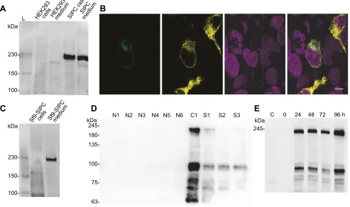
with our results where, upon expression of recombinant SIPC in HEK293 cells, we identified a secreted protein in the medium (Fig. 1A). The same was true for the Venus-containing recombinant SIPC (VSIPC), which was found to be secreted from HEK293 cells upon its expression (Fig. S1A). We observed that the expressed protein in HEK293 cells had a molecular mass>200 kDa, much higher than the calculated molecular mass of 171.45 kDa of secreted recombinant SIPC. The Venus-containing VSIPC had a molecular mass>230 kDa (Fig. S1A), much higher than the calculated molecular mass of 199.16 kDa of the secreted recombinant VSIPC. Fluorescence confocal microscopy revealed that recombinant VSIPC, expressed in HEK293 cells, had an endoplasmic-reticulum-like localisation distinct from the secretory-vesicle-localised PTPRN2 (Caromile et al., 2010; Fig. 1B), an indication that SIPC is secreted through transport vesicles.
Assuming that the less complex glycosylations of insect cells (Shi and Jarvis, 2007) will result in a protein of reduced molecular mass, we expressed recombinant SIPC in Sf9 cells and observed that it had a similar molecular mass (>200 kDa) to that observed in HEK293 cells (Fig. 1A,C). In this case, however, the protein was not secreted from Sf9 cells (Fig. 1C). To obtain a secreted form of SIPC from Sf9
SIPC medium
Sf9-SIPCmedium Sf9-SIPCcells
SIPC cell s
HEK293medium HEK293cells
N1 kDa kDa
kDa L
A
B
C
D
E
245-
230-
150-
100-
150-
100-kDa 245-
180-
135-
100-
75-
[image:7.612.61.554.317.609.2]63-N2 N3 N4 N5 N6 C1 S1 S2 S3 C 0 24 48 72 96 h
Fig. 1. Expression of recombinant SIPC fromAmphibalanus amphitrite.(A) Recombinant settlement-inducing protein complex (SIPC) is secreted by
HEK293 cells. Each lane contains 10μg of total cellular protein or 20μl of cell culture medium from untransfected cells (labelled as HEK293 cells and HEK293
medium) or cells transfected with the recombinant SIPC expression vector (labelled as SIPC cells and SIPC medium). Western blots were probed with an anti-myc
antibody and a typical image of several blots (n=10) is shown. L, protein ladder. (B) Recombinant VSIPC ( pENTR1α–SIPC–Myc–SphI–Venus–ORF–6×His) is
not localised in secretory vesicles. Fluorescence confocal imaging of recombinant VSIPC in HEK293 cells co-transfected with PTPRN2–mTurquoise. Image 1
(far left): PTPRN2–mTurquoise localisation in secretory vesicles. Image 2: VSIPC localisation in endoplasmic-reticulum-like structures. Image 3: DAPI
localisation in cell nuclei. Image 4 (far right): merge of images 1, 2 and 3 (scale bar: 10μm). (C) The endogenous signal peptide of SIPC does produce a secreted
recombinant SIPC in insect Sf9 cells. Gel loading and western blot conditions are identical to A. A typical image of several blots (n=3) is shown. (D) Detection of
native SIPC in extracts fromA. amphitriteat different developmental stages, with a mouse anti-SIPC monoclonal antibody. Each lane contains 10μg of total
cellular protein from different developmental stages of this species. A typical image of several blots (n=3) is shown. N1–N6, nauplii stage 1 to stage 6; C1, cyprid
day 1; S1–S3, settled day 1 to day 3. (E) Time course of secretion of recombinant SpSIPC ( pENTR1α–SpSIPC–Myc–SphI–ORF–6×His) from Sf9 cells after
infection with recombinant SpSIPC-expressing baculoviruses. Each lane contains 20μl of Sf9 cell culture medium. C, control Sf9 cell culture medium. Western
blots were probed with an anti-myc antibody and a typical image of several blots (n=3) is shown.
Journal
of
Experimental
cells, we engineered a construct that contained the signal peptide of
sarcotoxin 1A of the flesh flySarcophaga peregrinathat has been
shown to direct secretion of baculovirus-encoded proteins (O’Reilly
et al., 1995). This change in the signal peptide resulted in the expression and secretion of recombinant SIPC from Sf9 cells (Fig. S1B). These cells can be cultured in a serum-free and protein-free medium and this was convenient for the purification of recombinant SIPC from the medium. Upon purification of SIPC from Sf9 cell culture medium, to eliminate any possibility that the immunoreactive protein was not SIPC, we probed the overexpressed proteins with a polyclonal antibody generated against a peptide sequence in the C-terminus of SIPC (Fig. S1C). This antibody recognised a single band in western blots from Sf9 cell culture medium at a molecular mass>200 kDa, only when a different signal peptide replaced the endogenous signal peptide of SIPC, in
agreement with our previous results (Fig. 1A,C, Fig. S1A–C). As
shown in the western blots in Fig. S1A,B, bands of smaller molecular mass, similar to those described before (Matsumura et al., 1998b; Dreanno et al., 2006a), were immunoreactive with the N-terminally located myc-epitope but not with the anti-SIPC antibody (Fig. S1C).
We tested this antibody against lysates fromA. amphitritenauplii,
cyprids and adults of different developmental stages and observed that a protein of molecular mass much higher than the calculated molecular mass of endogenous SIPC immunoreacted with the anti-SIPC antibody (Fig. S1D). When probed in the same western blot, the antibody against SIPC recognised a band of identically high molecular mass (Fig. S1E). Because the western blot signals we were observing with the polyclonal antibody were generally weak
(see Fig. S1C–E), we generated a mouse monoclonal antibody
(mAb clone 1C4) against the same C-terminal epitope of SIPC and
used it in western blot assays against lysates fromA. amphitrite
nauplii, cyprids and adults of different developmental stages (Fig. 1D). The results showed that this mAb reacted strongly with
a band of∼96 kDa and less with a band of∼210 kDa, in addition to
several other bands of smaller molecular masses (Fig. 1D). Immunoreactive bands were recorded exclusively at the cyprid stage and not in the nauplii stage (Fig. 1D). This observation effectively ended all our preliminary attempts to purify from cyprid homogenates and use the native SIPC in settlement assays because it showed that native SIPC exists in multiple subunits in cyprids as previously described (Matsumura et al., 1998b; Dreanno et al., 2006c; Ferrier et al., 2016). Realising that settlement assay results
with the purified, native A. amphitrite SIPC can be potentially
marred by the biological role of each of the 3 reported SIPC subunits (Matsumura et al., 1998b) versus the biological role of the full-length protein, we expressed the full-full-length SIPC in heterologous expression systems. To obtain large amounts of the protein for
settlement assays withA. amphitritecyprids, we expressed the two
forms of the recombinant protein (SpSIPC and SpVSIPC) in Sf9 cells and observed that both were secreted from these cells (Fig. 1E,
Fig. S1F–H). Once again, bands of smaller molecular masses,
similar to those described before (Matsumura et al., 1998b; Dreanno et al., 2006a), were immunoreactive with the N-terminally located myc-epitope, in agreement with results from another study (Zhang et al., 2016).
Settlement assays with recombinant SIPC
We tested the biological activity of purified recombinant SIPC
(SpSIPC) in settlement assays with cyprids of A. amphitrite
(Fig. 2A) using an established protocol (Dreanno et al., 2007).
The results show that A. amphitrite cyprids behaved in a
bimodal fashion to the presence of SIPC, preferring to settle
and metamorphose at lower concentrations of SIPC
(EC50=3.73 nmol l−1) or preferring to avoid settlement at higher
concentrations of SIPC (IC50=101 nmol l−1) (Fig. 2B–D). The
settlement or avoidance response of the animals was evident within 24 h in the presence of SIPC (Fig. 2A), so the calculations of effective concentrations of SIPC were done with the 24 h data (Fig. 2C,D). Peak settlement preference responses were recorded at
a concentration of 25 nmol l−1 SpSIPC (Fig. 2A) and settlement
avoidance responses were evident from a concentration of
50 nmol l−1 SpSIPC onwards (Fig. 2A). Cyprids that did not
metamorphose to juveniles were actively seeking a substrate to settle even after 96 h in the wells of the culture dishes (Fig. S2A) and there was no increase in mortality of the animals neither time-wise nor SIPC-concentration-wise (Fig. S2B). By plotting the percentage of cyprids that settled and metamorphose to juveniles (Fig. 2B, Fig. S2C), it is obvious that SIPC does not curtail, in any way, the developmental progression of barnacles. To rule out that the observed settlement avoidance by the animals at the highest range of SpSIPC concentrations (Fig. 2A,D) was not a potential artefact of our assay design, we carried out the same assays in the
presence of BSA (Fig. S3A–D). The results showed that BSA at
concentrations as high as 1μmol l−1 had no settlement-inducing
effect on cyprids and neither affected metamorphosis nor mortality
(Fig. S3A–D). At a concentration of 10μmol l−1, BSA inhibited
settlement of cyprids (Fig. S3A,B) with a calculated EC50 of
3.69μmol l−1 at 24 h, a 36.5-fold higher concentration than the
settlement avoidance response of the cyprids to SpSIPC. Because native SIPC has been shown to be a cyprid cuticle-bound protein (Zhang et al., 2016; Dreanno et al., 2006a), we tested whether recombinant SIPC is recognised by the cyprids as a surface-adhered moiety. When polystyrene plates were incubated with or without recombinant SIPC, only the wells that contained SpSIPC showed immunostaining (Fig. 2E). This immunostaining (Fig. 2E) was evident when cyprids were incubated for 96 h in polystyrene plates whereupon immunoreactive traces of native SIPC deposited by the cyprids could be detected on the surface of the plates (Fig. 2F). Western blots of the ASW where cyprids were incubated for 96 h showed that native SIPC is not a diffusible molecule (Fig. S4A). Furthermore, bacterial growth in the presence of recombinant SIPC did not mediate the settlement preference or avoidance behaviour of the cyprids (Fig. S4B).
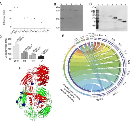
To clarify whether PTMs of SIPC and not SIPC itself are responsible for the behaviour of cyprids, in the presence of SIPC, and identify which of its multiple domains is responsible for its biological activity, we generated truncated fragments of SIPC, maintaining the endogenous signal peptide, and expressed and purified the fragments from culture medium of HEK293 cells
(Fig. 3A–D). The design of the fragments was based on a crystal
structure simulation that we generated through i-TASSER (Zhang, 2008) that aided the choice of truncated fragment length. The process was iterative and informed by the bioinformatics analysis on the PTMs of SIPC (see Table S5 and Fig. S4C). The cumulative
results (Fig. 3A–D) show that truncation of the first 212 amino acids
results in a poorly secreted protein (Fig. 3C,D), an effect that was remedied when further truncations were made to the full-length
SIPC (Fig. 3B–D). The apparent molecular masses of some of the
various truncated SIPC fragments differed from their calculated molecular masses substantially but, as the size of the truncated fragments decreased, the apparent and calculated molecular masses
became similar (Fig. 3A–D, Fig. S4D). As with the recombinant
SIPC expressed in Sf9 cells (Fig. 2E), we tested the ability of all the
Journal
of
Experimental
truncated SIPC fragments to adhere to polystyrene plates (Fig. S4E). The results revealed that truncated SIPC fragments Tr.1 to Tr.3 adhere to the surface of polystyrene plates, while Tr.6 to Tr.9 do not (Fig. S4E).
We tested the settlement of cyprids against the truncated fragments of SIPC and observed that removal of the N-terminal 482 amino acids made the cyprids avoid settlement (Table 1, Fig. S5), whereas truncated fragments up until the N-terminal 482 amino acids made cyprids exhibit settlement preference rather than avoidance (Table 1, Fig. S5). The settlement avoidance behaviour was consistent even for the small C-terminal fragment of SIPC (Table 1, Fig. S5), which contains the receptor-binding domain of
α2-macroglobulin [A2M_recep(PF07677.13)], the protein that is
phylogenetically related to SIPC (Dreanno et al., 2006c) (Fig. S4C).
PTMs of recombinant SIPC
We plotted the differences in the apparent and calculated molecular mass of recombinant SIPC and its truncated fragments from several western blots in an attempt to map the location of PTMs on the
primary structure of SIPC (Fig. 4A). Informed by a previous
analysis of theN-linked glycosylations on native SIPC (Pagett et al.,
2012), and by our bioinformatics analysis on the location of any PTMs on SIPC (see Table S5), we carried out deglycosylation assays on recombinant SIPC and selected truncated SIPC fragments
(Fig. 4B–D) that would allow us to determine where such PTMs
are located. We used a deglycosylation assay that provides more extensive removal of glycans (Fig. 4D) than previously reported
with PNGase F, which removes onlyN-linked glycans (Pagett et al.,
2012). Deglycosylation of the full-length SIPC reduced the molecular mass of the protein by 22.27±1.35 kDa (Fig. 4B, Table S6), while deglycosylation of Tr.5, Tr.6 and Tr.7 reduced the molecular mass of the fragments by 11.33±2.17, 13.91±1.83 and 7.04±1.24 kDa, respectively (Fig. 4C). We re-analysed the data presented in a previous study, in which the molecular nature and
relative abundance ofN-linked glycans, cleaved from native SIPC
with PNGase F, was determined (Pagett et al., 2012), and found that
N-linked glycans present on the native protein have a molecular
mass of 14.7 kDa (Table S6).
96 h 72 h 48 h 24 h
96 h
24 h, EC50=3.73 nmol l–1
24 h, EC50=101 nmol l–1
72 h 48 h 24 h 100
80
60
40
Settlement (%) Settlement (%)
Metamorphosis (%)
20
0
100
80
60
40
Settlement (%) 20
0
C 0.1 1 10
50 100 150
SIPC (nmol l–1)
Control
Control Cyprids Cyprids Cyprids myc-tag
mAb 10
µmol l–1
BSA 50 nmol l–1
SIPC
100 C 0.1 1 10
SIPC (nmol l–1)
100 C 0.1 1 10 100
100
A
B
C
D
E
F
80
60
40
20
0
100
80
60
40
20
[image:9.612.53.558.57.280.2]0
Fig. 2. Recombinant SIPC fromA. amphitriteinduces gregarious settlement preference or avoidance by cyprids of this species.(A) Recombinant SIPC
transduces gregarious settlement preference and settlement avoidance behaviour byA. amphitritecyprids in a dose-dependent manner. Values are expressed
as the percentage of animals that settled from the total number of animals placed in a well of a 24-well tissue culture plate. The cumulative results from 3
independent experiments with 5 replicates each is shown. Results of one-way ANOVA with the Tukey’s multiple comparisons test between matched observations
for the various time points revealed no statistical significance only between the datasets at 72 and 96 h (P>0.05). Results of one-way ANOVA with the Bonferonni’s
multiple comparisons test between control and the various concentrations of recombinant SIPC (SpSIPC) revealed statistical significance only between the
10 nmol l−1, 25 nmol l−1and 50 nmol l−1concentrations of SpSIPC and the control group (P<0.05) in all 4 tested time points. (B) Recombinant SIPC promotes
metamorphosis of the settledA. amphitritecyprids in a dose-dependent manner. Assay conditions and replicates are identical to A. For the percentage of animals
that underwent metamorphosis, results of one-way ANOVA with the Tukey’s multiple comparisons test between matched observations for the various time points
revealed no statistical significance only between the datasets at 72 and 96 h (P>0.05). (C) Effective concentration of recombinant SIPC that induces gregarious
settlement preference behaviour byA. amphitritecyprids within 24 h. Assay conditions and replicates are identical to A and plotted separately for clarity. Results
of one-way ANOVA with the Bonferonni’s multiple comparisons test between control and the various concentrations of recombinant SIPC (SpSIPC) revealed
statistical significance only between the 10 nmol l−1, 25 nmol l−1and 50 nmol l−1concentrations of SpSIPC and the control group (P<0.05). (D) Effective
concentration of recombinant SIPC that induces settlement avoidance behaviour byA. amphitritecyprids within 24 h. Assay conditions and replicates are
identical to A and plotted separately for clarity. Results of one-way ANOVA with the Bonferonni’s multiple comparisons test between control and the various
concentrations of recombinant SIPC (SpSIPC) revealed statistical significance only between the 50 nmol l−1and 150 nmol l−1concentrations of SpSIPC
(P<0.05). (E) Recombinant SIPC adheres to the surface of polystyrene tissue culture plates used in settlement bioassays. Sterile tissue culture plates were
incubated at 25°C empty (control) or with 0.3 ml of sterile artificial sea water (ASW) (Myc-Tag mAb) or with 0.3 ml of 10μmol l−1BSA in sterile ASW or with 0.3 ml
of 50 nmol l−1SpSIPC in sterile ASW. Solutions were then aspirated and western blotting carried out on the wells with a Myc-Tag (9B11) mAb. A typical image of
several experiments (n=3) is shown. (F) A mouse anti-SIPC monoclonal antibody can detect native SIPC deposited by cyprids ofA. amphitriteon the surface of
polystyrene tissue culture plates used in settlement bioassays. Sterile tissue culture plates were incubated at 25°C without (control) or with 10 cyprids each in 2 ml sterile ASW for 96 h. Solutions and settled and non-settled cyprids were then removed and western blots were carried out on the wells with a mouse anti-SIPC monoclonal antibody. The bright-field images on the lower row contain drawn circles that show the position of the settled cyprids on each well, whereas the upper
row shows the corresponding immunostained image. A typical image of several experiments (n=6) is shown.
Journal
of
Experimental
By plotting these data (Pagett et al., 2012) with our deglycosylation results, the apparent molecular masses on western
blots and the bioinformatic analysis, we estimate that N-linked
glycans present on full-length SIPC and Tr.1 (Figs 3A and 4A) as well as Tr.6 and Tr.7 (Figs 3A and 4A) actually contribute 15 kDa to the increased molecular mass of SIPC, and less to its truncated
fragments (see Table S6 for details). Numerous O-linked
glycosylation sites are predicted on truncated SIPC fragments that
are larger than Tr.4 (i.e. Tr.2 andΤr.3) and also in a region in the
middle of the primary structure of the protein (Fig. 3A). Such
O-linked glycans may contribute an additional 8 kDa cumulatively
to the full-length SIPC (see Tables S5 and S6 for details). The overall results, shown as a Circos figure (Krzywinski et al., 2009)
(Fig. 4E), depict the relative contribution of each putativeN-linked,
O-linked or other simple glycans on the increase in the molecular
mass of SIPC and its truncated fragments. The plot was created by a combination of bioinformatic analysis, western blots analysis, deglycosylation assays and previously published data (Pagett et al.,
2012) to show howN-linked orO-linked glycans alter the molecular
mass of full-length SIPC or its truncated fragments. The data table that was used to generate the Circos plot and the plot parameters are provided in Dataset 1.
DISCUSSION
SIPC (Dreanno et al., 2007; Matsumura et al., 1998b) serves as a mate-discerning (Dreanno et al., 2007) and mate-assessing (Matsumura et al., 1998b; Pagett et al., 2012) chemical cue but also serves as a predator-attracting chemical cue (Ferrier et al., 2016; Zimmer et al., 2016). In this context, and balancing the trade-off between reproduction and predation, having both settlement preference and avoidance cues conveyed by the same protein, i.e. SIPC, may be advantageous for barnacles because it will inform them of mate availability when the chemical cue is sparse enough to
1568 A. amphitrite SIPC
A
B
C
D
1353
1232
1183
1076
986
830 N-glycosylation
O-glycosylation
692
576
171 SIPC
Tr.1
Tr.2
Tr.3
Tr.4
Tr.5
Tr.6
Tr.7
Tr.8
Tr.9
kDa
245-
180-
135-
100-
75-kDa
245-
180-
135-
100-
75-kDa
245- 180- 135- 100- 75-
63- 48-
35-Sf9-SpSIPCVSIPCSIPC Tr.1 Tr.2 Tr.3 Tr.4 Tr.5 Tr.6 Tr.7
Sf9-SpSIPCVSIPCSIPC Tr.1 Tr.2 Tr.3 Tr.4 Tr.5 Tr.6 Tr.7
SIPC Tr.1Tr.2 Tr.3 Tr.4 Tr.5Tr.6 Tr.7 Tr.8 Tr.9 Myc-tag
Signal peptide
Signal peptide
Signal peptide
Signal peptide
Signal peptide
Signal peptide
Signal peptide
Signal peptide
Signal peptide
Signal peptide Myc-tag
Myc-tag
Myc-tag
Myc-tag
Myc-tag
Myc-tag
Myc-tag
Myc-tag
Myc-tag
17 Fig. 3. Design and expression of truncated SIPC fragments.(A) Graphical
representation of the truncated SIPC fragments showing the protein length and the post-translational modifications identified by our bioinformatic analysis.
Tr.1–Tr.9, truncated SIPC fragments 1 to 9. (B) Expression of recombinant
SIPC and its truncated fragments in HEK293 cells. Each lane contains
10μg of total cellular protein. Western blots were probed with an anti-myc
antibody and a typical image of several blots (n=3) is shown. (C) Secretion of
recombinant SIPC and its truncated fragments by HEK293 cells. Each lane
contains 20μl of cell culture medium. Western blots were probed with an
anti-myc antibody and a typical image, which corresponds to the samples shown in B, is shown. (D) Secretion of recombinant SIPC and its truncated
fragments by HEK293 cells. Each lane contains 20μl of cell culture medium.
Western blots were probed with an anti-myc antibody and proteins were resolved less than C to include truncated fragments Tr.8 and Tr.9. Sf9-SpSIPC, recombinant SIPC secreted from Sf9 cells; VSIPC, Venus-tagged recombinant SIPC secreted from HEK293 cells.
Table 1. Recombinant SIPC (rSIPC) or its truncated fragments transduce gregarious settlement preference or settlement avoidance behaviour byA. amphitritecyprids
rSIPC and truncated SIPC fragments (see Fig. 3A)
Settlement preference EC50(nmol l−1)
Settlement avoidance EC50(nmol l−1)
SIPC (rSIPC) 1.56 –
Tr.1 1.19 –
Tr.2 1.49 –
Tr.3 26.6 –
Tr.4 14.1 –
Tr.5 – 10.8
Tr.6 – 12.4
Tr.7 – 4.75
Tr.8 – 14.2
Tr.9 – 550
Settlement preference and avoidance EC50values are derived from dose–
response curves shown in Fig. S5 and were calculated using GraphPad Prism v.6. Values were computed from the percentage of animals that settled within 24 h from the total number of animals placed in a well of a 24-well tissue culture plate. The cumulative results from 2 independent experiments with 4 replicates
each is shown. See Fig. S5 for further details.