Pulmonary Perspective
Global Strategy for the Diagnosis, Management, and
Prevention of Chronic Obstructive Pulmonary Disease
GOLD Executive Summary
Jørgen Vestbo1,2, Suzanne S. Hurd3, Alvar G. Agustı´4, Paul W. Jones5, Claus Vogelmeier6, Antonio Anzueto7, Peter J. Barnes8, Leonardo M. Fabbri9, Fernando J. Martinez10,
Masaharu Nishimura11, Robert A. Stockley12, Don D. Sin13, and Roberto Rodriguez-Roisin4
1Manchester Academic Health Sciences Centre, South Manchester University Hospital NHS Foundation Trust, Manchester, United Kingdom; 2Odense University Hospital and University of Southern Denmark, Odense, Denmark;3Global Initiative for Chronic Obstructive Lung Disease, Vancouver, Washington;4Hospital Clı´nic, Universitat de Barcelona, Barcelona, Spain;5St George’s Hospital Medical School, London, United Kingdom;6University of Gießen and Marburg School of Medicine, Marburg Germany;7University of Texas Health Science Center, San Antonio, Texas;8National Heart and Lung Institute, London, United Kingdom;9University of Modena and Reggio Emilia, Modena, Italy;10University of Michigan School of Medicine, Ann Arbor, Michigan;11Hokkaido University School of Medicine, Sapporo, Japan;12University Hospitals Birmingham, Birmingham, United Kingdom; and13St Paul’s Hospital, Vancouver, Canada
Chronic obstructive pulmonary disease (COPD) is a global health problem, and since 2001, the Global Initiative for Chronic Obstruc-tive Lung Disease (GOLD) has published its strategy document for the diagnosis and management of COPD. This executive summary presents the main contents of the second 5-year revision of the GOLD document that has implemented some of the vast knowledge about COPD accumulated over the last years. Today, GOLD recommends that spirometry isrequiredfor the clinical diagnosis of COPD to avoid misdiagnosis and to ensure proper evaluation of severity of airflow lim-itation. The document highlights that the assessment of the patient with COPD should always include assessment of (1) symptoms, (2) severity of airflow limitation, (3) history of exacerbations, and (4) comorbidities. The first three points can be used to evaluate level of symptoms and risk of future exacerbations, and this is done in a way that splits patients with COPD into four categories—A, B, C, and D. Nonpharmacologic and pharmacologic management of COPD match this assessment in an evidence-based attempt to relieve symptoms and reduce risk of exacer-bations. Identification and treatment of comorbidities must have high priority, and a separate section in the document addresses manage-ment of comorbidities as well as COPD in the presence of comorbidities. The revised document also contains a new section on exacerbations of COPD. The GOLD initiative will continue to bring COPD to the attention of all relevant shareholders and will hopefully inspire future national and local guidelines on the management of COPD.
Keywords:COPD; clinical assessment; COPD management; exacerba-tions; comorbidities
CONTENTS Introduction
Summary of New Recommendations Levels of Evidence
1. Definition and Overview
Key Points Definition Burden of COPD
Factors That Influence Disease Development and Progression
Pathology, Pathogenesis, and Pathophysiology 2. Diagnosis and Assessment
Key Points Diagnosis Assessment of Disease Differential Diagnosis 3. Therapeutic Options Key Points
4. Management of Stable COPD Key Points
Introduction
Identify and Reduce Exposures Treatment of Stable COPD Nonpharmacologic Treatment Pharmacologic Treatment Monitoring and Follow-up 5. Management of Exacerbations Key Points Definition Diagnosis Assessment Treatment Options
Hospital Discharge and Follow-up Home Management of Exacerbations Prevention of COPD Exacerbations 6. COPD and Comorbidities
Key Points Introduction
Cardiovascular Diseases Osteoporosis
Anxiety and Depression Lung Cancer
Infections
Metabolic Syndrome and Diabetes INTRODUCTION
Chronic obstructive pulmonary disease (COPD) is a major public health problem. In 2020, COPD is projected to rank fifth worldwide
Author Contributions: All authors have contributed to this report; J.V. wrote the first draft. Correspondence and requests for reprints should be addressed to Jørgen Vestbo, D.M.Sc., Manchester Academic Sciences Health Centre, Respiratory Research Group, University of Manchester, University Hospital South Manchester, Southmoor Road, Manchester M23 9LT, UK. E-mail: [email protected] This article has an online supplement, which is accessible from this issue’s table of contents at www.atsjournals.org
Am J Respir Crit Care Med Vol 187, Iss. 4, pp 347–365, Feb 15, 2013 Copyrightª2013 by the American Thoracic Society
Originally Published in Press as DOI: 10.1164/rccm.201204-0596PP on August 9, 2012 Internet address: www.atsjournals.org
in terms of burden of disease and third in terms of mortality. Al-though COPD has received increasing attention from the medical community in recent years, it is still relatively unknown or ignored by the public as well as public health and government officials.
In 1998, the Global Initiative for Chronic Obstructive Lung Disease (GOLD) was formed to bring more attention to the management and prevention of COPD. Among the important objectives of GOLD are to increase awareness of COPD and to help the millions of people who suffer from this disease and die prematurely from it or its complications. In 2001, the GOLD pro-gram released a consensus report,Global Strategy for the Diag-nosis, Management, and Prevention of COPD; this document was revised in 2006, and now we present the 2011 version.
The GOLD document is a global document and for that reason alone should not be regarded a clinical guideline. It is impossible to make the same guidelines for developing countries as for, for ex-ample, Europe and North America. A strategy document provides advice on diagnosis and management that can be implemented in national guidelines. It can be expanded for rich countries and re-stricted for poorer ones. It provides guidance on principles and drug classes to be applied, and national guidelines can therefore build on the assessment and management principles suggested by GOLD—and then modify it to fit their country’s needs.
Based on multiple scientific and clinical achievements in the 10 years since the 2001 GOLD report was published, this revised edition provides a new paradigm for treatment of stable COPD. This major revision builds on the strengths from the original rec-ommendations and incorporates new knowledge to make three important new recommendations:
1. One of the strengths was the treatment objectives. These have stood the test of time, but are now organized into two groups: objectives that are directed toward immedi-ately relieving and reducing the impact of symptoms, and objectives that reduce the risk of adverse health events in the future. This emphasizes the need for clinicians to maintain a focus on both the short-term and long-term impact of COPD on their patients.
2. A second strength of the original strategy was the simple, intuitive system for classifying COPD severity. This was based on the FEV1and was called a staging system because it was believed, at the time, that the majority of patients followed a path of disease progression that tracked the severity of the airflow limitation. Much is now known about the characteristics of patients in the different GOLD stages—for example, their level of risk of exacerbations, hospitalization, and death. However at an individual patient level, the FEV1is an unreliable marker of the severity of breathlessness, exercise limitation, and health status impair-ment. This report retains the GOLD classification system of airflow limitation because it is a predictor of future adverse events, but the term “stage” is now replaced by “grade.” 3. At the time of the original report, improvement in both
symptoms and health status was a GOLD treatment objec-tive, but symptom assessment did not have a direct relation to the choice of management, and health status measure-ment was a complex process largely confined to clinical studies. Now, there are simple and reliable questionnaires designed for use in routine daily clinical practice. These have been validated in many languages, which has enabled the development of a new assessment system that integrates patient symptoms and their risk for serious adverse health events in the future. In turn, this new assessment system has led to the construction of a new approach to management— one that matches assessment to treatment objectives. The new management approach can be used in any clinical
setting anywhere in the world and moves COPD treatment toward individualized medicine—matching the patient’s therapy more closely to his or her needs. Whereas recom-mendations on treatment are evidence based, a novel assess-ment system will have to be consensus based, with the aim that future studies will test the value of this system.
Summary of New Recommendations
A summary of the new issues presented in this report follows: 1. This document has been considerably shortened in length
by limiting section 1 to the essential background data on COPD. Readers who wish to access more comprehensive information are referred to a variety of excellent text-books that have appeared in the last decade.
2. Section 2 includes information on diagnosis and assess-ment of COPD. The definition of COPD has not been significantly modified but has been reworded for clarity. 3. Assessment of COPD is based on the patient’s level of
symptoms, exacerbation history, the severity of the spiro-metric abnormality, and the identification of comorbid-ities. Whereas spirometry was previously used to support a diagnosis of COPD, spirometry is now required to make a confident diagnosis of COPD.
4. Airflow limitation as determined by spirometry is divided into four grades (GOLD 1, mild; GOLD 2, moderate; GOLD 3, severe; and GOLD 4, very severe) using the fixed ratio, post-bronchodilator FEV1/FVC,0.7, to de-fine airflow limitation. It is recognized that the use of the fixed ratio (FEV1/FVC) may lead to more frequent diag-noses of COPD in older adults with mild COPD as the normal process of aging affects lung volumes and flows, and may lead to underdiagnosis in adults younger than 45 years. The concept of staging has been abandoned be-cause a staging system based on FEV1alone was inade-quate and the evidence for an alternative staging system does not exist. The most severe spirometric grade, GOLD 4, does not include reference to respiratory failure as this seemed to be an arbitrary inclusion.
5. A new section (section 3) on therapeutic approaches has been added. This includes descriptive information on both pharmacologic and nonpharmacologic therapies, and iden-tifying any adverse effects.
6. Management of COPD is presented in three sections: M ANAGE-MENT OFSTABLECOPD (section 4); MANAGEMENT OFE XACER-BATIONS (section 5); and COPDANDCOMORBIDITIES(section
6), covering both management of comorbidities in patients with COPD and of COPD in patients with comorbidities.
7. In section 4, MANAGEMENT OFSTABLECOPD,
recommen-ded approaches to both pharmacologic and nonpharma-cologic treatment of COPD are presented. In previous GOLD documents, recommendations for management of COPD were based solely on spirometric category. However, there is considerable evidence that the level of FEV1 is a poor descriptor of disease status, and for this reason, the management of stable COPD based on a strategy considering both disease impact (determined mainly by symptom burden and activity limitation) and future risk of disease progression (especially of exacer-bations) is recommended.
8. Section 5, MANAGEMENT OF EXACERBATIONS, presents
9. Section 6, COMORBIDITIES ANDCOPD, focuses on cardiovas-cular diseases, osteoporosis, anxiety and depression, lung cancer, infections, and metabolic syndrome and diabetes.
Levels of Evidence
Levels of evidence are assigned to management recommenda-tions where appropriate with the system used in previous reports. Evidence levels are enclosed in parentheses after the relevant statement, for example, “(Evidence A).” Levels of evidence used in this document have not changed with respect to previous re-leases and are listed in the original document (www.goldcopd.org). 1. DEFINITION AND OVERVIEW
KEY POINTS
d COPD, a common preventable and treatable disease, is characterized by persistent airflow limitation that is usually progressive and associated with an enhanced chronic in-flammatory response in the airways and the lung to nox-ious particles or gases. Exacerbations and comorbidities contribute to the overall severity in individual patients. d COPD is a leading cause of morbidity and mortality
worldwide and results in an economic and social bur-den that is both substantial and increasing.
d Inhaled cigarette smoke and other noxious particles such as smoke from biomass fuels cause lung inflammation, a normal response that appears to be modified in pa-tients who develop COPD. This chronic inflammatory response may induce parenchymal tissue destruction (resulting in emphysema) and disrupt normal repair and defense mechanisms (resulting in small airway fibrosis). These pathological changes lead to air trapping and pro-gressive airflow limitation, and in turn to breathlessness and other characteristic symptoms of COPD.
Definition
COPD, a common preventable and treatable disease, is charac-terized by persistent airflow limitation that is usually progressive and associated with an enhanced chronic inflammatory response in the airways and the lung to noxious particles or gases. Exac-erbations and comorbidities contribute to the overall severity in individual patients.
The chronic airflow limitation characteristic of COPD is caused by a mixture of small airways disease (obstructive bron-chiolitis) and parenchymal destruction (emphysema), the relative contributions of which vary from person to person. Chronic in-flammation causes structural changes and narrowing of the small airways. Destruction of the lung parenchyma, also by inflamma-tory processes, leads to the loss of alveolar attachments to the small airways and decreases lung elastic recoil; in turn, these changes diminish the ability of the airways to remain open during expiration. Airflow limitation is best measured by spirometry, as this is the most widely available, reproducible test of lung function.
Burden of COPD
COPD prevalence, morbidity, and mortality vary across countries and across different groups within countries. COPD is the result of cumulative exposures over decades. Often, the prevalence of COPD is directly related to the prevalence of tobacco smoking, although in many countries, outdoor, occupational, and indoor
air pollution—the latter resulting from the burning of wood and other biomass fuels—are major COPD risk factors (1). The pre-valence and burden of COPD are projected to increase in the coming decades due to continued exposure to COPD risk fac-tors and the aging of the world’s population.
Prevalence. Existing COPD prevalence data show remarkable variation due to differences in survey methods, diagnostic crite-ria, and analytic approaches (2). Despite the complexities and the widespread underrecognition and underdiagnosis of COPD (3), data from the Latin American Project for the Investigation of Obstructive Lung Disease (PLATINO) and the Burden of Obstructive Lung Diseases program (BOLD) have documented more severe disease than previously found and a substantial prevalence (3–11%) of COPD among never-smokers (4).
Morbidity. Morbidity measures traditionally include physician visits, emergency department visits, and hospitalizations. Morbid-ity due to COPD increases with age (5–7) and may be affected by other comorbid chronic conditions (e.g., cardiovascular disease, musculoskeletal impairment, or diabetes mellitus) that are fre-quent in patients with COPD and may impact on the patient’s health status, as well as interfere with COPD management.
Mortality. Underrecognition and underdiagnosis of COPD still affect the accuracy of mortality data (8, 9) with COPD often listed as acontributorycause of death or omitted from the death cer-tificate entirely (10). The Global Burden of Disease Study pro-jected that COPD, which ranked sixth as a cause of death in 1990, will become the third leading cause of death worldwide by 2020; a newer projection estimated COPD will be the fourth leading cause of death in 2030 (11). This increased mortality is mainly driven by the expanding epidemic of smoking, reduced morta-lity from other common causes of death, and aging of the world population.
Economic and social burden. COPD is associated with signifi-cant economic burden. There is a direct relationship between the severity of COPD and the cost of care, and the distribution of costs changes as the disease progresses. For example, hospitalization and ambulatory oxygen costs soar as COPD severity increases. In developing countries, direct medical costs may be less important than the impact of COPD on workplace and home productivity. In 1990, COPD was the 12th leading cause of disability-adjusted life years (DALYs) lost in the world, responsible for 2.1% of the total. According to the projections, COPD will be the seventh leading cause of DALYs lost worldwide in 2030 (11).
Factors That Influence Disease Development and Progression
Although cigarette smoking is the best-studied COPD risk factor, there is consistent epidemiological evidence that nonsmokers may also develop chronic airflow limitation (5–7, 12). Besides, among people with the same smoking history, not all will develop COPD, for reasons that are still unclear but likely involve differences in genetic backgrounds and other exposures.
Across the world, cigarette smoking is the most commonly en-countered risk factor for COPD. Cigarette smokers have a higher prevalence of respiratory symptoms and lung function abnormal-ities, a greater annual rate of decline in FEV1, and a greater COPD mortality rate than nonsmokers (13). Other types of tobacco (e.g., pipe, cigar, water pipe [14]) and marijuana (15) are also risk fac-tors for COPD (16, 17). Passive exposure to cigarette smoke (also known as environmental tobacco smoke) may also contribute to respiratory symptoms (18) and COPD (19) by increasing the lung’s total burden of inhaled particles and gases (20, 21). Smok-ing durSmok-ing pregnancy may also pose a risk for the fetus, by af-fecting lung growth and development in uteroand possibly the priming of the immune system (22, 23).
Occupational exposures, including organic and inorganic dusts and chemical agents and fumes, are an underappreciated risk fac-tor for COPD (24–26). Wood, animal dung, crop residues, and coal, typically burned in open fires or poorly functioning stoves, may lead to very high levels of indoor air pollution. Evidence continues to grow that indoor pollution from biomass cooking and heating in poorly ventilated dwellings is an important risk factor for COPD (27–33). Almost 3 billion people worldwide use biomass and coal as their main source of energy for cooking, heating, and other household needs, so the population at risk worldwide is very large (30, 34).
Other factors associated with development and progression of COPD, such as genetics, lung development abnormalities, accel-erated aging, bronchial hyperreactivity, and socioeconomic sta-tus, among others, are listed in recent reviews and in the full document (www.goldcopd.org).
Pathology, Pathogenesis, and Pathophysiology1
Inhaled particles (from cigarette smoke or other sources) cause lung inflammation, a normal response that appears to be modi-fied in individuals who develop COPD. This chronic inflam-matory response may induce parenchymal tissue destruction (resulting in emphysema), and disrupt normal repair and defense mechanisms (resulting in small airway fibrosis), which in turn lead to air trapping and progressive airflow limitation.
Pathology. Chronic inflammatory changes with increased num-bers of inflammatory cell types, and structural changes resulting from repeated injury and repair, are found in the airways, lung parenchyma, and pulmonary vasculature of patients with COPD (35). In general, these changes increase with disease severity and persist despite smoking cessation.
Pathogenesis. The above-mentioned pathological changes ap-pears to be an enhancement of the normal, physiological, inflam-matory response of the respiratory tract to chronic irritants. The mechanisms for this amplified inflammation in COPD are not yet understood but may be genetically determined. Lung inflamma-tion persists after smoking cessainflamma-tion through unknown mech-anisms, although autoantigens and persistent microorganisms may play a role (36). Patients can clearly develop COPD with-out smoking, but the nature of the inflammatory response in these patients is unknown.
Pathophysiology. AIRFLOW LIMITATION AND GAS TRAPPING.
In-flammation and narrowing of peripheral airways leads to decreased FEV1. Parenchymal destruction due to emphysema also contrib-utes to airflow limitation due to reduced elastic recoil (37). In combination, both progressively lead to gas trapping during expi-ration, resulting in hyperinflation.
GAS EXCHANGE ABNORMALITIES. Gas exchange abnormalities
may result in hypoxemia and hypercapnia, and have several mech-anisms in COPD. The main one is ventilation–perfusion (VA/Q) abnormalities (38).
Reduced ventilatory drive may lead to carbon dioxide reten-tion, particularly when combined with reduced ventilation.
MUCUS HYPERSECRETION. Mucus hypersecretion, resulting in
a chronic productive cough, is a feature of chronic bronchitis and is not necessarily associated with airflow limitation. Conversely, not all patients with COPD have symptomatic mucus hyperse-cretion. When present, it is due to an increased number of goblet cells and enlarged submucosal glands in response to chronic airway irritation.
PULMONARY HYPERTENSION. Pulmonary hypertension may
de-velop late in the course of COPD. It can be due to hypoxic vaso-constriction of small pulmonary arteries, eventually resulting in
structural changes that include intimal hyperplasia and later smooth muscle hypertrophy/hyperplasia, and/or loss of pulmo-nary capillary bed due to emphysema (39). In pulmopulmo-nary vessels, an inflammatory response similar to that seen in the air-ways (and evidence of endothelial dysfunction) has been identi-fied. Severe pulmonary hypertension may lead to right ventricular hypertrophy and eventually to right-side cardiac failure.
EXACERBATIONS. Exacerbations of respiratory symptoms
of-ten occur in patients with COPD, triggered by infection with bac-teria or viruses (which may coexist), environmental pollutants, or unknown factors. During exacerbations, there is a flare-up of in-flammation, increased hyperinflation and gas trapping, reduced expiratory flow, and increased dyspnea (40). There is also wor-sening of VA/Q abnormalities, which can result in hypoxemia and hypercapnia (41). Other medical conditions (pneumonia, thromboembolism, and acute cardiac failure) may mimic or ag-gravate an exacerbation of COPD.
COMORBIDITIES. It is increasingly recognized that many
pa-tients with COPD have comorbidities and that these have a major impact on their quality of life and survival (42). The precise pathobiology of this association is under investigation but may involve mechanical as well as biological or genetic mechanisms. For instance, airflow limitation and hyperinflation affect cardiac function and gas exchange (43).
2. DIAGNOSIS AND ASSESSMENT
KEY POINTS
d A clinical diagnosis of COPD should be considered in any patient who has dyspnea, chronic cough and/or sputum production, and a history of exposure to risk factors for the disease.
d Spirometry is required to make the diagnosis in this clinical context; the presence of a post-bronchodilator FEV1/FVC less than 0.70 confirms the presence of per-sistent airflow limitation and thus of COPD.
d The goals of COPD assessment are to determine: (1) the impact of the disease on the patient’s health status, (2) the severity of airflow limitation, and (3) the risk of future exacerbations, in order to guide therapy. The risk of future exacerbations is estimated by the severity of airflow limitation and the history of previous exacerbations.
d Comorbidities, including cardiovascular disease, skele-tal muscle dysfunction, metabolic syndrome, osteopo-rosis, depression, and lung cancer, occur frequently in patients with COPD. Comorbidities should be actively looked for, and treated appropriately if present.
Diagnosis
A clinical diagnosis of COPD should be considered in any patient who has dyspnea, chronic cough and/or sputum production, and a history of exposure to risk factors for the disease. Spirometry is required to make the diagnosis in this clinical context; the pres-ence of a post-bronchodilator FEV1/FVC less than 0.70 confirms the presence of persistent airflow limitation and thus of COPD. The spirometric criterion for airflow limitation remains a post-bronchodilator fixed ratio of FEV1/FVC less than 0.70. This criterion is simple, independent of reference values, and has been used in numerous clinical trials forming the evidence base from which most of our treatment recommendations are drawn. Diagnostic simplicity and consistency are key for the busy nonspecialist clinician. Although post-bronchodilator spirometry 1
Illustrations of many of the topics covered in this section can be found in the Teaching Slide Set on the GOLD website: http://www.goldcopd.org.
is required for the diagnosis and assessment of severity of COPD, the degree of reversibility of airflow limitation (e.g., measuring FEV1before and after bronchodilator or corticosteroids) is no longer recommended.
Symptoms. The characteristic symptoms of COPD are chronic and progressive dyspnea, cough, and sputum production. Chro-nic cough and sputum production may precede the development of airflow limitation by many years. Individuals, particularly those exposed to COPD risk factors, who present with these symptoms should be examined to search for an underlying cause(s) and appropriate interventions taken. Conversely, signif-icant airflow limitation may develop without chronic cough and sputum production.
Medical history. A detailed medical history of a new patient known or thought to have COPD should assess:
d Exposure to risk factors d Past medical history
d Family history of COPD or other chronic respiratory disease
d Pattern of symptom development
d History of exacerbations or previous hospitalizations for respiratory disorder
d Presence of comorbidities
d Impact of the disease on the patient’s life d Social and family support available to the patient d Possibilities for reducing risk factors, especially smoking
cessation
Physical examination. Although an important part of patient care, a physical examination is rarely diagnostic in COPD. Physical signs of airflow limitation are usually not present un-til significant impairment of lung function has occurred (44, 45), and their detection has a relatively low sensitivity and specificity.
Spirometry. Spirometry is the most reproducible and objec-tive measurement of airflow limitation available. Peak expira-tory flow measurement alone cannot be reliably used as the only diagnostic test, despite its good sensitivity, because of its weak specificity (46). Good-quality spirometric measure-ment is possible in any health care setting, and all health care workers who care for patients with COPD should have access to spirometry.
Assessment of Disease
The goals of COPD assessment are to determine: (1) the impact of the disease on the patient’s health status, (2) the severity of airflow limitation, and (3) the risk of future events (such as ex-acerbations, hospital admissions, or death), to, eventually, guide therapy. To achieve these goals, COPD assessment must con-sider the following aspects of the disease separately:
d Current level of patient’s symptoms d Severity of airflow limitation d Exacerbation risk
d Presence of comorbidities
Assessment of symptoms. There are several validated ques-tionnaires to assess symptoms in patients with COPD that can be used to distinguish patients with less severe symptoms from patients with more severe symptoms. GOLD primarily recom-mends the use of the Modified British Medical Research Council (mMRC) questionnaire on breathlessness or the COPD Assessment
Test (CAT), the latter having a broader coverage of the impact of COPD on the patient’s daily life and well-being. Other symptoms scales can be used where available, for example, the Clinical COPD Questionnaire, and future GOLD updates are likely to expand in this area.
Assessment of airflow limitation severity. Table 1 shows the classification of airflow limitation severity in COPD. Specific spi-rometric cut points are used for purposes of simplicity. Spirom-etry should be performed after the administration of an adequate dose of a short-acting inhaled bronchodilator to minimize vari-ability. Worsening airflow limitation is associated with an increas-ing prevalence of exacerbations (see below) and risk of death.
Assessment of exacerbation risk. An exacerbation of COPD is defined as an acute event characterized by a worsening of the patient’s respiratory symptoms that is beyond normal day-to-day variations and leads to a change in medication (47–49). The rate at which exacerbations occur varies greatly between pa-tients (50). The best predictor of having frequent exacerbations (two or more exacerbations per year) is a history of previous treated events (51). Severity of exacerbations is usually clas-sified as mild when exacerbations of respiratory symptoms re-quire change of inhaled treatment by the patient, moderate when exacerbations of respiratory symptoms require medical intervention including a short course of antibiotic and/or oral steroids, and severe when exacerbations of respiratory symp-toms require hospitalization.
Assessment of comorbidities. Comorbidities occur frequently in COPD and include cardiovascular disease, skeletal muscle dysfunction, metabolic syndrome, osteoporosis, depression, and lung cancer. The existence of COPD may actually increase the risk for other concomitant diseases; this is particularly striking for COPD and lung cancer (52–55).
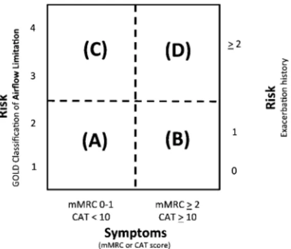
Combined COPD assessment. Figure 1 illustrates the pro-posed combined assessment of COPD. The MRC or CAT scale is recommended for assessing symptoms, with an mMRC grade greater than or equal to 2 or a CAT score greater than or equal to 10 indicating a high level of symptoms. These cutoffs should be used as indicators; the primary aim is to separate patients with a significant symptom burden from those with less symptoms. There are two methods of assessing exacerba-tion risk. One is a populaexacerba-tion-based method using the GOLD spirometric classification (Table 1), with GOLD 3 or GOLD 4 categories indicating high risk. The other is based on the indi-vidual patient’s history of exacerbations (51, 56), with two or more exacerbations in the preceding year indicating high risk. Given the significance of an exacerbation leading to hospital admission (57), hospitalization will often be an indicator of high risk as well. If there is a discrepancy between the risk category as assessed by spirometric classification and that de-rived from exacerbation history, the assessment pointing to the highest risk should be used.
To use Figure 1, first assess symptoms and determine if the patient belongs to the left side of the box—less symptoms (as indi-cated by mMRC grade 0–1 or CAT,10)—or the right side—more symptoms (as indicated by mMRC>2 or CAT>10). Next, assess the risk of exacerbations to determine if the patient belongs to the TABLE 1. GRADING OF SEVERITY OF AIRFLOW LIMITATION IN COPD (BASED ON POST-BRONCHODILATOR FEV1)
In patients with FEV1/FVC,0.70:
GOLD 1: Mild FEV1>80% predicted
GOLD 2: Moderate 50%<FEV1,80% predicted
GOLD 3: Severe 30%<FEV1,50% predicted
GOLD 4: Very severe FEV1,30% predicted
Definition of abbreviation: COPD ¼ chronic obstructive pulmonary disease; GOLD¼Global Initiative for Chronic Obstructive Lung Disease.
lower part of the box—low risk—or the upper part of the box—high risk. This can be done by either of two methods: (1) use spirometry to determine the GOLD grade of airflow limitation (GOLD 1 and 2 indicate low risk, whereas GOLD 3 and 4 indicate high risk); or (2) assess the number of exacerbations the patient has had within the previous 12 months (zero or one indicates low risk, whereas two or more exacerbations indicates high risk). In some patients, these two ways of assessing risk of exacerbations will not lead to the same level of risk; in this case, the risk should be determined by the method indicating high risk.
The groups can be summarized as follows:
Patient group A—low risk, less symptoms
GOLD 1–2 (mild or moderate airflow limitation)
and 0–1 exacerbation per yearandmMRC grade 0–1 or CAT score,10
Patient group B—low risk, more symptoms
GOLD 1–2 (mild or moderate airflow limitation)
and 0–1 exacerbation per year and mMRC grade > 2 or CAT score>10
Patient group C—high risk, less symptoms
GOLD 3–4 (severe or very severe airflow limitation) and/or> 2 exacerbations per year and/or>1 hospitalized exacerbation per yearandmMRC grade 0–1 or CAT score,10
Patient group D—high risk, more symptoms
GOLD 3–4 (severe or very severe airflow limitation) and/or>2 exacerbations per year /> 1 hospitalized exac-erbation per yearandmMRC grade>2 or CAT score>10
This approach, combined with an assessment of potential comorbidities, reflects the complexity of COPD better than the unidimensional analysis of airflow limitation previously used for staging the disease and forms the basis of the guide to indi-vidualized management provided in section 4.
Additional investigations. The following additional investigations may be considered as part of the diagnosis and assessment of COPD: IMAGING. A chest X-ray is not useful to establish a diagnosis
in COPD, but it is valuable in excluding alternative diagnoses and establishing the presence of significant comorbidities.
LUNG VOLUMES AND DIFFUSING CAPACITY. Patients with
COPD exhibit gas trapping (a rise in residual volume) from early in the disease, and as airflow limitation worsens, static hy-perinflation (an increase in total lung capacity) occurs. These changes can be documented by body plethysmography, or less accurately by helium dilution lung volume measurement. Dif-fusing capacity can be assessed by the uptake of carbon mon-oxide using the single-breath method. These measurements help characterize the severity of COPD but are not essential to patient management.
OXIMETRY AND ARTERIAL BLOOD GAS MEASUREMENT. Pulse oximetry can be used to evaluate a patient’s oxygen saturation and need for supplemental oxygen therapy. Pulse oximetry should be used to assess all stable patients with FEV1less than 35% predicted or with clinical signs suggestive of respiratory failure or right heart failure. If peripheral saturation is less than 92%, arterial blood gases should be assessed (58).
a1-ANTITRYPSIN DEFICIENCY SCREENING. The World Health Organization recommends that patients with COPD from areas with a particularly high prevalence ofa1-antitrypsin deficiency should be screened for this genetic disorder (59). The typical patient tends to present at a younger age (,45 yr) with lower lobe emphysema. A serum concentration ofa1-antitrypsin be-low 15 to 20% of the normal value is highly suggestive of ho-mozygousa1-antitrypsin deficiency.
EXERCISE TESTING. Objectively measured exercise
impair-ment, assessed by a reduction in self-paced walking distance (60) or during incremental exercise testing in a laboratory (61), is a powerful indicator of health status impairment and predictor of prognosis (62). Monitoring of physical activity may be more relevant regarding prognosis than evaluating exercise capacity (63).
COMPOSITE SCORES. Several variables, including age, dyspnea,
FEV1, body mass index, exercise tolerance assessed by walking distance or peak oxygen consumption, and/or arterial hypoxemia, identify patients at increased risk for mortality (64–66).
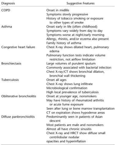
Differential Diagnosis
In some patients with chronic asthma, a clear distinction from COPD is not possible using current imaging and physiological testing techniques, and it is assumed that asthma and COPD co-exist in these patients. In these cases, current management will include use of antiinflammatory drugs, and other treatments need to be individualized. Other potential diagnoses are usually easier to distinguish from COPD (Table 2).
3. THERAPEUTIC OPTIONS
KEY POINTS
d In patients who continue to smoke, smoking cessation is a key therapeutic measure. Pharmacotherapy and nicotine replacement reliably increase long-term smok-ing abstinence rates.
d Appropriate pharmacologic therapy can reduce COPD symptoms, reduce the frequency and severity of exacer-bations, and improve health status and exercise tolerance. d To date, none of the existing medications for COPD has been shown conclusively to modify the long-term decline in lung function.
d Each pharmacological treatment regimen needs to be patient specific, guided by severity of symptoms, risk of exacerbations, comorbidities, drug availability, and the patient’s response. d Influenza and pneumococcal vaccination should be
of-fered to every patient with COPD; they appear to be more effective in older patients and those with more severe disease or cardiac comorbidity.
d All patients who get short of breath when walking on their own pace on level ground should be offered rehabilitation; it can improve symptoms, quality of life, and physical and emotional participation in everyday activities.
All text of this section can be found in the online supplement.
Figure 1. Combined COPD assessment. When assessing risk, choose the highest risk according to GOLD spirometric grade or exacerbation history.
4. MANAGEMENT OF STABLE COPD
KEY POINTS
d Identification and reduction of exposure to risk factors are important in the prevention and treatment of COPD. All individuals who smoke should be encouraged to quit. d The level of FEV1 is an inadequate descriptor of the impact of the disease on patients, and for this reason, individualized assessment of symptoms and future risk of exacerbation should also be incorporated into the management strategy for stable COPD.
d Regular physical activity is recommended for all patients with COPD.
d All patients with COPD with breathlessness when walk-ing at their own pace on level ground benefit from reha-bilitation and maintenance of physical activity, improving their exercise tolerance and quality of life, and reducing symptoms of dyspnea and fatigue.
d Pharmacologic therapy is used to reduce symptoms, reduce frequency and severity of exacerbations, and improve health status and exercise tolerance. Existing medications for COPD have not been conclusively shown to modify the long-term decline in lung function that is the hallmark of this disease.
d For both b2-agonists and anticholinergics, long-acting formulations are preferred over short-acting formula-tions. Based on efficacy and side effects, inhaled bron-chodilators are preferred over oral bronbron-chodilators. d Long-term treatment with inhaled corticosteroids added
to long-acting bronchodilators is recommended for pa-tients at high risk of exacerbations.
d Long-term monotherapy with oral or inhaled cortico-steroids is not recommended in COPD.
d The phospodiesterase-4 inhibitor roflumilast may be useful to reduce exacerbations for patients with FEV1 less than 50% predicted, chronic bronchitis, and fre-quent exacerbations.
d Influenza vaccines can reduce the risk of serious illness (such as hospitalization due to lower respiratory tract infections) and death in patients with COPD.
d The routine use of antibiotics is not indicated in patients with clinically stable COPD, other than for treating infec-tious exacerbations of COPD and other bacterial infections.
Introduction
Once COPD has been diagnosed, effective management should be based on an individualized assessment of the disease having two goals in mind:
1. Reduce current symptoms
2. Reduce the risk of future events (Table 3)
These goals should be reached with minimal side effects from treatment, a particular challenge in patients with COPD because they commonly have comorbidities that also need to be carefully identified and treated.
Identify and Reduce Exposures
Identification and reduction of exposure to risk factors are im-portant in the treatment (and prevention) of COPD. Since
cigarette smoking is the most commonly encountered and easily identifiable risk factor, smoking cessation should be encouraged for all individuals who smoke. Reduction of total personal expo-sure to occupational dusts, fumes, and gases and to indoor and out-door air pollutants may be more difficult but should be attempted.
Treatment of Stable COPD
In previous versions of the GOLD report, COPD treatment rec-ommendations were based on spirometry only. This is in keeping with the fact that most of the clinical trial evidence about treat-ment efficacy in COPD is oriented around baseline FEV1. How-ever, FEV1alone is a poor descriptor of disease status, and for this reason, the treatment strategy for stable COPD should con-sider also an individual patient’s symptoms and future risk of exacerbations as illustrated in Figure 1.
Nonpharmacologic Treatment
Physical activity. Regular physical activity is recommended for all patients with COPD.
Rehabilitation. Although more information is needed on cri-teria for patient selection for pulmonary rehabilitation programs, all patients with COPD appear to benefit from rehabilitation and maintenance of physical activity, improving their exercise toler-ance and experiencing decreased dyspnea and fatigue (67) (Ev-idence A).
Vaccination. Decisions about vaccination in patients with COPD depend on local policies, availability, and affordability.
Nonpharmacologic management of COPD according to the individualized assessment of symptoms and exacerbation risk (Figure 1) is shown in Table 4.
Pharmacologic Treatment
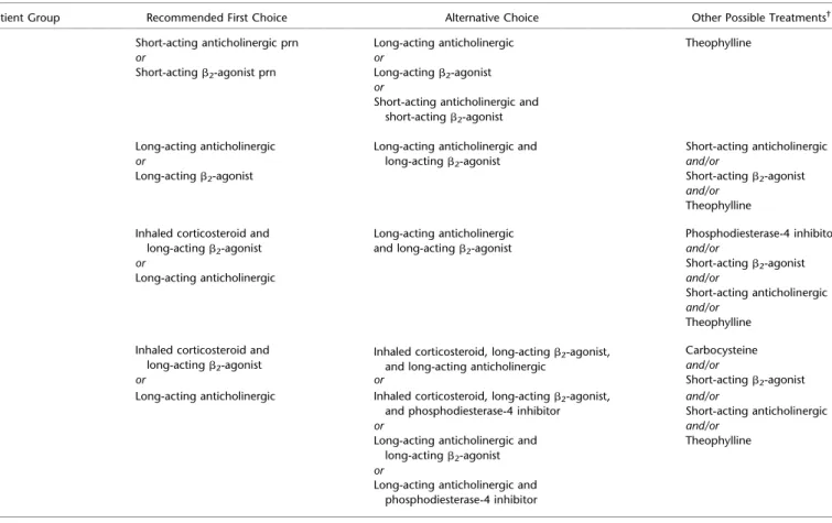
The classes of medications commonly used in treating COPD are shown in Table E1 in the online supplement, and a detailed de-scription of the effects of these medications is given in section 3 in the online supplement. The choice within each class depends on the availability of medication and the patient’s response. A pro-posed model for initial pharmacological management of COPD according to the individualized assessment of symptoms and exacerbation risk (Figure 1) is shown in Table 5.
Group A. Group A patients have few symptoms and a low risk of exacerbations. Specific evidence for the effectiveness of phar-macologic treatments is not available for patients with FEV1 greater than 80% predicted (GOLD 1). However, for all group A patients, a short-acting bronchodilator is recommended as first choice based on its effect on lung function and breathless-ness. Second choice is a combination of short-acting broncho-dilators or the introduction of a long-acting bronchodilator. The evidence for this step-up is weak; few studies of the combination exist (68, 69), and most trials of therapy with long-acting bron-chodilators have been performed in patients with more severe airflow limitation (70, 71).
Group B. Group B patients have more significant symptoms but still a low risk of exacerbations. Long-acting bronchodilators are superior to short-acting bronchodilators (taken as needed) and are therefore recommended (70, 71). There is no evidence to recommend one class of long-acting bronchodilators over another for initial treatment. In the individual patient, the choice should depend on the patient’s perception of symptom relief. For patients with severe breathlessness, the second choice is a combination of long-acting bronchodilators (72, 73). Only short-term studies of this treatment option have been reported, and patients on a combination of long-acting bronchodilators should be carefully followed and their treatment effect evaluated.
Alternative choices include short-acting bronchodilators and the-ophylline, the latter of which can be used if inhaled bronchodi-lators are unavailable or unaffordable.
Group C. Group C patients have few symptoms but a high risk of exacerbations. As first choice, a long-acting anticholinergic or a combination of inhaled corticosteroid/long-acting b2 -agonist is recommended (71, 74–79). Unfortunately, there is only one study directly comparing these treatments, which makes differentiation difficult (80). Both long-acting anticholinergic and long-actingb2-agonist reduce the risk of exacerbations (70, 71), and although good long-term studies are lacking, this principle of combination treatment seems sound (although in many countries expensive). The recommendation for a combi-nation of inhaled corticosteroid/long-acting anticholinergic is not evidence based. A phosphodiesterase-4 inhibitor may be
considered if the patient has chronic bronchitis (81, 82). Alter-native choices include short-acting bronchodilators and theoph-ylline if long-acting inhaled bronchodilators are unavailable or unaffordable.
Group D. Group D patients have many symptoms and a high risk of exacerbations. The rationale for the first choice of therapy is the same as that for patients in group C, as reduction of exacerbation risk seems most important. As second choice, a combination of all three classes of drugs (inhaled corticosteroid/ long-acting b2-agonist/long-acting anticholinergic) is recommen-ded (83), although there are conflicting findings concerning this treatment (84); support for it mainly comes from short-term stud-ies (85). It is also possible to add a phosphodstud-iesterase-4 inhibitor to the treatment chosen as first choice, provided the patient has chronic bronchitis (81). A phosphodiesterase-4 inhibitor is effec-tive when added to a long-acting bronchodilator (82), whereas evidence of its benefit when added to inhaled corticosteroid comes from less valid secondary analyses. Alternative choices include short-acting bronchodilators, and theophylline or carbocysteine (86) can be used if long-acting inhaled bronchodilators are un-available or unaffordable.
Bronchodilators—recommendations
d For bothb2-agonists and anticholinergics, long-acting for-mulations are preferred over short-acting forfor-mulations (Evidence A).
d The combined use of short- or long-actingb2-agonists and anticholinergics may be considered if symptoms are not improved with single agents (Evidence B).
d Based on efficacy and side effects, inhaled bronchodilators are preferred over oral bronchodilators (Evidence A). d Based on evidence of relatively low efficacy and more side
effects, treatment with theophylline is not recommended unless other long-term treatment bronchodilators are un-available or unaffordable (Evidence B).
Corticosteroids and phosphodiesterase-4 inhibitors—recom-mendations
d There is no evidence to recommend a short-term therapeu-tic trial with oral cortherapeu-ticosteroids in patients with COPD to identify those who will respond to inhaled corticosteroids or other medications.
d Long-term treatment with inhaled corticosteroids is recom-mended for patients with FEV1less than 50% of predicted and/or frequent exacerbations that are not adequately con-trolled by long-acting bronchodilators (Evidence A). d Long-term monotherapy with oral corticosteroids is not
recommended in COPD (Evidence A).
d Long-term monotherapy with inhaled corticosteroids is not recommended in COPD because it is less effective than the combination of inhaled corticosteroids with long-actingb2-agonists (Evidence A).
d The phosphodiesterase-4 inhibitor roflumilast may also be used to reduce exacerbations for patients with chronic bronchitis, FEV1less than 50% of predicted, and frequent exacerbations that are not adequately controlled by long-acting bronchodilators (Evidence B).
Monitoring and Follow-up
Routine up is essential in COPD. The frequency of follow-up visits and type of examinations needs to be individualized. In general, the following aspects need to be considered:
TABLE 3. GOALS FOR TREATMENT OF STABLE COPD
Reduce symptoms
d Relieve symptoms
d Improve exercise tolerance d Improve health status Reduce risk
d Prevent disease progression
d Prevent exacerbations
d Reduce mortality
Definition of abbreviation: COPD¼chronic obstructive pulmonary disease.
TABLE 2. COPD AND ITS DIFFERENTIAL DIAGNOSES
Diagnosis Suggestive Features
COPD Onset in midlife
Symptoms slowly progressive History of tobacco smoking or exposure
to other types of smoke Asthma Onset early in life (often childhood)
Symptoms vary widely from day to day Symptoms worse at night/early morning Allergy, rhinitis, and/or eczema also present Family history of asthma
Congestive heart failure Chest X-ray shows dilated heart, pulmonary edema
Pulmonary function tests indicate volume restriction, not airflow limitation Bronchiectasis Large volumes of purulent sputum
Commonly associated with bacterial infection Chest X-ray/CT shows bronchial dilation,
bronchial wall thickening Tuberculosis Onset all ages
Chest X-ray shows lung infiltrate Microbiological confirmation High local prevalence of tuberculosis Obliterative bronchiolitis Onset at younger age, nonsmokers
May have history of rheumatoid arthritis or acute fume exposure
Seen after lung or bone marrow transplantation CT on expiration shows hypodense areas Diffuse panbronchiolitis Predominantly seen in patients of Asian
descent
Most patients are male and nonsmokers Almost all have chronic sinusitis Chest X-ray and HRCT show diffuse small
centrilobular nodular opacities and hyperinflation
Definition of abbreviations: CT¼computer tomography; HRCT¼high-resolution computer tomography.
These features tend to be characteristic of the respective diseases, but are not mandatory. For example, a person who has never smoked may develop COPD (especially in the developing world where other risk factors may be more impor-tant than cigarette smoking); asthma may develop in adult and even in elderly patients.
Symptoms. At each visit, inquire about changes in symptoms since the last visit, including cough and sputum, breathless-ness, fatigue, activity limitation, and sleep disturbances. Ques-tionnaires such as the CAT (87) can be performed every 2 to 3 months; trends and changes are more valuable than single measurements.
Smoking status. At each visit, determine current smoking status and smoke exposure; strongly encourage participation in pro-grams to reduce and eliminate wherever possible exposure to COPD risk factors.
Lung function. It may worsen over time, even with the best available care. Decline in lung function is best tracked by spirom-etry performed at least once a year to identify patients whose lung function is declining quickly.
Pharmacotherapy and other medical treatment. To adjust ther-apy appropriately as the disease progresses, each follow-up visit should include a discussion of the current therapeutic regimen. Dosages of various medications, adherence to the regimen, inhaler technique, effectiveness of the current regime at con-trolling symptoms, and side effects of treatment should be mon-itored. Treatment modifications should be recommended as appropriate with a focus on avoiding unnecessary polypharmacy.
Exacerbation history. Evaluate the frequency, severity, and likely causes of any exacerbations (88). Specific inquiry into unscheduled visits to providers, telephone calls for assistance, and use of urgent or emergency care facilities is important. Se-verity of exacerbations can be estimated by the increased need for bronchodilator medication or corticosteroids, by the need for antibiotic treatment, or by documenting hospitalizations.
Comorbidities. Identification and manage them in line with lo-cal treatment guidance (seesection 6).
Surgery in the patient with COPD. General surgical risk increases in patients with COPD due to smoking, poor general health status, age, obesity, and COPD severity (89). Postoperative pulmonary complications include lung infections, atelectasis, and/or increased airflow limitation, which all potentially result in acute respiratory failure (90–93). The surgical site is the most important predictor of postoperative pulmonary complications and risk increases as the incision approaches the diaphragm (92). Epidural or spinal anes-thesia appears to have a lower risk than general anesanes-thesia. To prevent postoperative pulmonary complications, patients with COPD should be optimally treated before surgery. Surgery should be postponed if an exacerbation is present.
For lung resection, the individual patient’s risk factors should be identified by careful history, physical examination, chest radiogra-phy, and a complete battery of pulmonary function tests, including spirometry with bronchodilator response, static lung volumes, dif-fusing capacity, and arterial blood gases at rest (94, 95). The risk of postoperative complications is particularly high in patients with decreased predicted postoperative pulmonary function (FEV1or DLCO, 30–40% predicted). These patients should undergo fur-ther lung function assessment, for example, tests of regional dis-tribution of perfusion and exercise capacity (94, 95). Poor exercise capacity (peak VO2,10 ml/kg/min or 35% predicted) identifies a group of patients at very high risk. The final decision to pursue surgery should be made after discussion with the surgeon, pulmo-nary specialist, primary clinician, and the patient.
5. MANAGEMENT OF EXACERBATIONS
KEY POINTS
d An exacerbation of COPD is an acute event character-ized by a worsening of the patient’s respiratory symp-toms that is beyond normal day-to-day variations and leads to a change in medication.
d Exacerbations of COPD can be precipitated by several factors. The most common causes appear to be viral upper respiratory tract infections and infection of the tracheobronchial tree.
d The diagnosis of an exacerbation relies exclusively on the clinical presentation of the patient complaining of an acute change of symptoms (baseline dyspnea, cough, and/or sputum production) that is beyond normal day-to-day variation.
d The goal of treatment in COPD exacerbations is to min-imize the impact of the current exacerbation and to pre-vent the development of subsequent exacerbations. d Short-acting inhaledb2-agonists with or without
short-acting anticholinergics are usually the preferred bron-chodilators for treatment of an exacerbation.
d Systemic corticosteroids and antibiotics can shorten re-covery time, improve lung function (FEV1) and arterial hypoxemia (PaO2), and reduce the risk of early relapse, treatment failure, and length of hospital stay.
d COPD exacerbations can often be prevented. Smok-ing cessation, influenza and pneumococcal vaccina-tion, knowledge of current therapy including inhaler technique, and appropriate treatment are all interven-tions that reduce the number of exacerbainterven-tions and hospitalizations.
Definition
An exacerbation of COPD is an acute event characterized by a worsening of the patient’s respiratory symptoms that is be-yond normal day-to-day variations and leads to a change in medication (47–49).
Exacerbations of COPD are important events in the course of the disease because they:
d Negatively affect a patient’s quality of life (88, 96) d Have effects on symptoms and lung function that take
several weeks to recover (97)
d Accelerate the rate of decline of lung function (98, 99) d Are associated with significant mortality, particularly in
those requiring hospitalization d Have high socioeconomic costs (100)
In-hospital mortality of patients admitted for a hypercapnic exacerbation with acidosis is approximately 10% (101). Mortality TABLE 4. NONPHARMACOLOGIC MANAGEMENT OF COPD
Patient Group Essential Recommended Depending on Local Guidelines
A Smoking cessation (can include pharmacologic treatment) Physical activity Flu vaccination
Pneumococcal vaccination B–D Smoking cessation (can include pharmacologic treatment) Physical activity Flu vaccination
Pulmonary rehabilitation Pneumococcal vaccination
reaches 40% at 1 year after discharge in those needing mechan-ical ventilator support, and all-cause mortality 3 years after hospitalization is as high as 49% (100–104). Prevention, early detection, and prompt treatment of exacerbations are vital to reduce the burden of COPD (105).
Exacerbations of COPD can be precipitated by several fac-tors. The most common causes appear to be respiratory tract infections (viral or bacterial) (106–112). Air pollution can also precipitate exacerbations of COPD (113–115). However, the cause of about one-third of severe exacerbations of COPD can-not be identified. Some patients appear particularly prone to developing exacerbations of COPD, whereas others do not. Those reporting two or more exacerbations of COPD per year are often defined as “frequent exacerbators” (51, 56), a pheno-type that appears stable over time. Severity of exacerbations is usually classified as mild when exacerbations of respiratory symptoms require change of inhaled treatment by the patient, moderate when exacerbations of respiratory symptoms require medical intervention including a short course of antibiotic and/ or oral steroids, and severe when exacerbations of respiratory symptoms require hospitalization.
In addition to infections and exposure to pollutants, exacer-bations of respiratory symptoms (especially dyspnea) in patients with COPD may be due to different mechanisms that may overlap in the same patients. Conditions that may mimic and/or aggravate exacerbations, including pneumonia, pulmo-nary embolism, congestive heart failure, cardiac arrhythmia, pneumothorax, and pleural effusion, need to be considered in the differential diagnosis and treated if present (47, 90, 97, 116). Interruption of maintenance therapy has also been shown to lead to exacerbations.
Diagnosis
Currently, the diagnosis of an exacerbation relies exclusively on the clinical presentation of the patient complaining of an acute change of symptoms (baseline dyspnea, cough, and/or sputum production) that is beyond normal day-to-day variation. In the future, a biomarker or panel of biomarkers that allows a more precise etiologic diagnosis would be desirable.
Assessment
The assessment of an exacerbation is based on the patient’s medical history and clinical signs of severity and some labora-tory tests, if available. The following tests may be considered to assess the severity of an exacerbation:
d Pulse oximetry for tracking and/or adjusting supplemental oxygen therapy. The measurement of arterial blood gases is required if the coexistence of acute or acute-on-chronic respiratory failure is suspected (PaO2,8.0 kPa [60 mm Hg] with or without PaCO2 . 6.7 kPa [50 mm Hg] breathing
ambient air). Assessment of the acid–base status is neces-sary before initiating mechanical ventilation (90, 117). d Chest radiographs are useful in excluding alternative
diagnoses.
d An ECG may aid in the diagnosis of coexisting cardiac problems.
d Whole-blood count may identify polycythemia (hemato-crit.55%), anemia, or leukocytosis.
d The presence of purulent sputum during an exacerbation can be sufficient indication for starting empirical antibiotic treatment (118). Haemophilus influenzae, Streptococcus pneumoniae,andMoraxella catarrhalisare the most common
bacterial pathogens involved in an exacerbation (108); in GOLD 3 and GOLD 4 patients,Pseudomonas aeruginosa becomes important.
d Biochemical test abnormalities, including electrolyte dis-turbances and hyperglycemia, can be associated with exac-erbations. However, these abnormalities can also be due to associated comorbidities.
Spirometry is not recommended during an exacerbation be-cause it can be difficult to perform and measurements are not accurate enough.
Treatment Options
Treatment setting. The goals of treatment for COPD exacerbations are to minimize the impact of the current exacerbation and prevent the development of subsequent exacerbations (119). Depending on the severity of an exacerbation and/or the severity of the underly-ing disease, an exacerbation can be managed in an outpatient or inpatient setting. More than 80% of exacerbations can be managed on an outpatient basis (51, 79, 120) with pharmacologic therapies including bronchodilators, corticosteroids, and antibiotics.
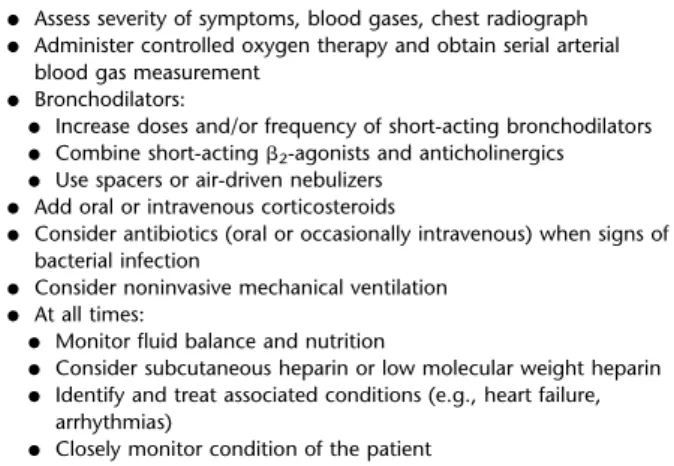
Table 6 shows the indications for hospital assessment and po-tential admission of a patient with a COPD exacerbation. When a patient comes to the emergency department, the first actions are to provide controlled oxygen therapy and to determine whether the exacerbation is life threatening (Table 7). If so, the patient should be admitted to the intensive care unit (ICU) immediately. Otherwise, the patient may be managed in the emergency depart-ment or hospital. In addition to pharmacologic therapy, hospital management of exacerbations includes respiratory support (oxy-gen therapy, ventilation).
Pharmacologic treatment. The three classes of medications most commonly used for exacerbations of COPD are broncho-dilators, corticosteroids, and antibiotics.
SHORT-ACTING BRONCHODILATORS. Although there are no controlled trials, short-acting inhaledb2-agonists with or without short-acting anticholinergics are usually the preferred bronchodi-lators for treatment of an exacerbation (90, 121) (Evidence C). A systematic review of the route of delivery of short-acting bron-chodilators found no significant differences in FEV1 between metered-dose inhalers (with or without a spacer device) and neb-ulizers (122), although the latter can be more convenient for sicker or frail patients. Intravenous methylxanthines (theophyl-line or aminophyl(theophyl-line) are only to be used in selected cases when there is insufficient response to short-acting bronchodilators (123–127) (Evidence B). Side effects of methylxanthines are sig-nificant, and their beneficial effects in terms of lung function and clinical endpoints are modest and inconsistent (128, 129).
CORTICOSTEROIDS. Data from studies in secondary health care indicate that systemic corticosteroids in COPD exacerbations shorten recovery time, improve lung function (FEV1) and arterial hypoxemia (PaO2) (130–133) (Evidence A), and reduce the risk of
early relapse, treatment failure, and length of hospital stay (130, 132, 134). A dose of 30–40 mg prednisolone per day for 10–14 days is recommended (Evidence D). Therapy with oral prednis-olone is preferable (135). Nebulized budesonide alone may be an alternative (although more expensive) to oral corticosteroids in the treatment of exacerbations (131, 136, 137).
ANTIBIOTICS. There is evidence supporting the use of
anti-biotics in exacerbations when patients have clinical signs of a bacterial infection, for example, increase in sputum purulence (118). A systematic review of the very few available placebo-controlled studies has shown that antibiotics reduce the risk of short-term mortality by 77%, treatment failure by 53%, and sputum purulence by 44%. This review supports antibiotics
for only moderately or severely ill patients with COPD exacer-bations with increased cough and sputum purulence (138, 139). Procalcitonin III, a marker that is specific for bacterial infec-tions, may be of value in the decision to use antibiotics (140), but this test is expensive and thus not widely established. A study in patients with COPD with exacerbations requiring me-chanical ventilation (invasive or noninvasive) indicated that not giving antibiotics was associated with increased mortality and a greater incidence of secondary nosocomial pneumonia (141). In summary, antibiotics should be given to patients with exacerba-tions of COPD who have three cardinal symptoms—increase in dyspnea, sputum volume, and sputum purulence (Evidence B); patients who have two of the cardinal symptoms, if increased purulence of sputum is one of the two symptoms (Evidence C); or require mechanical ventilation (invasive or noninvasive) (Ev-idence B) (142). The recommended length of antibiotic therapy is usually 5–10 days (Evidence D). The choice of the antibiotic should be based on the local bacterial resistance pattern.
ADJUNCT THERAPIES. Depending on the clinical condition of
the patient, an appropriate fluid balance with special attention to the administration of diuretics, anticoagulants, treatment of comorbidities, and nutritional aspects should be considered. At all times, health care providers should strongly enforce strin-gent measures against active cigarette smoking.
Respiratory support. OXYGEN THERAPY. Controlled oxygen
should be titrated to improve the patient’s hypoxemia with a target saturation of 88–92% (143). Once oxygen is started, arte-rial blood gases should be checked 30 to 60 minutes later to ensure satisfactory oxygenation without carbon dioxide retention or acidosis. Venturi masks (high-flow devices) offer more accurate
and controlled delivery of oxygen than do nasal prongs but are less likely to be tolerated by the patient (90).
VENTILATORY SUPPORT. Some patients need immediate
ad-mission to an ICU (Table 8). Adad-mission of patients with severe exacerbations to intermediate or special respiratory care units may be appropriate if personnel, skills, and equipment exist to identify and manage acute respiratory failure successfully.
Ventilatory support in an exacerbation can be provided by either noninvasive (by nasal or facial mask) or invasive (by oro-tracheal tube or tracheostomy) ventilation. Respiratory stimu-lants are not recommended for acute respiratory failure (121).
NONINVASIVE MECHANICAL VENTILATION. Noninvasive
mech-anical ventilation (NIV) has been studied in several randomized, controlled trials in acute respiratory failure, consistently pro-viding success rates of 80 to 85% (144–147). NIV improves respiratory acidosis (increases pH and decreases PaCO2) and decreases respiratory rate, severity of breathlessness, compli-cations such as ventilator-associated pneumonia, and length of hospital stay (Evidence A). More importantly, mortality and intubation rates are reduced by this intervention (145, 148–150) (Evidence A). Table 9 summarizes the indications for NIV (90, 144, 146, 151, 152).
INVASIVE MECHANICAL VENTILATION. The indications for
ini-tiating invasive mechanical ventilation during an exacerbation are shown in Table 10, and include failure of an initial trial of NIV (153). As experience is being gained with the generalized clinical use of NIV in COPD, several indications for invasive mechanical ventilation are being successfully treated with NIV, and in all but a few situations, there is nothing to be lost by a trial of noninvasive ventilation (153).
TABLE 5. INITIAL PHARMACOLOGIC MANAGEMENT OF COPD*
Patient Group Recommended First Choice Alternative Choice Other Possible Treatments† A Short-acting anticholinergic prn Long-acting anticholinergic Theophylline
or or
Short-actingb2-agonist prn Long-actingb2-agonist
or
Short-acting anticholinergic and short-actingb2-agonist
B Long-acting anticholinergic Long-acting anticholinergic and long-actingb2-agonist
Short-acting anticholinergic
or and/or
Long-actingb2-agonist Short-actingb2-agonist
and/or Theophylline C Inhaled corticosteroid and
long-actingb2-agonist
Long-acting anticholinergic and long-actingb2-agonist
Phosphodiesterase-4 inhibitor and/or Short-actingb2-agonist or and/or Long-acting anticholinergic Short-acting anticholinergic and/or Theophylline D Inhaled corticosteroid and
long-actingb2-agonist
Carbocysteine and/or and/or Inhaled corticosteroid, long-actingb2-agonist,
and long-acting anticholinergic
Short-actingb2-agonist or Long-acting anticholinergic or Short-acting anticholinergic and/or
Inhaled corticosteroid, long-actingb2-agonist,
and phosphodiesterase-4 inhibitor
Theophylline or
Long-acting anticholinergic and long-actingb2-agonist
or
Long-acting anticholinergic and phosphodiesterase-4 inhibitor Definition of abbreviation: COPD¼chronic obstructive pulmonary disease.
* Medications in each cell are mentioned in alphabetical order and therefore not necessarily in order of preference.
The use of invasive ventilation in patients with very severe COPD is influenced by the likely reversibility of the precipitating event, the patient’s wishes, and availability of intensive care facilities. When possible, a clear statement of the patient’s own treatment wishes—an advance directive or “living will”— makes these difficult decisions much easier to resolve. Major hazards include the risk of ventilator-acquired pneumonia (especially when multiresistant organisms are prevalent), baro-trauma, and failure to wean to spontaneous ventilation.
Contrary to some opinions, acute mortality among patients with COPD with respiratory failure is lower than mortality among patients ventilated for non-COPD causes (154). Despite this, there is evidence that patients who might otherwise survive may be denied admission to intensive care for intubation be-cause of unwarranted prognostic pessimism (155).
Hospital Discharge and Follow-up
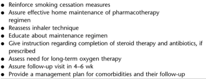
Insufficient clinical data exist to establish the optimal duration of hospitalization in individual patients with an exacerbation of COPD (156–158), although units with more respiratory consul-tants and better-organized care have lower mortality and reduced length of hospital stay after admission for an exacerbation (159). In the hospital prior to discharge, patients should start long-acting bronchodilators, either anticholinergics and/orb2-agonists with or without inhaled corticosteroids. Hospitalization offers a unique window of opportunity to reinforce smoking cessation measures if necessary. Table 11 provides a checklist of items to assess at time of discharge and Table 12 shows items to assess at follow-up 4 to 6 weeks after discharge from the hospital.
Home visits by a community nurse may permit earlier dis-charge of patients hospitalized with an exacerbation without
increasing readmission rates (90, 160–163). Use of a written action plan increases appropriate therapeutic interventions for an exacerbation, an effect that does not decrease health care resource use (164) (Evidence B) but may shorten recovery time (165).
For patients who are hypoxemic during an exacerbation, arte-rial blood gases and/or pulse oximetry should be evaluated prior to hospital discharge and in the following 3 months. If the patient remains hypoxemic, long-term supplemental oxygen therapy may be required.
Home Management of Exacerbations
Nurse-administered home care (also known as “hospital-at-home” care) represents an effective and practical alternative to hospitaliza-tion in selected patients with exacerbahospitaliza-tions of COPD without aci-dotic respiratory failure (160, 161) (Evidence A). However, the exact criteria for this approach as opposed to hospital treatment remain uncertain and will vary by health care setting. Treatment recommen-dations are the same as for hospitalized patients.
Prevention of COPD Exacerbations
COPD exacerbations can often be prevented. Smoking cessa-tion, influenza and pneumococcal vaccines, knowledge of current therapy including inhaler technique, and treatment with long-acting inhaled bronchodilators, with or without inhaled cortico-steroids, and phosphodiesterase-4 inhibitors are all therapies that reduce the number of exacerbations and hospitalizations (75, 79, 81, 82, 166, 167). Early outpatient pulmonary rehabil-itation after hospitalization for an exacerbation is safe and results in clinically significant improvements in exercise capacity and health status at 3 months (168). Patients should be encour-aged to maintain physical activity, and anxiety, depression, and social problems should be discussed. Principal caregivers should be identified if the patient has a significant persisting disability. 6. COPD AND COMORBIDITIES
KEY POINTS
d COPD often coexists with other diseases (comorbid-ities) that may have a significant impact on prognosis. d In general, the presence of comorbidities should not alter COPD treatment, and comorbidities should be treated as if the patient did not have COPD.
d Cardiovascular diseases are major comorbidities in COPD and probably both the most frequent and most important diseases coexisting with COPD.
d Osteoporosis and depression are also major comorbid-ities in COPD, are often underdiagnosed, and are as-sociated with poor health status and prognosis. d Lung cancer is frequently seen in patients with COPD
and has been found to be the most frequent cause of death in patients with mild COPD.
Introduction
COPD often coexists with other diseases (comorbidities) that may have a significant impact on prognosis (42, 169–171). Com-orbidities can occur at any COPD grade (50). Differential diag-nosis may be difficult because comorbidities may mimic COPD TABLE 6. POTENTIAL INDICATIONS FOR HOSPITAL ASSESSMENT
OR ADMISSION*
d Marked increase in intensity of symptoms, such as sudden development of resting dyspnea
d Severe underlying COPD
d Onset of new physical signs (e.g., cyanosis, peripheral edema) d Failure of an exacerbation to respond to initial medical management
d Presence of serious comorbidities (e.g., heart failure or newly occurring
arrhythmias)
d Frequent exacerbations d Older age
d Insufficient home support
Definition of abbreviation: COPD¼chronic obstructive pulmonary disease. * Local resources need to be considered.
TABLE 7. MANAGEMENT OF SEVERE BUT NOT LIFE-THREATENING EXACERBATIONS*
d Assess severity of symptoms, blood gases, chest radiograph d Administer controlled oxygen therapy and obtain serial arterial
blood gas measurement
d Bronchodilators:
d Increase doses and/or frequency of short-acting bronchodilators
d Combine short-actingb2-agonists and anticholinergics d Use spacers or air-driven nebulizers
d Add oral or intravenous corticosteroids
d Consider antibiotics (oral or occasionally intravenous) when signs of bacterial infection
d Consider noninvasive mechanical ventilation
d At all times:
d Monitor fluid balance and nutrition
d Consider subcutaneous heparin or low molecular weight heparin d Identify and treat associated conditions (e.g., heart failure,
arrhythmias)
d Closely monitor condition of the patient * Local resources need to be considered.