STRUCTURE, FUNCTION AND REGULATION OF THE COATED
VESICLE V-ATPase
BY MICHAEL FORGAC
Department of Cellular and Molecular Physiology, Tufts University School of Medicine, 136 Harrison Avenue, Boston, MA 02111, USA
Summary
The coated vesicle V-ATPase plays an important role in both receptor-mediated endocytosis and intracellular membrane traffic by providing the acidic environment required for ligand-receptor dissociation and receptor recycling. The coated vesicle V-ATPase is a macromolecular complex of relative molecular mass 750000 composed of nine subunits arranged in two structural domains. The peripheral Vi domain, which has a relative molecular mass of 500000, has the subunit structure 73358340i34i33i and possesses all the nucleotide binding sites of the V-ATPase. The integral Vo domain of relative molecular mass 250000 has a subunit composition of 100i38i 19i 176 and possesses the pathway for proton conduction across the membrane. Reassembly studies have allowed us to probe the role of specific subunits in the V-ATPase complex while chemical labeling studies have allowed us to identify specific residues which play a critical role in catalysis. From both structural analysis and sequence homology, the vacuolar-type H+-ATPases resemble the F-type H+-ATPases. Unlike the Fi and F
o domains of the F-type ATPases, however, the Vi and Vo domains do not appear to function independently. The possible relevance of these observations to the regulation of vacuolar acidification is discussed.
Function of the coated vesicle V-ATPase
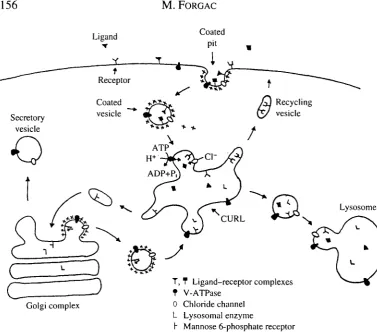
Clathrin-coated vesicles play a critical role in both receptor-mediated endocytosis and intracellular membrane traffic (for a review, see Forgac, 1989). As illustrated in Fig. 1, during endocytosis ligand—receptor complexes become concentrated in clathrin-coated pits, which pinch off from the cell surface to form clathrin-coated vesicles. Among the ligands internalized by this pathway are low-density lipoprotein, transferrin, insulin and epidermal growth factor. Rapid uncoating of the vesicle and subsequent membrane fusion events lead to delivery of the ligand-receptor complexes to CURL (compartment of uncoupling of receptor and ligand, Geuze et al. 1983). Acidification of CURL by the V-ATPases causes dissociation of ligand-receptor complexes, allowing recycling of unoccupied receptors to the cell surface and transport of the dissociated ligands to lysosomes for degradation.
Clathrin-coated vesicles play a similar role in the intracellular targeting of newly synthesized lysosomal enzymes from the Golgi apparatus to lysosomes (Fig. 1). By
156
M . FORGACLigand
c
J
Golgi complex
T, T Ligand-receptor complexes t V-ATPase
0 Chloride channel L Lysosomal enzyme
[image:2.451.35.413.47.379.2]h Mannose 6-phosphate receptor
Fig. 1. Functions of vacuolar acidification in eukaryotic cells. Receptor-mediated endocytosis involves the concentration of ligand-receptor complexes in clathrin-coated pits at the cell surface, followed by their invagination to form clathrin-coated vesicles. Ligand-receptor complexes are then delivered to CURL (compartment of uncoupling of receptor and ligand) where exposure to low pH activates ligand dissociation and recycling of receptors to the cell surface. Acidification of CURL requires the activity of both a vacuolar H+-ATPase and a chloride channel. A similar mechanism is employed in the delivery of newly synthesized lysosomal enzymes from the Golgi to lysosomes via the mannose 6-phosphate receptor. V-ATPases maintain the low pH within lysosomes and secretory vesicles required for processing and degrading macromolecules contained within these organelles; they also establish the electrochemical gradient employed in the coupled transport of various small molecules. Monoclonal antibodies that recognize the coated vesicle V-ATPase immunolabel endosomes, lysosomes, the Golgi complex and the plasma membrane as well as coated vesicles in MDBK cells (Marquez-Sterling etat. 1991).
virtue of the mannose 6-phosphate recognition marker, lysosomal enzymes bind to the mannose 6-phosphate receptor in the trans-Golgi, where they become concentrated in clathrin-coated pits that pinch off to form clathrin-coated vesicles. Vesicle uncoating and delivery to an acidic compartment results in dissociation and recycling of the receptors to the trans-Go\g\ and targeting of the lysosomal enzymes to lysosomes.
contain a V-ATPase which provides the electrochemical driving force for neurotransmitter uptake, it is likely that coated vesicles retrieve this pump from the plasma membrane and deliver it to synaptic vesicles.
Transport and inhibitor properties of the coated vesicle V-ATPase
The coated vesicle V-ATPase transports protons from the cytoplasm across the vesicle membrane uncoupled to the countertransport of other cations (Forgac and Cantley, 1984). This pump is therefore electrogenic and requires the movement of another ion for proton transport to occur. As illustrated in Fig. 1, the membrane potential generated by the proton pump is dissipated by the activity of a parallel chloride channel (Arai et al. 1989; Xie et al. 1983). We have recently shown that this chloride channel is controlled by protein-kinase-A-dependent phosphorylation (Mulberg et al. 1991). Because acidification is dependent upon chloride conductance, modulation of channel activity represents an important mechanism for controlling vacuolar acidification (see below).
The coated vesicle H+-ATPase, like other V-ATPases, is sensitive to micromolar concentrations of /V-ethylmaleimide (NEM) and 7-chloro-4-nitrobenz-2-oxa-l,3-diazole (NBD-CI) (Arai et al. 19876). Protection of activity by ATP suggests that both reagents react with groups present at the catalytic site. We have recently identified the cysteine residue responsible for NEM-sensitivity of the coated vesicle V-ATPase (Feng and Forgac, 1992) (see below). In addition, protection of the NEM-reactive cysteine by NBD-CI indicates that both reagents inhibit activity by reacting with the same residue.
Unlike the P-ATPases (Pedersen and Carafoli, 1987), the coated vesicle V-ATPase is resistant to vanadate (Forgac et al. 1983) and does not form a phosphorylated intermediate during turnover (Forgac and Cantley, 1984). In addition, the coated vesicle V-ATPase is resistant to the F-ATPase inhibitors oligomycin and aurovertin (Forgac et
al. 1983), but is sensitive to dicyclohexylcarbodiimide (DCCD), a carboxyl reagent
which inhibits proton flux (Arai et al. 1987a). As with other V-ATPases (E. J. Bowman et
al. 1988a), the coated vesicle H+-ATPase is sensitive to nanomolar concentrations of bafilomycin (M. Myers and M. Forgac, unpublished observations).
Structure of the coated vesicle H+-ATPase
Structural model
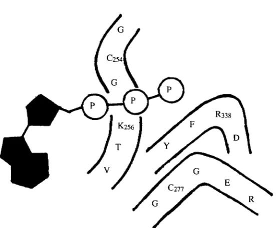
Our current model of the coated vesicle V-ATPase (Adachi et al. 19906), based on the structural data described below, is shown in Fig. 2. The V-ATPase complex is divided into a peripheral V| domain containing subunits of relative molecular mass 73 (A), 58 (B), 40, 34 and 33xl03 and an integral Vo domain composed of the 100, 38, 19 and 17 (c)
158
M. FORGAC33xlCPMr 'accessory' subunits. The Vo domain is responsible for proton translocation
whereas the HxlCPMr c subunit is responsible for the DCCD-sensitivity of proton transport. The 100x1 (PA/r subunit is a transmembrane glycoprotein of as yet unknown
ATP
Cytoplasm
Membrane
Lumen
- D C C D Vo
[image:4.451.99.354.136.389.2]19
Fig. 2. Structural model of the coated vesicle H+-ATPase. The coated vesicle V-ATPase is composed of two domains. The peripheral Vi domain (unshaded) contains the A, B, 40, 34 and 33x\(fiMr subunits in the stoichiometry Ajfi340i34i33i and is oriented towards the
cytoplasmic side of the membrane (Arai et al. 1988; Adachi et al. 1990a,ft). The A subunits possess the catalytic nucleotide binding sites (filled triangles) (Arai et al. 1987ft; Feng and Forgac, 1992), whereas the B subunits also appear to possess nucleotide binding sites (open triangles) (Adachi et al. 1990a) of unknown function. The Vi domain appears to be attached to the integral Vosector via the 40, 34 and 33X1O3Mr 'accessory' subunits (Adachi et al. 1990ft),
with the 40x103 MT subunit contributing to the stability of the complex but not essential for
function. The V-ATPases thus closely resemble the F-ATPases of mitochondria, chloroplasts and bacteria, with which they share sequence homology. Unlike the Fi and Fo domains of the F-ATPases, however, the Vi and Vo domains do not function
independently. The possible significance of this finding to the mechanisms regulating the activity of the V-ATPases is discussed below.
Subunit composition, stoichiometry and domain structure
The coated vesicle V-ATPase is composed of nine subunits of relative molecular mass 100, 73 (A), 58 (£), 40, 38, 34, 33, 19 and 17 (c) xlO3, which are immunoprecipitated as a single macromolecular complex by monoclonal antibodies directed against the native enzyme (Arai et al. 1987ft). Using quantitative amino acid analysis, we have shown that these subunits are present in a stoichiometry of 1 O O I A 3 / ? 3 4 O | 3 8 I 3 4 I 3 3 | 1 9 I 176 (Arai etal. 1988). A similar subunit composition is observed for the V-ATPases of kidney (Gluck and Caldwell, 1987), chromaffin granules (Moriyama and Nelson, 1989), Neurospora
crassa (Bowman etal. 1989), yeast (Kane etal. 1989) and plants (Parry etal. 1989; Lai et al. 1988).
The V-ATPase complex is divided into two structural domains. The peripheral Vi domain, which has a relative molecular mass of 500X103, has the structure A3fi340i34|33i (Arai et al. 1988) and is removed from the membrane by chaotropic agents such as KI and KNO3 (Arai etal. 1989; Adachi etal. 1990i>). Dissociation of Vi is activated by submicromolar concentrations of ATP (Arai et al. 1989). The Vi subunits are released from the membrane as monomers which are capable of reassembling into various subcomplexes (Puopolo and Forgac, 1990; Puopolo et al. \992b), as discussed below. The integral Vo domain contains four subunits in a stoichiometry of 100i38i 19ic6
(Arai et al. 1988). These polypeptides remain assembled as a complex of 250xl03Mr
following removal of Vi and detergent solubilization (Zhang etal. 1992).
Topographical analysis
Topographical studies indicate that the Vi domain is oriented towards the cytoplasmic side of the membrane (Arai etal. 1988; Adachi etal. 1990a). Thus, all the Vi subunits are labeled by membrane-impermeant reagents in intact coated vesicles where only the cytoplasmic surface is exposed (Arai et al. 1988). In addition, all the Vi subunits are cleaved by trypsin in reconstituted vesicles containing the purified V-ATPase oriented with the cytoplasmic surface facing out (Adachi etal. 1990a). Of the Vo subunits, the 100
and 3&xl(fiMr subunits are exposed on the cytoplasmic side of the membrane based on
their labeling by impermeant reagents and their sensitivity to trypsin added from the cytoplasmic surface (Arai et al. 1988; Adachi et al. 1990a). The 100, 19 and 1 7 x 1 0 ^ subunits also possess luminal domains since they show a significant increase in labeling by impermeant reagents upon detergent permeabilization of intact coated vesicles (Arai et
al. 1988). Lectin binding studies indicate that the 100xl03Mr subunit possesses
160 M.FORGAC
Membrane-embedded domains
Both the c and the 19xl03jWr subunits are highly hydrophobic proteins extensively
buried in the bilayer, based upon their extraction by organic solvents (Arai et al. 1987a) and their high proportion of nonpolar amino acids (Arai et al. 1988). Heavy labeling of the c subunit with the photoactivated hydrophobic reagent [125I]TED confirms that a large proportion of this protein is in contact with the lipid bilayer (Arai et al. 1988). Interestingly, [125I]TID does not label the 38 and \9y.\QP Mr subunits while a small
amount of labeling of the 100xl03Mr subunit is observed (Arai etal. 1988). This result is
consistent with the transmembrane orientation of the IOOXICPMI- polypeptide and suggests that the 19xl03Mr subunit is shielded from contact with the lipid bilayer through
interaction with other membrane-spanning domains, probably contributed by the c and lOOxK^Mr subunits. The absence of [125]TTD labeling of the 38xl03Mr polypeptide is
also consistent with the sequence of the corresponding chromaffin granule protein, which shows no putative transmembrane helices (Wang et al. 1988). The 38x1 (P/Wr subunit therefore appears to be a peripheral polypeptide, which remains tightly bound through protein-protein contacts to the integral Vo domain.
Subunit interactions and the structure of subcomplexes
Crosslinking studies of the coated vesicle V-ATPase using the reversible crosslinking reagent 3,3'-dithiobis(sulfosuccinimidylpropionate) (DTSSP) have revealed extensive contact between the A and B subunits as well as between the c subunit and the 40, 34 and 33x103Mr subunits (Adachi et al. 19906). These results have led to the model shown in
Fig. 2 where the A and B subunits form a hexameric complex attached to the Vo sector via
a bridge formed by the accessory subunits.
As mentioned above, dissociation of the V| subunits from the membrane by treatment with KI and ATP results in their initial release as monomers. Removal of the chaotropic agents in the presence of membranes containing Vo results in the reassembly of functional
Vi Vo complexes (Puopolo and Forgac, 1990). If KI and ATP are removed in the absence of Vo and reassembly is monitored by sedimentation on glycerol density gradients, the following results are obtained. A subcomplex of approximately 50Ox\(fi Mx is formed
which has the structure AiB?,3A\331 (Puopolo etal. 19926). This subcomplex completely lacks the 40xl03A/r subunit, which appears in the 'monomeric' fraction, together with
approximately 5 0 % of the A, B and 33xl(flMT subunits. Interestingly, the monomeric
fraction is almost completely devoid of the 34xlO3Mr subunit (Puopolo etal. 19926).
Evidence for interaction between the 40 and 33xl(fiMr subunits derives from
immunoprecipitation experiments using the monoclonal antibody 1C-11G (Puopolo etal. 19926). This antibody immunoprecipitates both the 40 and 33x\(fiMT subunits from the
'monomeric' fraction, suggesting that these two subunits are complexed. That the antibody is directed against an epitope expressed on the native 33xlO3yVfr subunit is
indicated by its ability to immunoprecipitate the Vi ( ^ O x ^ M r ) subcomplex (Puopolo
et al. 19926). Thus, during in vitro reassembly, the 33x103 Mr subunit assembles with
either the 40xl03Afr polypeptide or with the remaining Vi subunits to form the
The Vo subunits, unlike the Vi subunits, remain together as a complex of 250x1 CPMr
following removal of Vi with KI and ATP (Zhang et al. 1992). The Vo subunits are
present in the same stoichiometry in the free Vo domain as in Vi Vo and possess many of
the same properties. Thus, the c subunit in the free Vo domain is extracted by organic
solvents and is labeled by [I4C]DCCD. In addition, a monoclonal antibody has been isolated which recognizes the 100x103Mr subunit in both complexes. Both the 100 and
38x1 (fi MT subunits show the same tryptic cleavage pattern in the free Vo complex as in
ViV0, although the sensitivity of these polypeptides to proteolysis is increased,
suggesting that they are more exposed to the aqueous phase on removal of Vi (Zhang et
al. 1992).
ATP binding and covalent labeling of the catalytic site
The existence of multiple nucleotide binding sites on the coated vesicle V-ATPase is suggested by several lines of evidence. First, the dependence of ATP hydrolysis on the concentration of ATP indicates the existence of two ATP binding sites with Kd values of 80 and 800/i,moll~' (Arai et al. 1989). Saturation of the higher-affinity site leads to increased proton transport, whereas occupation of the low-affinity site appears to decrease proton transport, resulting in a decreased H+/ATP stoichiometry. ATP binding to a still higher affinity site {Kd 200nmoll~') activates dissociation of the peripheral Vi domain from the integral Vo domain (Arai et al. 1989), suggesting a decrease in stability
of the V-ATPase complex.
Further evidence for the existence of multiple nucleotide binding sites has come from studies of the nucleotide analog 2',3'-0-(2,4,6-trinitrophenyl)adenosine 5'-triphosphate (TNP-ATP) (Adachi et al. 1990a). TNP-ATP inhibits activity of the purified V-ATPase with A"d values of 50 nmol 1~' and 3 yu,mol 1~' and protects both the A and B subunits from tryptic cleavage with a K<i of l ^ m m o l l "1 (Adachi et al. 1990a). Finally, the coated vesicle V-ATPase contains multiple copies of both the A and B subunits which have been shown to possess ATP binding sites either by direct chemical labeling or by sequence homology (see below).
Evidence that the catalytic nucleotide binding site is located on the A subunit comes from its labeling by [3H]NEM and [I4C]NBD-C1 in an ATP-protectable manner (Arai et
al. 1987£>). Similar results have been obtained with V-ATPases from a variety of sources
(Forgac, 1989). Labeling of the B subunit of the plant V-ATPase using an ATP analog has also been reported (Manolson et al. 1985). Moreover, as first demonstrated by Zimniak et
al. (1988), E. J. Bowman et al. (19886) and B. J. Bowman et al. (1988), the A and B
162 M. FORGAC
binding subunits of the V-ATPases and F-ATPases have been derived from a common evolutionary ancestor.
Like the V-ATPases from other sources, the A and B subunits of the coated vesicle H+ -ATPase show similar sequence homology (Puopolo et al. 1991, 1992a; Sudhof et al. 1989). The bovine A subunit appears to be encoded by a single gene, which gives rise to a single transcript in all tissues tested (Puopolo et al. 1991). The bovine B subunit, in contrast, is encoded by at least two and possibly three, genes, which give rise to multiple transcripts in a tissue-specific manner (Puopolo et al. 1992a). Expression of both a 3.2 and 2.0 kb mRNA can be detected in all tissues examined except brain, where expression of only the 3.2 kb message is detected. Southern analysis is also consistent with the existence of multiple genes encoding the B subunit (Puopolo et al. 1992a).
We have recently identified the cysteine residue responsible for the sensitivity of the V-ATPases to sulfhydryl reagents (Feng and Forgac, 1992). We demonstrated that, in addition to NEM, the coated vesicle V-ATPase is also inhibited by cystine in an ATP-protectable manner. Unlike NEM, however, inhibition by cystine is reversible upon treatment with reducing agents, such as dithiothreitol (DTT). That cystine inhibits activity by forming a disulfide bond with the same cysteine residue that reacts with NEM was demonstrated by the fact that extensive treatment of the cystine-reacted enzyme with NEM followed by treatment with DTT restores activity of the coated vesicle V-ATPase. We then took advantage of this protection of the NEM-reactive cysteine by cystine to label this residue selectively. The catalytic cysteine was first protected with cystine, the enzyme was reacted extensively with NEM, the disulfide bond was reduced with DTT and the catalytic cysteine residue was then labeled with fluorescein maleimide. Under these conditions, fluorescein maleimide labels only the 73X103 MT A subunit. Proteolytic
cleavage of the labeled A subunit gives rise to a single 3.9xl03Afr fragment, which is
labeled with fluorescein maleimide on cyteine residue 254 (Feng and Forgac, 1992). This residue is conserved as cysteine in all the V-ATPase A subunit sequences obtained thus far (Puopolo et al. 1991) and is located in the Walker consensus 'A' sequence GXGKTV (Walker et al. 1985). Moreover, the corresponding residue is valine in the F-ATPases and serine in the archaebacterial H+-ATPases (Penefsky and Cross, 1991), consistent with the insensitivity of the latter two classes to sulfhydryl reagents. As discussed above, we have demonstrated that this same cysteine residue is responsible for the sensitivity of the V-ATPases to NBD-C1. Based upon identification of this cyteine residue at the catalytic site of the A subunit, the existence of highly conserved consensus sequences involved in nucleotide binding (Walker et al. 1985) and the structures proposed for the ATP binding site of Fi (Penefsky and Cross, 1991; Garboczi et al. 1988; Duncan et al. 1986) and adenylate kinase (Kim et al. 1990), we propose the model for the structure of the catalytic site of the V-ATPases shown in Fig. 3. Much additional work will be required to determine how accurately this model describes the true structure.
Proton conduction through Vo
The 17x 103 Afr c subunit is responsible for the DCCD-sensitivity of proton transport by
Fig. 3 Proposed model for the catalytic site of the V-ATPases. This model is based on identification of cys254 as the catalytic site residue responsible for sensitivity of the V-ATPases to sulfyhdryl reagents (Feng and Forgac, 1992), the existence in the V-ATPase A subunits (Zimniak et al. 1988; E. J. Bowman et al. 1988b; Puopolo et al. 1991) of highly conserved consensus sequences believed to be involved in nucleotide binding (Walker et al. 1985) and the proposed structures of the catalytic site of Fi (Penefsky and Cross, 1991; Garboczi et al. 1988; Duncan et al. 1986) and adenylate kinase (Kim et at. 1990).
results in complete loss of proton transport activity, although ATP hydrolysis is only inhibited if the H+-ATPase is embedded in the membrane. Comparison of the stoichiometry of DCCD labeling with the degree of inhibition suggests that complete blockage of proton conduction occurs after reaction of only one-sixth of the c subunits with DCCD (Arai et al. 1987a). As with the c subunit of Fo, this result suggests that
formation of a proton channel requires the cooperative interaction of six copies of the c subunit, in agreement with the measured subunit stoichiometry (Arai et al. 1988).
Cloning and sequence analysis of the c subunit of the chromaffin granule V-ATPase (Mandel et al. 1988) has confirmed that this polypeptide is the vacuolar counterpart of the Fo c subunit, which is present in a stoichiometry of 10-12 copies per Fo complex (Foster
and Fillingame, 1982). Despite being twice the size of the Fo protein, the vacuolar c
subunit has only a single buried aspartate (the likely site of reaction with DCCD), suggesting that the number of transmembrane helices contributed by the c subunit has been conserved while the number of buried carboxyl groups has been reduced by a factor of two. This observation has suggested a mechanism in which the H+/ATP stoichiometry of the V-type and F-type H+-ATPases was altered during the course of their evolution (Cross and Taiz, 1990).
164 M.FORGAC
(Sun et al. 1987). Based on the homology between the vacuolar and Fo c subunits
(Mandel et al. 1988) and on the extensive genetic and biochemical evidence indicating that the c subunit of Fo is not sufficient to form a proton pore (Cain and Simoni, 1986;
Schneider and Altendorf, 1985), it seems most likely that additional subunits are also required for proton conduction through Vo. One possible candidate for die vacuolar
counterpart to the a subunit, which is critical for proton conduction through Fo, is the \9xl(fiMr polypeptide. Like the a subunit, this protein is present as one copy per
complex and is extensively buried in the bilayer (Arai et al. 1988).
We have investigated the proton conductance properties of the Vo domain of the coated
vesicle V-ATPase (Zhang et al. 1992). Proton conductance was measured both in native membranes from which Vi had been removed and in reconstituted vesicles containing the isolated Vo complex by uptake of the fluorescent dye
9-amino-6-chloro-2-methoxyacridine (ACMA) in response to a K+- and valinomycin-induced membrane potential. In neither case was any DCCD-inhibitable proton conduction detected, although proton conduction could readily be detected upon addition of the proton ionophore carbonyl cyanide /?-chlorophenylhydrazone (CCCP) and although the Vo
domain was still competent to reassemble with the Vi subunits to give a functional V-ATPase. These results suggest that one or more of the Vo subunits may be suppressing
proton conduction through Vo (Zhang et al. 1992). The possible role of such suppression in vivo is discussed below.
Activity of V-ATPase subcomplexes
To address the role of the accessory subunits of the coated vesicle V-ATPase, we have developed a protocol for reassembly of the V-ATPase from the dissociated Vi and Vo
domains that restores both ATP hydrolysis and proton translocation (Puopolo and Forgac, 1990). Reassembly involves attachment of the complete complement of Vi subunits to the Vo sector, is time-dependent and protein-concentration-dependent and gives rise to a reassembled V-ATPase with inhibitor sensitivities identical to those of the native enzyme.
Recently, we have employed this reassembly system to address the role of the 40 and 34xlO3Mr subunits (Puopolo et al. \992b). As explained above, we have isolated a
Vi (-40xl03Mr) subcomplex which contains the A, B, 34 and 33x\(fiMT subunits. This
subcomplex, although devoid of ATPase activity, is able reassemble onto the Vo domain
to give a complex possessing approximately 5 0 % of the proton transport activity obtained using the complete complement of Vi and Vo subunits (Puopolo et al. [992b).
Reassembly requires that the Vi (-40xl(P MT) subcomplex first be dissociated with KI
and MgATP, followed by reassembly in the presence of Vo. Thus, the assembled
subcomplex appears to be unable to bind to the membrane sector. Similarly, addition of the isolated 40xl03Mr subunit to the reassembly mixture is without effect on activity
unless the Vi (-40xl03Afr) subcomplex is first dissociated, under which conditions
addition of the 40xl(fi MT subunit restores activity to maximal levels. The binding sites
for both Voand for the 40x103Afr subunit thus appear to be inaccessible in the assembled
The monomeric Vi fraction lacking the 34xlO3Mr subunit obtained by sedimentation
(see above) is also competent to reassemble with Vo to give a partially active complex
(Puopolo et al. \992b). The absence of either the 40 or 34xlO3/V/r subunits, however,
makes the reassembled complexes unstable to detergent solubilization and immunoprecipitation. These results, summarized in Table 1, suggest that, although not absolutely required for coupling of ATP hydrolysis and proton transport, both the 40 and 34xl03yVfr subunits are required for stability and maximal activity of the coated vesicle
V-ATPase. These subunits may therefore play some role in regulation of either assembly or activity of the V-ATPases in vivo.
It is important to note that, unlike Fi and Fo, the V| and Vo domains of the coated
vesicle V-ATPase do not appear to function independently. Thus the V| (-^xlCPMr) subcomplex lacks ATPase activity (Puopolo et al. \992b) and no conditions have yet been identified that allow binding of the 40x103Mr subunit to this subcomplex in vitro.
Moreover, preliminary results suggest that a Vi complex containing the 40x1 (fiMT
subunit formed in vivo is still unable to hydrolyze ATP (M. Myers and M. Forgac, in preparation). Similarly, as discussed above, the Vo domain, unlike the Fo domain, is
unable to carry out passive, DCCD-inhibitable proton conduction (Zhang et al. 1992).
Regulation of coated vesicle acidification
Although it is clear that cells are able to modulate the pH of the various intracellular compartments differentially (Forgac, 1989), the mechanisms of modulation remain uncertain. For example, clathrin-coated vesicles have been demonstrated to contain a V-ATPase capable of acidifying the lumen of the vesicle (Forgac et al. 1983; Stone et al. 1983), yet numerous data suggest that ligand-receptor complexes do not become exposed to an acidic pH until delivery to CURL. Thus, ligand-receptor complexes remain associated in endocytic coated vesicles and small peripheral endosomes (Geuze et al. 1983) and endocytic coated vesicles appear to be neutral organelles within the cell, based upon their failure to accumulate the electron microscopic probe
3-(2,4-dinitroanilino)-3'-Table 1. Structure and activity of reassembled H+-ATPase complexes Subunit composition
Vi subunits
A-i Bj, 40 34 33 A3 S3 34 33 A3 S3 40 33
Vo subunits
100 38 19 c6 100 38 19 ce 100 38 19 c6
Relative proton transport activity*
1.0 0.52 0.80
Relative
stability!"
1.0 <0.05 <0.05
•Proton transport activity of reassembled complexes is expressed relative to that obtained following reassembly employing the complete complement of V| and Vo subunits (Puopolo et al. 1992b). Reassembled complexes containing all of the V-ATPase subunits had activity values of 70-80% relative to those obtained for the undissociated V-ATPase (Puopolo and Forgac, 1990).
"("Stability corresponds to the amount of V-ATPase complex detected by SDS—PAGE following
[image:11.451.41.412.463.549.2]166 M.FORGAC
amino-N-methyldipropylamine (DAMP) (Anderson and Orci, 1988). Finally, rat liver endocytic coated vesicles loaded with the pH-sensitive fluorescence probe fluorescein-5'-isothiocyanate (FITC)-dextran appear to be incapable of ATP-dependent acidification (Fuchs etal. 1987).
To determine whether the failure of endocytic coated vesicles to acidify was due to the absence of the proton pump, we have carried out immunocytochemical studies of MDBK cells, a bovine epithelial cell line, using monoclonal antibodies directed against the coated vesicle V-ATPase (Marquez-Sterling et al. 1991). We have demonstrated that, in addition to staining endosomes, lysosomes and portions of the Golgi complex, these antibodies label the plasma membrane and vesicles just beneath the plasma membrane. Moreover, significant co-localization of the V-ATPase with clathrin was observed at both the cell surface and in peripheral vesicles, indicating that the vacuolar proton pump is present in endocytic coated vesicles (Marquez-Sterling et al. 1991). These results, together with those described above, suggest that endocytic coated vesicles are not acidified because the activity of the coated vesicle H+-ATPase is suppressed within the cell.
A number of possible mechanisms may be involved in controlling vacuolar acidification. Changes in subunit composition of the V-ATPase represent one important regulatory mechanism which may be employed. One candidate for such a modulatory subunit is the \OOxKftMT polypeptide, which is present in the coated vesicle V-ATPase
and other V-ATPases but appears to be absent from the V-ATPase found in the plasma membrane of kidney cells (Gluck and Caldwell, 1987). Alternatively, regulation may be accomplished through substitution of organelle-specific isoforms of particular V-ATPase subunits. The existence of multiple isoforms of the human (Bernasconi et al. 1990) and bovine (Puopolo et al. 1992a) B subunit has recently been demonstrated. The degree of vacuolar acidification may in some cases be controlled by changes in the number of V-ATPases in a particular compartment. This mechanism operates in regulating acid secretion across the luminal membrane of the intercalated cells of the mammalian kidney (Gluck et al. 1982). In this case, the density of pumps in the luminal membrane is controlled by reversible fusion with the plasma membrane of intracellular vesicles containing a high density of vacuolar proton pumps.
A third possible mechanism for regulating vacuolar acidification involves control of coupling between ATP hydrolysis and proton translocation. As discussed above, there are several experimental conditions that have been shown to alter the tightness of this coupling for the coated vesicle V-ATPase, including high concentrations of ATP (Arai et
al. 1989), partial proteolysis (Adachi etal. 1990a) and detergent solubilization (Arai etal.
1987a). These results suggest that the enzyme may be poised in a state in which the stoichiometry of proton transport can readily be altered in response to the appropriate intracellular signal. A fourth possible regulatory mechanism is suggested by the apparent inactivity of the separated Vi and Vo domains with respect to ATP hydrolysis and proton
conduction (Puopolo etal. 1992b; Zhang etal. 1992) (see above). This mechanism would involve control of ATP-driven proton transport by controlling attachment of the Vi and Vo domains. Such a mechanism would necessitate that at least the Vo domain was not
multiple copies of Vo, no significant pH gradient could be achieved until all the Vo sites had been occupied. Further evidence consistent with this model comes from the observation that coated vesicles appear to contain an excess of Vo domains over those required to form functional ViVocomplexes (Zhang etal. 1992) and from the observation that a soluble pool of V| domains exists free in the cytoplasm (M. Myers and M. Forgac, in preparation).
A final mechanism of controlling vacuolar pH involves control of the chloride channel required for acidification. We have recently demonstrated that both chloride conductance and ATP-dependent acidification in coated vesicles are modulated by a protein-kinase-A-dependent phosphorylation and that this effect is the result of alteration in the activity of the chloride channel (Mulberg et al. 1991). It is possible that several of the mechanisms described above may be employed in regulating vacuolar acidification in vivo.
This work was supported by National Institutes of Health Grant GM 34478 and GM 44828 and American Heart Grant-in-Aid 890772. M.F. is an American Heart Association Established Investigator.
References
ADACHI, I., ARAI, H., PIMENTAL, R. AND FORGAC, M. (1990a). Proteolysis and orientation on
reconstitution of the coated vesicle proton pump. J. biol. Chem. 265, 960-966.
ADACHI, I., PUOPOLO, K., MARQUEZ-STERLING, N., ARAI, H. AND FORGAC, M. (1990/?). Dissociation, crosslinking and glycosylation of the coated vesicle proton pump. J. biol. Chem. 265, 967-973. ANDERSON, R. G. AND ORCI, L. (1988). A view of acidic intracellular compartments. J. Cell Biol. 106,
539-543.
ARAI, H., BERNE, M. AND FORGAC, M. (1987a). Inhibition of the coated vesicle proton pump and labeling of a 17,000 daltonpolypeptide by DCCD. J. biol. Chem. 262, 11006-11011.
ARAI, H., BERNE, M., TERRES, G., TERRES, H., PUOPOLO, K. AND FORGAC, M. (1987i). Subunit composition and ATP site labeling of the coated vesicle (H+)-ATPase. Biochemistry N.Y. 26, 6632-6638.
ARAI, H., PINK, S. AND FORGAC, M. (1989). Interaction of anions and ATP with the coated vesicle proton pump. Biochemistry N. Y. 28, 3075-3082.
ARAI, H., TERRES, G., PINK, S. AND FORGAC, M. (1988). Topography and subunit stoichiometry of the coated vesicle proton pump. J. biol. Chem. 263, 8796-8802.
BERNASCONI, P., RAUSCH, T., STRUVE, I., MORGAN, L. AND TAIZ, L. (1990). An mRNA from human brain encodes an isoform of the B subunit of the vacuolar H+-ATPase. J. biol.Chem. 265, 17428-17431. BOWMAN, B. J., ALLEN, R., WECHSER, M. A. AND BOWMAN, E. J. (1988). Isolation of the genes encoding
the Neurospora vacuolar ATPase: analysis of vma-2 encoding the 57 kDa polypeptide and comparison to vma-1. J. biol. Chem. 263, 14002-14007.
BOWMAN, B. J., DSCHIDA, W. J., HARRIS, T. AND BOWMAN, E. J. (1989). The vacuolar ATPase of
Neurospora crassa contains an Fi-like structure. J. biol. Chem. 264, 15606-15612.
BOWMAN, E. J., SIEBERS, A. AND ALTENDORF, K. (1988a). Bafilomycins: A class of inhibitors of membrane ATPases from microorganisms, animal cells and plant cells. Proc. natn. Acad. Sci. U.S.A. 85, 7972-7976.
BOWMAN, E. J., TENNEY, K. AND BOWMAN, B. (1988fc). Isolation of the genes encoding the Neurospora vacuolar ATPase: analysis of vma-1 encoding the 66 kDa subunit reveals homolgy to other ATPases. J. biol. Chem. 263, 13994-14001.
CAIN, B. D. AND SIMONI, R. D. (1986). Impaired proton conductivity resulting from mutations in the a subunit of FiFoATPase in E. coli. J. biol.Chem. 261, 10043-10050.
168 M.FORGAC
DUNCAN, T. M., PARSONAGE, D. AND SENIOR, A. E. (1986). Structure of the nucleotide-binding domain in the beta-subunit of E. coli F|-ATPase. FEBS Lett. 208, 1-6.
FENG, Y. AND FORGAC, M. (1992). Cysteine 254 of the 73-kDa A subunit is responsible for inhibition of the coated vesicle (H+)-ATPase upon modification by sulfhydryl reagents. J. biol. Client. 267, 5817-5822.
FORGAC, M. (1989). Structure and function of the vacuolar class of ATP-driven proton pumps. Physiol. Rev. 69, 765-796.
FORGAC, M. AND CANTLEY, L. (1984). Characterization of the ATP-dependent proton pump of clathrin coated vesicles. J. biol. Chem. 259, 8101-8105.
FORGAC, M , CANTLEY, L., WIEDENMANN, B., ALTSTIEL, L. AND BRANTON, D. (1983). Clathrin-coated vesicles contain an ATP-dependent proton pump. Proc. natn, Acad. Sci. U.S.A. 80, 1300-1303. FOSTER, D. L. AND FILLINGAME, R. H. (1982). Stoichiometry of subunits in the H+-ATPase complex of
E. coli. J. biol. Chem. 257, 2009-2015.
FUCHS, R., ELLINGER, A., PAVELEKA, M., PETERLIK, M. AND MELLMAN, I. (1987). Endocytic coated
vesicles do not exhibit ATP-dependent acidification in vitro. J. Cell Biol. 105, 91 a.
GARBOCZI, D. N., SHENBAGAMURTHI, P., KIRK, W., HULLIHEN, J. AND PEDERSEN, P. L. (1988). Mitochondrial ATP synthase: Interaction of a synthetic 50-amino acid, beta-subunit peptide with ATP. J. biol. Chem. 263, 812-816.
GEUZE, H. J., SLOT, J. W., STROUS, G. J., LODISH, H. F. AND SCHWARTZ, A. L. (1983). Intracellular site of
asialoglycoprotein receptor-ligand uncoupling: double label immunoelectron microscopy during receptor-mediated endocytosis. Cell 32, 277-287.
GLUCK, S. AND CALDWELL, J. (1987). Immunoaffinity purification and characterization of vacuolar H+ -ATPase from bovine kidney. J. biol. Chem. 262, 15780-15789.
GLUCK, S., CANNON, C. AND AL-AWQATI, Q. (1982). Exocytosis regulates urinary acidification in turtle bladder by rapid insertion of proton pumps in the luminal membrane. Proc. natn. Acad. Sci. U.S.A. 79, 4327-4331.
KANE, P. M , YAMASHIRO, C. T. AND STEVENS, T. H. (1989). Biochemical characterization of the yeast vacuolar H+-ATPase. / biol. Chem. 264, 19236-19244.
KJM, H. J., NISHIKAWA, S., TOKUTAMI, Y., TAKENAKA, H., HAMADA, M., KUBY, S. A. AND UESUGI, S. (1990). In vitro mutagenesis studies at the arginine residues of adenylate kinase. A revised binding site for AMP in the X-ray deduced model. Biochemistry N. K 29, 1107—1 111.
LAI, S., RANDALL, S. K. AND SZE, H. (1988). Peripheral and integral subunits of the tonoplast H+-ATPase from oat roots. J. biol. Chem. 263, 16731-16737.
MANDEL, M., MORIYAMA, Y., HULMES, J. D., PAN, Y. C , NELSON, H. AND NELSON, N. (1988). cDNA sequence encoding the 16-kDa proteolipid of chromaffin granules implies gene duplication in the evolution of H+-ATPases. Proc. natn. Acad. Sci. U.S.A. 85, 5521-5524.
MANOLSON, M. F., REA, P. A. AND POOLE, R. J. (1985). Identification of BzATP and DCCD-binding subunits of a higher plant H+-translocating ATPase. J. biol. Chem. 260, 12273-12279.
MARQUEZ-STERLING, N., HERMAN, I. M., PESECRETA, T., ARAI, H., TERRES, G. AND FORGAC, M. (1991). Immunolocalization of the vacuolar-type (H+)-ATPase from clathrin-coated vesicles. Eur. J. Cell Biol. 56, 19-33.
MORIYAMA, Y. AND NELSON, N. (1989). Cold inactivation of vacuolar proton-ATPases. J. biol. Chem. KA, 3577-3582.
MULBERG, A. E., TULK, B. M. AND FORGAC, M. (1991). Modulation of coated vesicle chloride channel activity and acidification by reversible protein kinase A-dependent phosphorylation. J. biol. Chem.
266, 20590-20593.
PARRY, P. V., TURNER, J. C. AND REA, P. (1989). High purity preparations of higher plant vacuolar H+ -ATPase reveal additional subunits. J. biol. Chem. 264, 20025-20032.
PEDERSON, P. L. AND CARAFOLI, E. (1987). Ion motive ATPases. I. Ubiquity, properties and significance to cell function. Trends biochem. Sci. 12, 146-150.
PENEFSKY, H. S. AND CROSS, R. L. (1991). Structure and mechanism of FoFi-type ATP synthases and ATPases. Adv. Enzymol. 64, 173-214.
PFEFFER, S. R. AND KELLY, R. B. (1985). The subpopulation of brain coated vesicles that carries synaptic vesicle proteins carries two unique polypeptides. Cell 40, 949—957.
PUOPOLO, K., KUMAMOTO, C , ADACHI, I. AND FORGAC, M. (1991). A single gene encodes the catalytic 'A' subunit of the bovine vacuolar H+-ATPase. J. biol. Chem. 266, 24564-24572.
PUOPOLO, K., KUMAMOTO, C , ADACHI, I., MAGNER, R. AND FORGAC, M. (1992a). Differential expression
of the 'B' subunit of the vacuolar H+-ATPase in bovine tissues. J. biol. Chem. 161, 3696-3706.
PUOPOLO, K., SCZEKAN, M., MAGNER, R. AND FORGAC, M. (1992b). The 40 kDa subunit enhances but is
not required for activity of the coated vesicle proton pump. J. biol. Chem. 267, 5171-5176.
SCHNEIDER, E. AND ALTENDORF, K. (1985). All three subunits are required for the reconstitution of an active proton channel (Fo) of £ colt ATP synthase (FiFo). EMBOJ. 4, 515-518.
SENIOR, A. E. (1988). ATP synthesis by oxidative phosphorylation. Physiol. Rev. 68, 177-231. STONE, D. K., XlE, X. S. AND RACKER, E. (1983). An ATP-driven proton pump from clathrin-coated
vesicles. J. biol. Chem. 258, 4059^062.
SUDHOF, T. C , FRIED, V. A., STONE, D. K., JOHNSTON, P. A. AND XIE, X. S. (1989). Human endomembrane H+ pump strongly resembles the ATP-synthase of Archaebacteria. Proc. natn. Acad. Sci. U.S.A. 86, 6067-6071.
SUN, S. Z., XIE, X. S. AND STONE, D. K. (1987). Isolation and reconstitution of the DCCD-sensitive proton pore of the clathrin-coated vesicle proton translocating complex. J. biol. Chem. 262,
14790-14794.
WALKER, J. E., FEARNLEY, 1. M , GAY, N. J., GIBSON, B. W., NORTHROP, F. D., POWELL, S. J., RUNSWICK, M. J., SARASTE, M. AND TYBULEWICZ, V. L. (1985). Primary structure and subunit stoichiometry of Fi-ATPase from bovine mitochondria. J. molec. Biol. 184, 677-701.
WANG, S. Y., MORIYAMA, Y., MANDEL, M., HULMES, J. D., PAN, Y. C , DANHO, W., NELSON, H. AND NELSON, N. (1988). Cloning of cDNA encoding a 32-kDa protein; An accessory polypeptide of the H+-ATPase from chromaffin granules. J. biol. Chem. 263, 17638-17642.
XIE, X. S., STONE, D. K. AND RACKER, E. (1983). Determinants of clathrin-coated vesicle acidification. J. biol. Chem. 258, 14834-14838.
YSERN, X., AMZEL, L. M. AND PEDERSEN, P. L. (1988). ATP-synthases-structure of the F|-moiety and its relationship to function and mechanism. J. Bioenerg. Biomembr. 20, 423-450.
ZHANG, J., MYERS, M. AND FORGAC, M. (1992). Characterization of the Vodomain of the coated vesicle (H+)-ATPase. J. biol. Chem. 267, 9773-9778.
ZIMNIAK, L., DITTRICH, P., GOGARTEN, J. P., KIBAK, H. AND TAIZ, L. (1988). The cDNA sequence of the
![Fig. 2. Structural model of the coated vesicle H125I]iodophenyldiazirine) (Arai et al](https://thumb-us.123doks.com/thumbv2/123dok_us/1166793.638541/4.451.99.354.136.389/fig-structural-model-coated-vesicle-h-iodophenyldiazirine-arai.webp)