2,4-Dichloropyrimidine
Yan Chen,aZheng Fangb* and Ping Weia
aCollege of Biotechnology and Pharmaceutical Engineering, Nanjing University of Technolgy, Xinmofan Road No. 5 Nanjing, Nanjing 210009, People’s Republic of China, andbSchool of Pharmaceutical Sciences, Nanjing University of Technolgy, Xinmofan Road No. 5 Nanjing, Nanjing 210009, People’s Republic of China Correspondence e-mail: [email protected]
Received 21 May 2009; accepted 24 May 2009
Key indicators: single-crystal X-ray study;T= 294 K; mean(C–C) = 0.008 A˚; Rfactor = 0.069;wRfactor = 0.180; data-to-parameter ratio = 15.6.
The molecule of the title compound, C4H2Cl2N2, is almost planar [maximum deviation = 0.013 (3) A˚ for a Cl atom]. In the crystal structure, intermolecular C—H N interactions link the molecules into chains.
Related literature
For a related structure, see: Bhasin et al. (2009). For bond-length data, see: Allenet al.(1987).
Experimental
Crystal data
C4H2Cl2N2
Mr= 148.98
Monoclinic,P21=c a= 7.5090 (15) A˚
b= 10.776 (2) A˚
c= 7.1980 (14) A˚
= 92.92 (3)
V= 581.7 (2) A˚3
MoKradiation
= 0.99 mm1
0.300.200.20 mm
Data collection
Enraf–Nonius CAD-4 diffractometer
Absorption correction: scan (Northet al., 1968)
Tmin= 0.755,Tmax= 0.826
1223 measured reflections
1139 independent reflections 733 reflections withI> 2(I)
Rint= 0.084
3 standard reflections frequency: 120 min intensity decay: 1%
Refinement
R[F2> 2(F2)] = 0.069
wR(F2) = 0.180
S= 1.01 1139 reflections
73 parameters
H-atom parameters constrained
max= 0.39 e A˚3
min=0.32 e A˚3
Table 1
Hydrogen-bond geometry (A˚ ,).
D—H A D—H H A D A D—H A
C1—H1B N2i
0.93 2.62 3.548 (7) 174
Symmetry code: (i)xþ2;yþ1 2;zþ
1 2.
Data collection: CAD-4 Software (Enraf–Nonius, 1989); cell refinement: CAD-4 Software; data reduction: XCAD4 (Harms & Wocadlo, 1995); program(s) used to solve structure: SHELXS97 (Sheldrick, 2008); program(s) used to refine structure:SHELXL97 (Sheldrick, 2008); molecular graphics: ORTEP-3 for Windows (Farrugia, 1997) and PLATON (Spek, 2009); software used to prepare material for publication:SHELXL97.
The authors thank the Center of Testing and Analysis, Nanjing University, for support.
Supplementary data and figures for this paper are available from the IUCr electronic archives (Reference: HK2698).
References
Allen, F. H., Kennard, O., Watson, D. G., Brammer, L., Orpen, A. G. & Taylor, R. (1987).J. Chem. Soc. Perkin Trans. 2, pp. S1–19.
Bhasin, K. K., Arora, E., Kaur, K., Kang, S. K., Gobel, M., Klapoetke, T. M. & Mehta, S. K. (2009).Tetrahedron,65, 247–252.
Enraf–Nonius (1989).CAD-4 Software. Enraf–Nonius, Delft, The Nether-lands.
Farrugia, L. J. (1997).J. Appl. Cryst.30, 565.
Harms, K. & Wocadlo, S. (1995).XCAD4. University of Marburg, Germany. North, A. C. T., Phillips, D. C. & Mathews, F. S. (1968).Acta Cryst.A24, 351–
359.
Sheldrick, G. M. (2008).Acta Cryst.A64, 112–122. Spek, A. L. (2009).Acta Cryst.D65, 148–155.
Structure Reports Online
supporting information
Acta Cryst. (2009). E65, o1438 [doi:10.1107/S1600536809019667]
2,4-Dichloropyrimidine
Yan Chen, Zheng Fang and Ping Wei
S1. Comment
Some derivatives of pyrimidine are important chemical materials. We report herein the crystal structure of the title
compound.
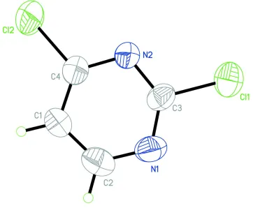
In the molecule of the title compound (Fig 1), the bond lengths (Allen et al., 1987) and angles are within normal ranges.
Ring A (N1/N2/C1-C4) is, of course, planar. Atoms Cl1 and Cl2 are 0.012 (3) and 0.013 (3) Å away from the ring plane,
respectively. So, the molecule is planar.
In the crystal structure, intermolecular C-H···N interactions (Table 1) link the molecules into chains (Fig. 2), in which
they may be effective in the stabilization of the structure.
S2. Experimental
For the preparation of the title compound, uracil (100 g, 0.82 mol) was dissolved in phosphorous oxychloride (400 ml) in
a two-necked round-bottom flask (500 ml) equipped with a condenser. The solution was refluxed with stirring for 3.5 h at
383 K. The residual phosphorous oxychloride was removed in vacuo at 323 K, and the remaining oil was poured into ice
(50 g) followed by extraction with chloroform (3 × 50 ml). The combined organic extract was washed with dilute sodium
carbonate solution and dried over anhydrous sodium sulfate. The title compound was obtained by evaporation of solvent
(Bhasin et al., 2009). Crystals suitable for X-ray analysis were obtained by slow evaporation of a methanol solution.
S3. Refinement
H atoms were positioned geometrically, with C-H = 0.93 Å for aromatic H and constrained to ride on their parent atoms,
Figure 1
The molecular structure of the title molecule, with the atom-numbering scheme. Displacement ellipsoids at the 30%
Figure 2
A partial packing diagram of the title compound. Hydrogen bonds are shown as dashed lines.
2,4-Dichloropyrimidine
Crystal data
C4H2Cl2N2
Mr = 148.98
Monoclinic, P21/c
Hall symbol: -P 2ybc a = 7.5090 (15) Å b = 10.776 (2) Å c = 7.1980 (14) Å β = 92.92 (3)° V = 581.7 (2) Å3
Z = 4
F(000) = 296 Dx = 1.701 Mg m−3
Mo Kα radiation, λ = 0.71073 Å Cell parameters from 25 reflections θ = 10–13°
µ = 0.99 mm−1
T = 294 K Block, colorless 0.30 × 0.20 × 0.20 mm
Data collection
Enraf–Nonius CAD-4 diffractometer
Radiation source: fine-focus sealed tube Graphite monochromator
ω/2θ scans
Absorption correction: ψ scan (North et al., 1968)
1139 independent reflections 733 reflections with I > 2σ(I) Rint = 0.084
θmax = 26.0°, θmin = 2.7°
Refinement on F2
Least-squares matrix: full R[F2 > 2σ(F2)] = 0.069
wR(F2) = 0.180
S = 1.01 1139 reflections 73 parameters 0 restraints
Primary atom site location: structure-invariant direct methods
Secondary atom site location: difference Fourier map
Hydrogen site location: inferred from neighbouring sites
H-atom parameters constrained w = 1/[σ2(F
o2) + (0.07P)2 + 1.45P]
where P = (Fo2 + 2Fc2)/3
(Δ/σ)max < 0.001
Δρmax = 0.39 e Å−3
Δρmin = −0.32 e Å−3
Special details
Geometry. All e.s.d.'s (except the e.s.d. in the dihedral angle between two l.s. planes) are estimated using the full covariance matrix. The cell e.s.d.'s are taken into account individually in the estimation of e.s.d.'s in distances, angles and torsion angles; correlations between e.s.d.'s in cell parameters are only used when they are defined by crystal symmetry. An approximate (isotropic) treatment of cell e.s.d.'s is used for estimating e.s.d.'s involving l.s. planes.
Refinement. Refinement of F2 against ALL reflections. The weighted R-factor wR and goodness of fit S are based on F2,
conventional R-factors R are based on F, with F set to zero for negative F2. The threshold expression of F2 > σ(F2) is used
only for calculating R-factors(gt) etc. and is not relevant to the choice of reflections for refinement. R-factors based on F2
are statistically about twice as large as those based on F, and R- factors based on ALL data will be even larger.
Fractional atomic coordinates and isotropic or equivalent isotropic displacement parameters (Å2)
x y z Uiso*/Ueq
Cl1 0.5768 (2) 0.63809 (14) 0.0975 (2) 0.0762 (6) Cl2 1.1679 (2) 0.83010 (16) 0.3510 (3) 0.0862 (7) N1 0.6273 (6) 0.8744 (4) 0.0912 (7) 0.0612 (12) N2 0.8628 (5) 0.7492 (4) 0.2211 (6) 0.0555 (11) C1 0.8975 (7) 0.9681 (5) 0.2072 (8) 0.0652 (15) H1B 0.9665 1.0384 0.2321 0.078* C2 0.7272 (8) 0.9751 (5) 0.1252 (9) 0.0689 (16) H2B 0.6808 1.0526 0.0927 0.083* C3 0.7027 (6) 0.7699 (5) 0.1403 (7) 0.0485 (12) C4 0.9577 (7) 0.8520 (5) 0.2490 (8) 0.0562 (14)
Atomic displacement parameters (Å2)
U11 U22 U33 U12 U13 U23
Geometric parameters (Å, º)
Cl1—C3 1.725 (5) N2—C4 1.327 (6) Cl2—C4 1.723 (5) C1—C2 1.383 (8) N1—C2 1.334 (7) C1—C4 1.358 (7) N1—C3 1.302 (6) C1—H1B 0.9300 N2—C3 1.327 (6) C2—H2B 0.9300
C3—N1—C2 114.9 (5) C1—C2—H2B 118.9 C4—N2—C3 113.2 (4) N1—C3—N2 129.5 (5) C4—C1—C2 115.8 (5) N1—C3—Cl1 115.9 (4) C4—C1—H1B 122.1 N2—C3—Cl1 114.6 (4) C2—C1—H1B 122.1 N2—C4—C1 124.4 (5) N1—C2—C1 122.2 (5) N2—C4—Cl2 115.0 (4) N1—C2—H2B 118.9 C1—C4—Cl2 120.5 (4)
C3—N1—C2—C1 −0.2 (9) C4—N2—C3—Cl1 178.8 (4) C4—C1—C2—N1 0.7 (10) C3—N2—C4—C1 2.5 (9) C2—N1—C3—N2 1.0 (9) C3—N2—C4—Cl2 −179.2 (4) C2—N1—C3—Cl1 −179.9 (4) C2—C1—C4—N2 −2.0 (10) C4—N2—C3—N1 −2.0 (9) C2—C1—C4—Cl2 179.8 (5)
Hydrogen-bond geometry (Å, º)
D—H···A D—H H···A D···A D—H···A
C1—H1B···N2i 0.93 2.62 3.548 (7) 174