8-Methoxy-2
H
-chromene-3-carbaldehyde
Dongsoo Koh
Department of Applied Chemistry, Dongduk Women’s University, Seoul 136-714, Republic of Korea
Correspondence e-mail: [email protected]
Received 14 November 2012; accepted 17 November 2012
Key indicators: single-crystal X-ray study;T= 200 K; mean(C–C) = 0.003 A˚;
Rfactor = 0.051;wRfactor = 0.158; data-to-parameter ratio = 17.8.
In the title molecule, C11H10O3, the fused dihydropyran ring is in a half-chair conformation with the O atom and the methylene C atom positioned 0.1318 (13) and 0.143 (2) A˚ , respectively, on either side of the mean plane formed by the other four atoms. In the crystal, weak C—H O hydrogen bonds link molecules along [001].
Related literature
For the synthesis and biological properties of chromene derivatives, see: Munet al.(2012); Kallikatet al.(2011); Zhang
et al.(2009); Gebhardtet al.(2007); Yoonet al.(2012). For the chromene group in natural products, see: Escando´n-Riveraet al. (2012); Chen et al. (2008). For related structures, see: Yusufzaiet al.(2012); Betzet al.(2011); Bardajeeet al.(2007).
Experimental
Crystal data
C11H10O3 Mr= 190.19
Orthorhombic,Pbca a= 6.8940 (6) A˚ b= 13.2079 (11) A˚ c= 20.0964 (16) A˚ V= 1829.9 (3) A˚3
Z= 8
MoKradiation
= 0.10 mm1 T= 200 K
0.230.210.19 mm
Data collection
Bruker SMART CCD diffractometer
12690 measured reflections
2276 independent reflections 1194 reflections withI> 2(I) Rint= 0.056
Refinement
R[F2> 2(F2)] = 0.051
wR(F2) = 0.158
S= 0.92 2276 reflections
128 parameters
H-atom parameters constrained
max= 0.28 e A˚3
min=0.28 e A˚
3
Table 1
Hydrogen-bond geometry (A˚ ,).
D—H A D—H H A D A D—H A
C6—H6B O1i
0.98 2.49 3.340 (3) 145
Symmetry code: (i)x;yþ1 2;zþ
1 2.
Data collection:SMART(Bruker, 2000); cell refinement:SAINT
(Bruker, 2000); data reduction:SAINT; program(s) used to solve structure:SHELXS97(Sheldrick, 2008); program(s) used to refine structure: SHELXL97 (Sheldrick, 2008); molecular graphics:
PLATON (Spek, 2009); software used to prepare material for publication:SHELXTL(Sheldrick, 2008).
Supplementary data and figures for this paper are available from the IUCr electronic archives (Reference: LH5559).
References
Bardajee, G. R., Winnik, M. A. & Lough, A. J. (2007).Acta Cryst.E63, o1269– o1270.
Betz, R., McCleland, C. & Marchand, H. (2011).Acta Cryst.E67, o1151. Bruker (2000).SMARTandSAINT. Bruker AXS Inc., Madison, Wisconsin,
USA.
Chen, J.-J., Wang, T.-Y. & Hwang, T.-L. (2008).J. Nat. Prod.71, 212–217. Escando´n-Rivera, S., Gonza´lez-Andrade, M., Bye, R., Linares, E., Navarrete,
A. & Mata, R. (2012).J. Nat. Prod.75, 968–974.
Gebhardt, P., Dornberger, K., Gollmick, F. A., Grafe, U., Hartl, A., Gorls, H., Schlegela, B. & Hertweck, C. (2007).Bioorg. Med. Chem. Lett.17, 2558– 2560.
Kallikat, A. J., Agnes, B., Nath, A. R. & Atta, R. (2011).Synlett,15, 2223–2227. Mun, J., Jabbar, A. A., Devi, N. S., Liu, Y., Meir, E. G. V. & Goodman, M. M.
(2012).Bioorg. Med. Chem. Lett.20, 4590–4597. Sheldrick, G. M. (2008).Acta Cryst.A64, 112–122. Spek, A. L. (2009).Acta Cryst.D65, 148–155.
Yoon, H., Ahn, S., Hwang, D., Jo, G., Kim, D., Kim, S. H., Koh, D. & Lim, Y. (2012).Magn. Reson. Chem.50, 759–764.
Yusufzai, S. K., Osman, H., Rahim, A. S. A., Arshad, S. & Razak, I. A. (2012). Acta Cryst.E68, o2416–o2417.
Zhang, J.-M., Lou, C.-L., Hu, Z.-P. & Yan, M. (2009).ARKIVOC,14, 362–375.
Acta Crystallographica Section E Structure Reports
Online
supporting information
Acta Cryst. (2012). E68, o3419 [doi:10.1107/S1600536812047319]
8-Methoxy-2
H
-chromene-3-carbaldehyde
Dongsoo Koh
S1. Comment
Chromenes have been important heterocyclic components in biologically active pharmaceuticals which show
anti-inflammatory (Gebhardt et al. 2007) and anticancer (Mun et al., 2012) activities. The 2H-chromene skeleton is a core
structure of oxygen heterocycles in many natural products having biological activities (Escandón-Rivera et al., 2012;
Chen et al., 2008). In a continuation of our research interest to develop novel chalcone derivatives containing
heterocycles (Yoon et al., 2012) the crystal structure of the title compound was determined.
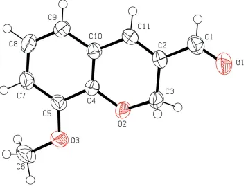
The molecular structure of the title compound is shown in Fig. 1. The fused dihydropyran ring is in a half-chair
conformation with atoms O2 and C3 positioned 0.1318 (13) and 0.143 (2)Å respectively, either side of the mean plane of
the other four atoms (C2/C4/C10/C11). In the crystal, weak C—H···O hydrogen bonds link molecules along [001] (Fig.
2). Examples of structures of chromene compounds have been published (Yusufzai et al., 2012; Betz et al., 2011;
Bardajee et al., 2007).
S2. Experimental
To a solution of 2-hydroxy-3-methoxy-benzaldehyde (1.52 g, 10 mmol) in 20 ml of 1,4-dioxane was added excess
amount of acrolein (840 mg, 15 mmol) and potassium carbonate (1.4 g, 10 mmol) at room temperature. The reaction
mixture was refluxed for 8 h and TLC showed no starting material of 2-hydroxy-3-methoxy-benzaldehyde. After cooling
to room temperature, the mixture was poured into iced water (40 ml) and extracted with diethylether (3 × 30 ml) and
combined organic layers were dried under MgSO4. Filtration, evaporation of filtrate gave residue which was purified by
flash chromatography to give the title compound (1.21 g, 82%). Recrystallization of a solution of the title compound in
ethanol gave pale yellow crystals (mp: 352-353K).
S3. Refinement
Figure 1
The molecular structure of the title compound, showing displacement ellipsoids drawn at the 50% probability level.
Figure 2
Part of the crystal structure with weak intermolecular C—H···O hydrogen bonds shown as dashed lines.
8-Methoxy-2H-chromene-3-carbaldehyde
Crystal data
C11H10O3
Mr = 190.19
Orthorhombic, Pbca
Hall symbol: -P 2ac 2ab
a = 6.8940 (6) Å
b = 13.2079 (11) Å
c = 20.0964 (16) Å
V = 1829.9 (3) Å3
Z = 8
F(000) = 800
Dx = 1.381 Mg m−3
Mo Kα radiation, λ = 0.71073 Å Cell parameters from 3190 reflections
θ = 3.1–28.2°
µ = 0.10 mm−1
T = 200 K
[image:3.610.127.482.385.525.2]Data collection
Bruker SMART CCD diffractometer
Radiation source: fine-focus sealed tube Graphite monochromator
φ and ω scans
12690 measured reflections 2276 independent reflections
1194 reflections with I > 2σ(I)
Rint = 0.056
θmax = 28.3°, θmin = 2.0°
h = −9→9
k = −17→16
l = −26→19
Refinement
Refinement on F2 Least-squares matrix: full
R[F2 > 2σ(F2)] = 0.051
wR(F2) = 0.158
S = 0.92 2276 reflections 128 parameters 0 restraints
Primary atom site location: structure-invariant direct methods
Secondary atom site location: difference Fourier map
Hydrogen site location: inferred from neighbouring sites
H-atom parameters constrained
w = 1/[σ2(F
o2) + (0.0862P)2] where P = (Fo2 + 2Fc2)/3 (Δ/σ)max = 0.001
Δρmax = 0.28 e Å−3 Δρmin = −0.28 e Å−3
Special details
Geometry. All e.s.d.'s (except the e.s.d. in the dihedral angle between two l.s. planes) are estimated using the full covariance matrix. The cell e.s.d.'s are taken into account individually in the estimation of e.s.d.'s in distances, angles and torsion angles; correlations between e.s.d.'s in cell parameters are only used when they are defined by crystal symmetry. An approximate (isotropic) treatment of cell e.s.d.'s is used for estimating e.s.d.'s involving l.s. planes.
Refinement. Refinement of F2 against ALL reflections. The weighted R-factor wR and goodness of fit S are based on F2, conventional R-factors R are based on F, with F set to zero for negative F2. The threshold expression of F2 > σ(F2) is used only for calculating R-factors(gt) etc. and is not relevant to the choice of reflections for refinement. R-factors based on F2 are statistically about twice as large as those based on F, and R- factors based on ALL data will be even larger.
Fractional atomic coordinates and isotropic or equivalent isotropic displacement parameters (Å2)
x y z Uiso*/Ueq
O1 0.1026 (2) 0.40157 (12) 0.04444 (7) 0.0583 (5) C1 0.1052 (3) 0.47227 (17) 0.08309 (9) 0.0456 (5)
H1 0.1013 0.5387 0.0651 0.055*
C2 0.1139 (2) 0.46201 (14) 0.15445 (8) 0.0351 (4) C3 0.1208 (3) 0.35793 (14) 0.18379 (8) 0.0381 (5)
H3A 0.0257 0.3148 0.1601 0.046*
H3B 0.2512 0.3291 0.1757 0.046*
O2 0.08111 (19) 0.35301 (9) 0.25332 (6) 0.0438 (4) C4 0.1089 (2) 0.43707 (13) 0.29215 (8) 0.0318 (4) C5 0.1082 (2) 0.42242 (14) 0.36068 (9) 0.0340 (4) O3 0.09121 (18) 0.32461 (10) 0.38224 (6) 0.0450 (4) C6 0.0786 (3) 0.30827 (18) 0.45209 (9) 0.0530 (6)
H6A 0.1980 0.3320 0.4735 0.080*
H6B 0.0616 0.2358 0.4609 0.080*
H6C −0.0324 0.3458 0.4700 0.080*
C7 0.1241 (2) 0.50626 (15) 0.40230 (9) 0.0394 (5)
C8 0.1403 (3) 0.60283 (15) 0.37558 (9) 0.0449 (5)
H8 0.1496 0.6597 0.4043 0.054*
C9 0.1428 (3) 0.61667 (15) 0.30801 (9) 0.0400 (5)
H9 0.1565 0.6829 0.2902 0.048*
C10 0.1255 (2) 0.53375 (13) 0.26518 (8) 0.0325 (4) C11 0.1184 (2) 0.54390 (14) 0.19384 (9) 0.0351 (4)
H11 0.1170 0.6095 0.1745 0.042*
Atomic displacement parameters (Å2)
U11 U22 U33 U12 U13 U23
O1 0.0741 (11) 0.0674 (11) 0.0334 (8) 0.0080 (8) −0.0032 (7) −0.0087 (7) C1 0.0500 (12) 0.0538 (14) 0.0330 (11) 0.0056 (9) −0.0001 (9) 0.0023 (9) C2 0.0338 (10) 0.0450 (12) 0.0266 (10) −0.0007 (8) 0.0004 (7) 0.0008 (8) C3 0.0505 (12) 0.0364 (11) 0.0274 (10) −0.0032 (8) 0.0029 (8) −0.0036 (7) O2 0.0709 (10) 0.0331 (8) 0.0275 (7) −0.0052 (6) 0.0046 (6) −0.0021 (5) C4 0.0335 (10) 0.0324 (10) 0.0294 (10) 0.0004 (7) 0.0009 (7) −0.0034 (7) C5 0.0367 (10) 0.0368 (11) 0.0285 (10) 0.0030 (8) 0.0018 (7) 0.0038 (7) O3 0.0620 (9) 0.0394 (8) 0.0335 (8) 0.0043 (6) 0.0047 (6) 0.0069 (6) C6 0.0680 (14) 0.0563 (14) 0.0347 (11) 0.0109 (11) 0.0086 (9) 0.0144 (9) C7 0.0438 (11) 0.0480 (12) 0.0264 (9) 0.0043 (9) −0.0004 (7) −0.0029 (8) C8 0.0536 (12) 0.0419 (12) 0.0392 (11) −0.0009 (9) 0.0001 (9) −0.0124 (9) C9 0.0501 (12) 0.0333 (11) 0.0365 (10) 0.0001 (8) 0.0021 (8) −0.0017 (8) C10 0.0313 (9) 0.0359 (11) 0.0304 (10) 0.0011 (7) 0.0012 (7) −0.0011 (7) C11 0.0379 (10) 0.0359 (11) 0.0315 (10) 0.0001 (8) 0.0004 (7) 0.0058 (7)
Geometric parameters (Å, º)
O1—C1 1.215 (2) O3—C6 1.423 (2)
C1—C2 1.442 (2) C6—H6A 0.9800
C1—H1 0.9500 C6—H6B 0.9800
C2—C11 1.341 (3) C6—H6C 0.9800
C2—C3 1.497 (3) C7—C8 1.388 (3)
C3—O2 1.425 (2) C7—H7 0.9500
C3—H3A 0.9900 C8—C9 1.370 (2)
C3—H3B 0.9900 C8—H8 0.9500
O2—C4 1.370 (2) C9—C10 1.398 (2)
C4—C5 1.391 (2) C9—H9 0.9500
C4—C10 1.392 (2) C10—C11 1.441 (2)
C5—O3 1.368 (2) C11—H11 0.9500
C5—C7 1.392 (3)
O1—C1—C2 124.4 (2) O3—C6—H6B 109.5
O1—C1—H1 117.8 H6A—C6—H6B 109.5
C2—C1—H1 117.8 O3—C6—H6C 109.5
C11—C2—C1 120.83 (18) H6A—C6—H6C 109.5
C11—C2—C3 120.50 (16) H6B—C6—H6C 109.5
O2—C3—C2 114.96 (14) C8—C7—H7 119.8
O2—C3—H3A 108.5 C5—C7—H7 119.8
C2—C3—H3A 108.5 C9—C8—C7 120.46 (18)
O2—C3—H3B 108.5 C9—C8—H8 119.8
C2—C3—H3B 108.5 C7—C8—H8 119.8
H3A—C3—H3B 107.5 C8—C9—C10 120.29 (18)
C4—O2—C3 119.63 (13) C8—C9—H9 119.9
O2—C4—C5 116.79 (16) C10—C9—H9 119.9
O2—C4—C10 122.21 (16) C4—C10—C9 119.08 (17)
C5—C4—C10 120.89 (16) C4—C10—C11 118.04 (16)
O3—C5—C4 116.44 (16) C9—C10—C11 122.86 (17)
O3—C5—C7 124.61 (16) C2—C11—C10 120.88 (17)
C4—C5—C7 118.95 (17) C2—C11—H11 119.6
C5—O3—C6 117.45 (15) C10—C11—H11 119.6
O3—C6—H6A 109.5
O1—C1—C2—C11 179.25 (18) C4—C5—C7—C8 0.1 (2)
O1—C1—C2—C3 0.2 (3) C5—C7—C8—C9 −0.7 (3)
C11—C2—C3—O2 15.8 (2) C7—C8—C9—C10 1.2 (3)
C1—C2—C3—O2 −165.19 (15) O2—C4—C10—C9 176.48 (15)
C2—C3—O2—C4 −23.3 (2) C5—C4—C10—C9 0.4 (2)
C3—O2—C4—C5 −166.65 (15) O2—C4—C10—C11 −1.8 (2)
C3—O2—C4—C10 17.1 (2) C5—C4—C10—C11 −177.88 (14)
O2—C4—C5—O3 3.7 (2) C8—C9—C10—C4 −1.1 (3)
C10—C4—C5—O3 179.99 (15) C8—C9—C10—C11 177.14 (16)
O2—C4—C5—C7 −176.19 (15) C1—C2—C11—C10 179.49 (15)
C10—C4—C5—C7 0.1 (2) C3—C2—C11—C10 −1.5 (2)
C4—C5—O3—C6 −176.38 (15) C4—C10—C11—C2 −6.0 (2)
C7—C5—O3—C6 3.5 (2) C9—C10—C11—C2 175.80 (17)
O3—C5—C7—C8 −179.85 (16)
Hydrogen-bond geometry (Å, º)
D—H···A D—H H···A D···A D—H···A
C6—H6B···O1i 0.98 2.49 3.340 (3) 145