research communications
Acta Cryst.(2019). E75, 423–427 https://doi.org/10.1107/S2056989019000021
423
Received 26 October 2018 Accepted 2 January 2019
Edited by A. V. Yatsenko, Moscow State University, Russia
Keywords:crystal structure; amide; X-ray diffraction; Hirshfeld surface; hydrogen bonding.
CCDC reference:1584572
Supporting information:this article has supporting information at journals.iucr.org/e
Synthesis, crystal structure, spectroscopic features
and Hirshfeld surfaces of
2-methyl-3-[(2-methyl-phenyl)carbamoyl]phenyl acetate
Mavis¸e Yaman,aS¸ukriye Cakmak,bNecmi Dege,aMustafa Odabas¸og˘lu,cVadim A. Pavlenkod* and Halil Kutuke
aOndokuz Mayıs University, Faculty of Arts and Sciences, Department of Physics, 55139, Samsun, Turkey,bVocational School of Health Services, Environmental Health Programme, Sinop, University TR-57000, Sinop, Turkey,cPamukkale University, Department of Chemistry and Chemical Processing Technologies, 20070 Kınıklı-Denizli, Turkey,dTaras Shevchenko National University of Kyiv, Department of Chemistry, 64, Vladimirska Str., Kiev 01601, Ukraine, and eOndokuz Mayıs University, Faculty of Arts and Sciences, Department of Chemistry, 55139, Samsun, Turkey. *Correspondence e-mail: [email protected]
The title compound, C17H17NO3, was synthesized, characterized by IR
spectroscopy and its crystal structure was determined from single-crystal diffraction data. The asymmetric unit contains two molecules, which adopt different conformations. In one molecule, the acetoxy and the terminal 2-methylphenyl groups are positioned on opposite sides of the plane formed by the central benzene ring, whereas in the other molecule they lie on the same side of this plane. In the crystal, the molecules are linked through strong N— H O hydrogen bonds into chains along [010]. Hirshfeld surface analysis and fingerprint plots were used to investigate the intermolecular interactions in the solid state.
1. Chemical context
Amides and their derivatives are extremely important biolo-gically active compounds. Amide groups are present in a number of natural products, polymers and pharmaceuticals (Valeur & Bradley, 2009; Xianget al., 2012). Amide derivatives have been found to exhibit biological and pharmacological activities such as antitumor, antimicrobial, antibacterial, antifungal, HSV, analgesic, inflammatory and anti-cancer (Carbonnelle et al., 2005). Moreover, amide-based compounds represent an important group of efficient chelating ligands (Strotmeyer et al., 2003; Sliva et al., 1997; Pavlishchuk et al., 2011; Gumienna-Kontecka et al., 2007). Recently, we synthesized and studied some new substituted secondary benzamide derivatives obtained as a result of the interaction of aniline-based compounds with acyl chlorides (C¸ akmak et al., 2016; Kırca et al., 2018; Demir et al., 2015; Kansız, C¸ akmak et al., 2018). Among them, 3-acetoxy-2-methyl-N-(4-methoxyphenyl) benzamide was found to exhibit good antioxidant activity (Demiret al., 2015). As a continua-tion of this work, we prepared the title compound and studied its spectroscopic and structural features.
2. Structural commentary
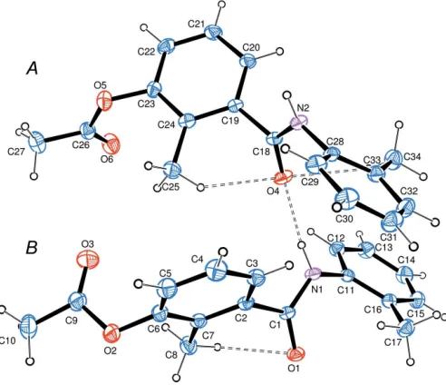
The asymmetric unit of the title compound (Fig. 1) contains two molecules,AandB, which adopt different conformations that can be characterized by the mutual arrangement of the acetoxy and terminal 2-methylphenyl groups with respect to
the plane of the central benzene ring: in moleculeAthey lie on different sides of this plane, whereas in molecule Bthey are positioned on the same side. The torsion angles characterizing the conformation details are summarized in Table 1. The dihedral angles subtended by the aromatic rings are 54.33 (12) and 66.68 (11)in moleculesAandB, respectively. The
mol-ecular conformations are stabilized by weak intramolmol-ecular C—H O contacts (Table 2). All bond lengths and angles are typical of similar compounds, bearing in mind the effect of intermolecular hydrogen bonds on the geometry of the amido groups.
3. Supramolecular features
[image:2.610.82.259.210.279.2]The packing diagram of the title compounds is presented in Fig. 2. In the crystal, the molecules are linked through strong N—H O hydrogen bonds (Table 2) into chains along [010].
They are further linked by C—H O and C—H contacts
(Table 2).
4. Database survey
A search in the Cambridge Structural Database (CSD version 5.39, update of August 2018; Groomet al., 2016) for
3-acetoxy-N-phenylbenzamide derivatives gave three hits: 3-acetoxy-2-methyl-N-(4-methylphenyl)benzamide (HEJBIK; Kırcaet al., 2018), 3-acetoxy-2-methyl-N-phenylbenzamide and
3-acetoxy-2-methyl-N-(4-methoxyphenyl)benzamide (HEJBOQ and
JUMCEB, respectively; both Demiret al., 2015). The structure of HEJBIK is especially close to that of the title compound: it also contains two molecules in an asymmetric unit and is isostructural to the title compound with the exception of one methyl group (2-Me in the title compound and 4-Me in HEJBIK). The two independent molecules in HEJBIK have different conformations in the same manner, as in the title structure. In the two structures HEJBOQ and JUMCEB, the acetoxy groups and the terminal benzene rings are positioned on opposite sides of the planes formed by the central benzene rings. In all these structures, the molecules are linked into
chains by N—H O hydrogen bonds.
424
Yamanet al. C17H17NO3 Acta Cryst.(2019). E75, 423–427
research communications
Table 1
Selected geometric parameters (A˚ ,).
O1—C1 1.222 (3) O3—C9 1.186 (4)
O4—C18 1.224 (3) O6—C26 1.188 (4)
N1—C1 1.348 (3) N2—C18 1.344 (4)
O1—C1—N1 123.5 (3) C1—N1—C11 123.4 (2)
O4—C18—N2 123.6 (3) C18—N2—C28 124.2 (2)
[image:2.610.315.566.255.341.2]C9—O2—C6—C7 100.0 (3) C26—O5—C23—C24 83.7 (3) N1—C1—C2—C7 129.1 (3) C24—C19—C18—N2 113.6 (3) C2—C1—N1—C11 172.4 (2) C28—N2—C18—C19 166.2 (2) C1—N1—C11—C16 66.4 (4) C18—N2—C28—C33 66.0 (4)
Figure 2
[image:2.610.48.294.510.722.2]Packing diagram of the title compound showing the short intermolecular contacts.Cg1 is the centroid of the C28–C33 benzene ring.
Figure 1
The asymmetric unit of the title compound, with displacement ellipsoids drawn at the 50% probability level.
Table 2
Hydrogen-bond geometry (A˚ ,).
Cg1 is the centroid of the C28–C33 ring.
D—H A D—H H A D A D—H A
C5—H5 O6i 0.93 2.49 3.402 (4) 167
N2—H2 O1ii 0.88 (3) 1.96 (3) 2.813 (3) 164 (2)
N1—H1 O4 0.91 (3) 1.91 (3) 2.804 (3) 166 (2)
C25—H25B O4 0.96 2.76 3.117 (4) 103
C34—H34A O4 0.96 2.59 3.100 (4) 114
C8—H8B O1 0.96 2.75 2.986 (4) 95
C3—H3 Cg1 0.93 2.81 3.666 (3) 153
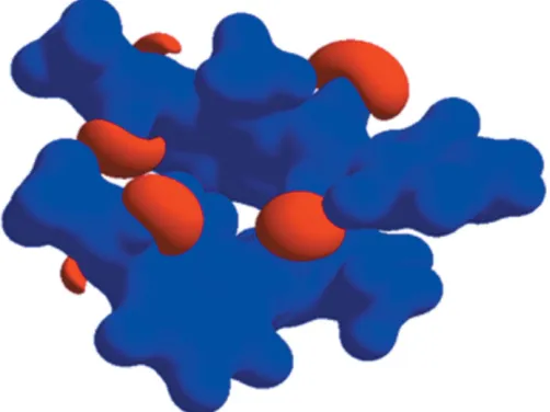
[image:2.610.316.561.539.716.2]5. Hirshfeld surface analysis
The molecular Hirshfeld surfaces (dnorm) for moleculesAand
B of the title compound generated usingCrystalExplorer3.1
(Wolffet al., 2012) and are presented in Fig. 3. Thednormvalues
are mapped on the Hirshfeld surfaces using a red–blue–white colour scheme (Spackman & Jayatilaka, 2009) as follows: the dark-red spots indicate the closest contacts related to the N—
H O hydrogen bonds, the other short intermolecular
contacts appear as light-red spots, blue regions depict positive
dnorm values, and in the white regions the lengths of the
contacts are exactly equal to the sum of van der Waals radii (dnorm = 0). Analogous dark-red spots related to the N—
H O interactions were observed on the Hirshfeld surfaces of similar molecules (S¸enet al., 2017; Gu¨mu¨s¸et al., 2018; Kansız & Dege, 2018). Figs. 4 and 5 show the two-dimensional fingerprint plots for moleculesAandB, respectively. For both molecules, the contributions from the H H/ H H contacts are the largest (55.3 and 53.9% forAand B, respectively). The
contributions of the other intermolecular contacts are as follows: C H/H C (22.5%) and O H/H O (20.7%) for
Aand C H/H C (23.8%) and O H/H O (21.7%) forB. The Hirshfeld surface mapped over the electrostatic potential
research communications
Acta Cryst.(2019). E75, 423–427 Yamanet al. C
[image:3.610.313.567.68.318.2]17H17NO3
425
Figure 5 [image:3.610.50.293.310.728.2]The fingerprint plots for moleculeB: (a) all atomsinside all atomsoutside (100%), (b) Hinside Houtside/Houtside Hinside (53.9%), (c) Cinside Houtside/Houtside Cinside (23.8%) and (d) Oinside Houtside/Houtside Oinside(21.7%).
Figure 3
Hirsfeld surfaces of 3-acetoxy-2-methyl-N-(3-methylphenyl) benzamide (three-dimensionaldnormsurface): (a) moleculeAand (b) moleculeB.
Figure 4
[image:3.610.314.566.450.695.2]n (0.25 a.u.) is shown in Fig. 6 where blue regions correspond to positive electrostatic potential and red spots related to the oxygen atoms represent the areas of negative electrostatic potential; the distribution is analogous to that in a similar compound (Yamanet al., 2018).
6. Vibrational spectrum
The IR spectrum of the title compound (KBr, cm1) shown in Fig. 7 exhibits the following characteristic bands: 3210 (N—H),
1761 (acetoxy C O), 1651 (amide C O). Because of the
interaction of the aromatic group with the acetoxy carbonyl moiety, the frequency of the acetoxy C O stretching vibra-tion is larger compared to the normal frequency of the stretching vibrations in esters (1740 cm1).
7. Synthesis and crystallization
The synthesis was performed according to the reaction scheme presented in Fig. 8 and applied earlier for the synthesis of analogous compounds (Cakmaket al., 2016; Kırcaet al., 2018, Demir et al., 2015). A solution of 3-acetoxy-2-methylbenzoyl
chloride (11 mmol) in THF (10 mL) was added dropwise to a solution of 2-methylaniline (10 mmol) and triethylamine (10 mmol) in THF (10 mL) at room temperature. After the reaction mixture had been stirred at room temperature for 15 h, the resulting white precipitate was filtered off and then 100 ml of water was added dropwise to the filtrate. The precipitate was filtered off and washed several times with water to remove the unreacted reagents and triethylamine hydrochloride. The crude product was recrystallized from acetonitrile (1.82 g, 58%; m.p. 435-438 K). Single crystals were obtained from an acetonitrile solution after incubation in the fridge for 20 days.
8. Refinement
Crystal data, data collection and structure refinement details are summarized in Table 3. The N-bound H atoms were freely refined. C-bound hydrogen atoms were positioned
geom-426
Yamanet al. C17H17NO3 Acta Cryst.(2019). E75, 423–427
research communications
Figure 7
IR spectrum of the title compound.
Figure 8 Reaction scheme. Figure 6
[image:4.610.46.297.74.262.2] [image:4.610.311.567.86.378.2]Electrostatic potential mapped on the Hirshfeld surface (0.25 a.u.).
Table 3
Experimental details.
Crystal data
Chemical formula C17H17NO3
Mr 283.31
Crystal system, space group Triclinic,P1
Temperature (K) 296
a,b,c(A˚ ) 7.7842 (5), 8.8802 (5), 22.2112 (15)
,,() 94.791 (5), 97.620 (5), 90.043 (5)
V(A˚3) 1516.37 (17)
Z 4
Radiation type MoK
(mm1) 0.09
Crystal size (mm) 0.420.370.21
Data collection
Diffractometer StoeIPDS2
Absorption correction Integration (X-RED32; Stoe & Cie, 2002)
Tmin,Tmax 0.958, 0.993
No. of measured, independent and observed [I> 2(I)] reflections
21781, 5950, 3029
Rint 0.086
(sin/ )max(A˚
1) 0.617
Refinement
R[F2> 2(F2)],wR(F2),S 0.057, 0.159, 0.90
No. of reflections 5950
No. of parameters 393
H-atom treatment H atoms treated by a mixture of independent and constrained refinement
max,min(e A˚
3) 0.17,0.14
etrically and refined as riding with C—H = 0.93 A˚ andUiso(H)
= 1.2Ueq(C) for aromatic C atoms and C—H = 0.96 A˚ and
Uiso(H) = 1.5Ueq(C) for methyl groups. Each methyl group was
allowed to rotate about its parent C—C bond.
Acknowledgements
The authors acknowledge the Faculty of Arts and Sciences, Ondokuz Mayıs University, Turkey, for the use of the Stoe IPDS 2 diffractometer (purchased under grant F.279 of the University Research Fund).
References
Cakmak, S., Kutuk, H., Odabasoglu, M., Yakan, H. & Buyukgungor, O. (2016).Lett. Org. Chem.13, 181–194.
Carbonnelle, D., Ebstein, F., Rabu, C., Petit, J. Y., Gregoire, M. & Lang, F. (2005).Eur. J. Immunol.35, 546–556.
Demir, S., Cakmak, S., Dege, N., Kutuk, H., Odabasoglu, M. & Kepekci, R. A. (2015).J. Mol. Struct.1100, 582–591.
Farrugia, L. J. (2012).J. Appl. Cryst.45, 849–854.
Groom, C. R., Bruno, I. J., Lightfoot, M. P. & Ward, S. C. (2016).Acta Cryst.B72, 171–179.
Gumienna-Kontecka, E., Golenya, I. A., Dudarenko, N. M., Dobosz, A., Haukka, M., Fritsky, I. O. & S´wia˛tek-Kozłowska, J. (2007).New J. Chem.31, 1798–1805.
Gu¨mu¨s¸, M. K., Kansız, S., Aydemir, E., Gorobets, N. Y. & Dege, N. (2018).J. Mol. Struct.1168, 280–290.
Kansız, S., C¸ akmak, S¸., Dege, N., Meral, G. & Ku¨tu¨k, H. (2018). X-Ray Struct. Anal. Online,34, 17–18.
Kansız, S. & Dege, N. (2018).J. Mol. Struct.1173, 42–51.
Kırca, B. K., C¸ akmak, S¸., Ku¨tu¨k, H., Odabas¸og˘lu, M. & Bu¨yu¨kgu¨ngo¨r, O. (2018).J. Mol. Struct.1151, 191–197.
Pavlishchuk, A. V., Kolotilov, S. V., Zeller, M., Shvets, O. V., Fritsky, I. O., Lofland, S. E., Addison, A. W. & Hunter, A. D. (2011).Eur. J. Inorg. Chem.pp. 4826–4836.
S¸en, F., Kansiz, S. & Uc¸ar, I˙. (2017).Acta Cryst.C73, 517–524. Sheldrick, G. M. (2015a).Acta Cryst.A71, 3–8.
Sheldrick, G. M. (2015b).Acta Cryst.C71, 3–8.
Sliva, T. Yu., Duda, A. M., Głowiak, T., Fritsky, I. O., Amirkhanov, V. M., Mokhir, A. A. & Kozłowski, H. (1997).J. Chem. Soc. Dalton Trans.pp. 273–276.
Spackman, M. A. & Jayatilaka, D. (2009).CrystEngComm,11, 19–32. Spek, A. L. (2009).Acta Cryst.D65, 148–155.
Stoe & Cie (2002). X-AREAand X-RED32. Stoe & Cie GmbH, Darmstadt, Germany.
Strotmeyer, K. P., Fritsky, I. O., Ott, R., Pritzkow, H. & Kra¨mer, R. (2003).Supramol. Chem.15, 529–547.
Valeur, E. & Bradley, M. (2009).Chem. Soc. Rev.38, 606–631. Westrip, S. P. (2010).J. Appl. Cryst.43, 920–925.
Wolff, S. K., Grimwood, D. J., McKinnon, J. J., Turner, M. J., Jayatilaka, D. & Spackman, M. A. (2012). CrystalExplorer3.1. University of Western Australia.
Xiang, Y.-F., Qian, C.-W., Xing, G.-W., Hao, J., Xia, M. & Wang, Y.-F. (2012).Bioorg. Med. Chem. Lett.22, 4703–4706.
Yaman, M., Almarhoon, Z. M., C¸ akmak, S¸., Ku¨tu¨k, H., Meral, G. & Dege, N. (2018).Acta Cryst.E74, 41–44.
research communications
Acta Cryst.(2019). E75, 423–427 Yamanet al. C
supporting information
sup-1 Acta Cryst. (2019). E75, 423-427
supporting information
Acta Cryst. (2019). E75, 423-427 [https://doi.org/10.1107/S2056989019000021]
Synthesis, crystal structure, spectroscopic features and Hirshfeld surfaces of
2-methyl-3-[(2-methylphenyl)carbamoyl]phenyl acetate
Mavi
ş
e Yaman,
Ş
ukriye Cakmak, Necmi Dege, Mustafa Odaba
ş
o
ğ
lu, Vadim A. Pavlenko and
Halil Kutuk
Computing details
Data collection: X-AREA (Stoe & Cie, 2002); cell refinement: X-AREA (Stoe & Cie, 2002); data reduction: X-RED32
(Stoe & Cie, 2002); program(s) used to solve structure: SHELXT (Sheldrick, 2015a); program(s) used to refine structure:
SHELXL2017 (Sheldrick, 2015b); molecular graphics: ORTEP-3 for Windows (Farrugia, 2012); software used to prepare
material for publication: WinGX (Farrugia, 2012), PLATON (Spek, 2009) and publCIF (Westrip, 2010).
2-Methyl-3-[(2-methylphenyl)carbamoyl]phenyl acetate
Crystal data
C17H17NO3
Mr = 283.31
Triclinic, P1
a = 7.7842 (5) Å
b = 8.8802 (5) Å
c = 22.2112 (15) Å
α = 94.791 (5)°
β = 97.620 (5)°
γ = 90.043 (5)°
V = 1516.37 (17) Å3
Z = 4
F(000) = 600
Dx = 1.241 Mg m−3
Mo Kα radiation, λ = 0.71073 Å
Cell parameters from 19688 reflections
θ = 1.9–27.5°
µ = 0.09 mm−1
T = 296 K
Prism, colorless 0.42 × 0.37 × 0.21 mm
Data collection
Stoe IPDS 2 diffractometer
Radiation source: sealed X-ray tube, 12 x 0.4 mm long-fine focus
Detector resolution: 6.67 pixels mm-1
rotation method scans
Absorption correction: integration (X-RED32; Stoe & Cie, 2002)
Tmin = 0.958, Tmax = 0.993
21781 measured reflections 5950 independent reflections 3029 reflections with I > 2σ(I)
Rint = 0.086
θmax = 26.0°, θmin = 1.9°
h = −9→9
k = −10→10
l = −27→27
Refinement
Refinement on F2
Least-squares matrix: full R[F2 > 2σ(F2)] = 0.057
wR(F2) = 0.159
S = 0.90
5950 reflections
393 parameters 0 restraints
Hydrogen site location: mixed
supporting information
sup-2 Acta Cryst. (2019). E75, 423-427
w = 1/[σ2(F
o2) + (0.0763P)2]
where P = (Fo2 + 2Fc2)/3
(Δ/σ)max < 0.001
Δρmax = 0.17 e Å−3
Δρmin = −0.13 e Å−3
Special details
Geometry. All esds (except the esd in the dihedral angle between two l.s. planes) are estimated using the full covariance matrix. The cell esds are taken into account individually in the estimation of esds in distances, angles and torsion angles; correlations between esds in cell parameters are only used when they are defined by crystal symmetry. An approximate (isotropic) treatment of cell esds is used for estimating esds involving l.s. planes.
Fractional atomic coordinates and isotropic or equivalent isotropic displacement parameters (Å2)
x y z Uiso*/Ueq
O1 0.5356 (3) 0.3278 (2) 0.28533 (9) 0.0605 (6)
N2 0.6781 (3) 1.0533 (3) 0.32141 (11) 0.0482 (6)
O2 0.2232 (3) 0.5083 (3) 0.07897 (10) 0.0717 (6)
C1 0.5516 (3) 0.4634 (3) 0.28130 (12) 0.0448 (7)
N1 0.6771 (3) 0.5500 (3) 0.31509 (11) 0.0486 (6)
O5 0.7722 (3) 1.0738 (3) 0.07181 (9) 0.0662 (6)
C28 0.5682 (4) 0.9934 (3) 0.36083 (12) 0.0468 (7)
O4 0.7921 (3) 0.8326 (2) 0.28774 (10) 0.0608 (6)
C19 0.8414 (3) 1.0503 (3) 0.23666 (12) 0.0438 (6)
C18 0.7690 (3) 0.9681 (3) 0.28425 (13) 0.0464 (7)
C11 0.8180 (3) 0.4903 (3) 0.35334 (12) 0.0463 (7)
C16 0.7951 (4) 0.4184 (3) 0.40468 (13) 0.0537 (7)
C2 0.4280 (3) 0.5450 (3) 0.23801 (13) 0.0452 (7)
C3 0.3430 (4) 0.6702 (3) 0.26034 (15) 0.0567 (8)
H3 0.368858 0.706558 0.301043 0.068*
C12 0.9848 (4) 0.5130 (3) 0.33845 (14) 0.0572 (8)
H12 0.999896 0.565609 0.304998 0.069*
C7 0.3944 (4) 0.4892 (3) 0.17655 (14) 0.0516 (7)
C17 0.6204 (4) 0.4057 (4) 0.42552 (15) 0.0705 (9)
H17A 0.558650 0.319989 0.403650 0.106*
H17B 0.555999 0.495828 0.417841 0.106*
H17C 0.634656 0.393102 0.468398 0.106*
C20 0.9676 (4) 1.1600 (3) 0.25396 (14) 0.0560 (8)
H20 1.006541 1.184171 0.295057 0.067*
C6 0.2705 (4) 0.5654 (4) 0.14029 (14) 0.0585 (8)
C24 0.7774 (4) 1.0137 (3) 0.17529 (14) 0.0532 (7)
C34 0.8224 (4) 0.8962 (4) 0.42816 (16) 0.0729 (9)
H34A 0.864042 0.813155 0.403828 0.109*
H34B 0.842987 0.876968 0.470381 0.109*
H34C 0.882266 0.987300 0.422249 0.109*
C23 0.8470 (4) 1.0938 (3) 0.13335 (13) 0.0545 (8)
O6 0.9365 (4) 0.8729 (3) 0.05401 (12) 0.0885 (8)
C33 0.6320 (4) 0.9139 (3) 0.40945 (14) 0.0550 (7)
C13 1.1258 (4) 0.4580 (4) 0.37307 (17) 0.0728 (10)
H13 1.236428 0.472783 0.362982 0.087*
supporting information
sup-3 Acta Cryst. (2019). E75, 423-427
H22 1.020909 1.250648 0.119977 0.077*
C26 0.8233 (5) 0.9545 (4) 0.03635 (15) 0.0646 (9)
C4 0.2194 (4) 0.7415 (4) 0.22218 (18) 0.0727 (10)
H4 0.161175 0.824827 0.237254 0.087*
C32 0.5113 (5) 0.8545 (4) 0.44225 (15) 0.0713 (9)
H32 0.550319 0.797517 0.474527 0.086*
C29 0.3907 (4) 1.0207 (3) 0.34776 (15) 0.0608 (8)
H29 0.350054 1.078584 0.315939 0.073*
C5 0.1834 (4) 0.6884 (4) 0.16207 (18) 0.0736 (10)
H5 0.100416 0.735476 0.136156 0.088*
C15 0.9407 (4) 0.3628 (4) 0.43830 (15) 0.0678 (9)
H15 0.928027 0.311797 0.472381 0.081*
C21 1.0363 (4) 1.2343 (4) 0.21012 (16) 0.0680 (9)
H21 1.123516 1.306624 0.221625 0.082*
C9 0.2956 (5) 0.5755 (5) 0.03514 (16) 0.0724 (10)
C31 0.3375 (5) 0.8771 (4) 0.42858 (17) 0.0758 (10)
H31 0.260317 0.834551 0.451079 0.091*
C14 1.1044 (5) 0.3818 (4) 0.42210 (17) 0.0739 (10)
H14 1.200093 0.342190 0.444874 0.089*
C25 0.6354 (5) 0.8978 (4) 0.15608 (16) 0.0806 (11)
H25A 0.568073 0.923610 0.119042 0.121*
H25B 0.562053 0.895667 0.187499 0.121*
H25C 0.685593 0.800058 0.149429 0.121*
O3 0.4029 (4) 0.6720 (3) 0.04766 (13) 0.1018 (9)
C30 0.2768 (4) 0.9620 (4) 0.38199 (17) 0.0752 (10)
H30 0.158693 0.979873 0.373513 0.090*
C8 0.4864 (5) 0.3554 (4) 0.15108 (15) 0.0709 (9)
H8A 0.432986 0.264201 0.160420 0.106*
H8B 0.605821 0.358900 0.168815 0.106*
H8C 0.479238 0.357183 0.107663 0.106*
C10 0.2217 (5) 0.5106 (5) −0.02652 (16) 0.0945 (13)
H10A 0.118483 0.563994 −0.040154 0.142*
H10B 0.193802 0.405789 −0.025063 0.142*
H10C 0.304914 0.520111 −0.054284 0.142*
C27 0.7180 (5) 0.9428 (5) −0.02461 (16) 0.0866 (11)
H27A 0.778041 0.882794 −0.052970 0.130*
H27B 0.699550 1.042040 −0.038207 0.130*
H27C 0.608231 0.896027 −0.022119 0.130*
H2 0.653 (3) 1.146 (3) 0.3128 (11) 0.043 (7)*
H1 0.696 (3) 0.644 (3) 0.3039 (12) 0.050 (8)*
Atomic displacement parameters (Å2)
U11 U22 U33 U12 U13 U23
O1 0.0662 (13) 0.0394 (12) 0.0731 (14) −0.0031 (10) −0.0063 (10) 0.0143 (10)
N2 0.0520 (14) 0.0373 (13) 0.0590 (15) 0.0039 (11) 0.0162 (12) 0.0120 (12)
O2 0.0670 (14) 0.0830 (16) 0.0625 (15) −0.0198 (12) −0.0088 (11) 0.0188 (12)
supporting information
sup-4 Acta Cryst. (2019). E75, 423-427
N1 0.0473 (14) 0.0361 (13) 0.0616 (15) −0.0058 (11) −0.0006 (11) 0.0121 (12)
O5 0.0718 (14) 0.0768 (15) 0.0500 (13) 0.0150 (12) 0.0071 (11) 0.0076 (11)
C28 0.0541 (17) 0.0373 (15) 0.0503 (17) −0.0026 (13) 0.0124 (13) 0.0025 (13)
O4 0.0714 (14) 0.0373 (11) 0.0799 (15) 0.0040 (10) 0.0271 (11) 0.0141 (10)
C19 0.0424 (15) 0.0389 (15) 0.0523 (18) 0.0006 (12) 0.0119 (13) 0.0075 (13)
C18 0.0422 (15) 0.0405 (17) 0.0565 (18) −0.0036 (13) 0.0057 (13) 0.0064 (14)
C11 0.0478 (16) 0.0380 (15) 0.0510 (17) −0.0028 (13) 0.0001 (13) 0.0021 (13)
C16 0.0569 (18) 0.0495 (17) 0.0538 (18) −0.0046 (14) 0.0028 (14) 0.0064 (14)
C2 0.0387 (15) 0.0391 (15) 0.0582 (19) −0.0023 (12) 0.0039 (13) 0.0111 (14)
C3 0.0539 (18) 0.0506 (18) 0.065 (2) 0.0044 (15) 0.0038 (15) 0.0091 (15)
C12 0.0500 (18) 0.0579 (19) 0.0633 (19) −0.0053 (15) 0.0046 (15) 0.0080 (15)
C7 0.0455 (16) 0.0484 (17) 0.0611 (19) −0.0051 (13) 0.0033 (14) 0.0117 (15)
C17 0.068 (2) 0.080 (2) 0.067 (2) −0.0040 (18) 0.0158 (17) 0.0151 (18)
C20 0.0559 (18) 0.0562 (18) 0.0562 (18) −0.0131 (15) 0.0101 (14) 0.0026 (15)
C6 0.0532 (18) 0.062 (2) 0.058 (2) −0.0064 (16) −0.0075 (15) 0.0158 (16)
C24 0.0503 (17) 0.0479 (17) 0.062 (2) 0.0002 (14) 0.0069 (15) 0.0078 (15)
C34 0.065 (2) 0.085 (2) 0.068 (2) 0.0039 (19) 0.0013 (17) 0.0160 (19)
C23 0.0582 (18) 0.0571 (18) 0.0504 (18) 0.0051 (15) 0.0124 (15) 0.0093 (15)
O6 0.0905 (18) 0.0892 (18) 0.0804 (17) 0.0270 (16) −0.0028 (14) −0.0017 (14)
C33 0.0586 (18) 0.0497 (17) 0.0571 (19) −0.0006 (14) 0.0090 (15) 0.0047 (15)
C13 0.0478 (19) 0.084 (2) 0.085 (3) −0.0015 (17) 0.0010 (17) 0.009 (2)
C22 0.069 (2) 0.061 (2) 0.068 (2) −0.0093 (17) 0.0290 (17) 0.0134 (17)
C26 0.060 (2) 0.075 (2) 0.059 (2) −0.0054 (18) 0.0066 (17) 0.0032 (19)
C4 0.060 (2) 0.067 (2) 0.090 (3) 0.0220 (17) 0.0032 (19) 0.009 (2)
C32 0.085 (3) 0.071 (2) 0.063 (2) −0.0019 (19) 0.0214 (18) 0.0162 (18)
C29 0.0547 (19) 0.0576 (19) 0.073 (2) 0.0033 (15) 0.0166 (16) 0.0071 (16)
C5 0.056 (2) 0.071 (2) 0.091 (3) 0.0117 (18) −0.0102 (18) 0.024 (2)
C15 0.069 (2) 0.071 (2) 0.062 (2) 0.0009 (18) −0.0044 (17) 0.0197 (17)
C21 0.070 (2) 0.066 (2) 0.070 (2) −0.0283 (17) 0.0181 (17) 0.0029 (18)
C9 0.059 (2) 0.084 (3) 0.074 (2) −0.0010 (19) −0.0024 (18) 0.022 (2)
C31 0.071 (2) 0.085 (3) 0.079 (2) −0.004 (2) 0.034 (2) 0.014 (2)
C14 0.061 (2) 0.079 (2) 0.079 (2) 0.0102 (18) −0.0053 (18) 0.013 (2)
C25 0.074 (2) 0.091 (3) 0.073 (2) −0.036 (2) −0.0032 (18) 0.013 (2)
O3 0.097 (2) 0.108 (2) 0.101 (2) −0.0409 (18) 0.0092 (16) 0.0158 (17)
C30 0.054 (2) 0.083 (2) 0.093 (3) 0.0012 (18) 0.0241 (19) 0.009 (2)
C8 0.080 (2) 0.068 (2) 0.063 (2) 0.0065 (18) 0.0068 (17) 0.0001 (17)
C10 0.082 (3) 0.135 (4) 0.064 (2) −0.010 (3) −0.0021 (19) 0.020 (2)
C27 0.075 (2) 0.119 (3) 0.063 (2) −0.005 (2) 0.0047 (18) −0.008 (2)
Geometric parameters (Å, º)
O1—C1 1.222 (3) C24—C25 1.504 (4)
O4—C18 1.224 (3) C34—C33 1.497 (4)
N1—C1 1.348 (3) C34—H34A 0.9600
O3—C9 1.186 (4) C34—H34B 0.9600
O6—C26 1.188 (4) C34—H34C 0.9600
N2—C18 1.344 (4) C23—C22 1.372 (4)
supporting information
sup-5 Acta Cryst. (2019). E75, 423-427
N2—H2 0.88 (3) C13—C14 1.357 (5)
O2—C9 1.365 (4) C13—H13 0.9300
O2—C6 1.414 (4) C22—C21 1.371 (4)
C1—C2 1.499 (4) C22—H22 0.9300
N1—C11 1.427 (3) C26—C27 1.483 (5)
N1—H1 0.91 (3) C4—C5 1.371 (5)
O5—C26 1.359 (4) C4—H4 0.9300
O5—C23 1.409 (4) C32—C31 1.365 (5)
C28—C33 1.378 (4) C32—H32 0.9300
C28—C29 1.400 (4) C29—C30 1.372 (4)
C19—C20 1.378 (4) C29—H29 0.9300
C19—C24 1.399 (4) C5—H5 0.9300
C19—C18 1.502 (4) C15—C14 1.383 (5)
C11—C16 1.384 (4) C15—H15 0.9300
C11—C12 1.400 (4) C21—H21 0.9300
C16—C15 1.387 (4) C9—C10 1.482 (5)
C16—C17 1.501 (4) C31—C30 1.365 (5)
C2—C3 1.384 (4) C31—H31 0.9300
C2—C7 1.404 (4) C14—H14 0.9300
C3—C4 1.385 (4) C25—H25A 0.9600
C3—H3 0.9300 C25—H25B 0.9600
C12—C13 1.369 (4) C25—H25C 0.9600
C12—H12 0.9300 C30—H30 0.9300
C7—C6 1.387 (4) C8—H8A 0.9600
C7—C8 1.497 (4) C8—H8B 0.9600
C17—H17A 0.9600 C8—H8C 0.9600
C17—H17B 0.9600 C10—H10A 0.9600
C17—H17C 0.9600 C10—H10B 0.9600
C20—C21 1.383 (4) C10—H10C 0.9600
C20—H20 0.9300 C27—H27A 0.9600
C6—C5 1.374 (5) C27—H27B 0.9600
C24—C23 1.382 (4) C27—H27C 0.9600
O1—C1—N1 123.5 (3) C32—C33—C34 120.9 (3)
O4—C18—N2 123.6 (3) C14—C13—C12 120.1 (3)
C1—N1—C11 123.4 (2) C14—C13—H13 120.0
C18—N2—C28 124.2 (2) C12—C13—H13 120.0
C18—N2—H2 118.5 (17) C21—C22—C23 119.4 (3)
C28—N2—H2 113.7 (17) C21—C22—H22 120.3
C9—O2—C6 117.7 (2) C23—C22—H22 120.3
O1—C1—C2 121.1 (2) O6—C26—O5 122.6 (3)
N1—C1—C2 115.4 (2) O6—C26—C27 126.8 (4)
C1—N1—H1 118.2 (17) O5—C26—C27 110.6 (3)
C11—N1—H1 114.9 (17) C5—C4—C3 119.6 (3)
C26—O5—C23 118.4 (2) C5—C4—H4 120.2
C33—C28—C29 121.0 (3) C3—C4—H4 120.2
C33—C28—N2 122.5 (3) C31—C32—C33 122.1 (3)
supporting information
sup-6 Acta Cryst. (2019). E75, 423-427
C20—C19—C24 121.4 (2) C33—C32—H32 118.9
C20—C19—C18 119.9 (3) C30—C29—C28 119.8 (3)
C24—C19—C18 118.7 (2) C30—C29—H29 120.1
O4—C18—C19 121.1 (3) C28—C29—H29 120.1
N2—C18—C19 115.3 (2) C4—C5—C6 119.6 (3)
C16—C11—C12 120.3 (3) C4—C5—H5 120.2
C16—C11—N1 122.5 (2) C6—C5—H5 120.2
C12—C11—N1 117.1 (2) C14—C15—C16 121.3 (3)
C11—C16—C15 117.9 (3) C14—C15—H15 119.4
C11—C16—C17 121.8 (3) C16—C15—H15 119.4
C15—C16—C17 120.3 (3) C22—C21—C20 119.8 (3)
C3—C2—C7 121.3 (3) C22—C21—H21 120.1
C3—C2—C1 119.0 (3) C20—C21—H21 120.1
C7—C2—C1 119.6 (2) O3—C9—O2 121.8 (3)
C4—C3—C2 120.1 (3) O3—C9—C10 127.5 (4)
C4—C3—H3 119.9 O2—C9—C10 110.7 (3)
C2—C3—H3 119.9 C30—C31—C32 120.2 (3)
C13—C12—C11 120.1 (3) C30—C31—H31 119.9
C13—C12—H12 119.9 C32—C31—H31 119.9
C11—C12—H12 119.9 C13—C14—C15 120.2 (3)
C6—C7—C2 116.1 (3) C13—C14—H14 119.9
C6—C7—C8 121.5 (3) C15—C14—H14 119.9
C2—C7—C8 122.4 (3) C24—C25—H25A 109.5
C16—C17—H17A 109.5 C24—C25—H25B 109.5
C16—C17—H17B 109.5 H25A—C25—H25B 109.5
H17A—C17—H17B 109.5 C24—C25—H25C 109.5
C16—C17—H17C 109.5 H25A—C25—H25C 109.5
H17A—C17—H17C 109.5 H25B—C25—H25C 109.5
H17B—C17—H17C 109.5 C31—C30—C29 119.7 (3)
C19—C20—C21 119.9 (3) C31—C30—H30 120.2
C19—C20—H20 120.0 C29—C30—H30 120.2
C21—C20—H20 120.0 C7—C8—H8A 109.5
C5—C6—C7 123.1 (3) C7—C8—H8B 109.5
C5—C6—O2 118.2 (3) H8A—C8—H8B 109.5
C7—C6—O2 118.6 (3) C7—C8—H8C 109.5
C23—C24—C19 116.4 (3) H8A—C8—H8C 109.5
C23—C24—C25 121.7 (3) H8B—C8—H8C 109.5
C19—C24—C25 121.8 (3) C9—C10—H10A 109.5
C33—C34—H34A 109.5 C9—C10—H10B 109.5
C33—C34—H34B 109.5 H10A—C10—H10B 109.5
H34A—C34—H34B 109.5 C9—C10—H10C 109.5
C33—C34—H34C 109.5 H10A—C10—H10C 109.5
H34A—C34—H34C 109.5 H10B—C10—H10C 109.5
H34B—C34—H34C 109.5 C26—C27—H27A 109.5
C22—C23—C24 122.9 (3) C26—C27—H27B 109.5
C22—C23—O5 118.5 (3) H27A—C27—H27B 109.5
C24—C23—O5 118.4 (3) C26—C27—H27C 109.5
supporting information
sup-7 Acta Cryst. (2019). E75, 423-427
C28—C33—C34 122.1 (3) H27B—C27—H27C 109.5
C9—O2—C6—C7 −100.0 (3) C20—C19—C24—C23 −0.3 (4)
N1—C1—C2—C7 129.1 (3) C18—C19—C24—C23 179.3 (3)
C2—C1—N1—C11 −172.4 (2) C20—C19—C24—C25 −177.9 (3)
C1—N1—C11—C16 −66.4 (4) C18—C19—C24—C25 1.7 (4)
C26—O5—C23—C24 −83.7 (3) C19—C24—C23—C22 1.8 (4)
C24—C19—C18—N2 −113.6 (3) C25—C24—C23—C22 179.4 (3)
C28—N2—C18—C19 166.2 (2) C19—C24—C23—O5 −172.5 (2)
C18—N2—C28—C33 66.0 (4) C25—C24—C23—O5 5.1 (4)
N1—C1—C2—C3 −53.9 (3) C26—O5—C23—C22 101.7 (3)
O1—C1—N1—C11 8.5 (4) C29—C28—C33—C32 3.9 (4)
C18—N2—C28—C29 −114.4 (3) N2—C28—C33—C32 −176.5 (3)
C28—N2—C18—O4 −13.4 (4) C29—C28—C33—C34 −173.8 (3)
C20—C19—C18—O4 −114.4 (3) N2—C28—C33—C34 5.8 (4)
C24—C19—C18—O4 66.0 (4) C11—C12—C13—C14 −0.3 (5)
C20—C19—C18—N2 66.0 (3) C24—C23—C22—C21 −1.6 (5)
C1—N1—C11—C12 116.4 (3) O5—C23—C22—C21 172.7 (3)
C12—C11—C16—C15 −3.6 (4) C23—O5—C26—O6 −5.3 (5)
N1—C11—C16—C15 179.2 (3) C23—O5—C26—C27 174.0 (3)
C12—C11—C16—C17 173.8 (3) C2—C3—C4—C5 −0.8 (5)
N1—C11—C16—C17 −3.3 (4) C28—C33—C32—C31 −2.0 (5)
O1—C1—C2—C3 125.3 (3) C34—C33—C32—C31 175.7 (3)
O1—C1—C2—C7 −51.7 (4) C33—C28—C29—C30 −3.0 (5)
C7—C2—C3—C4 1.3 (4) N2—C28—C29—C30 177.4 (3)
C1—C2—C3—C4 −175.6 (3) C3—C4—C5—C6 −0.1 (5)
C16—C11—C12—C13 3.0 (4) C7—C6—C5—C4 0.6 (5)
N1—C11—C12—C13 −179.7 (3) O2—C6—C5—C4 176.4 (3)
C3—C2—C7—C6 −0.8 (4) C11—C16—C15—C14 1.6 (5)
C1—C2—C7—C6 176.1 (2) C17—C16—C15—C14 −175.9 (3)
C3—C2—C7—C8 179.1 (3) C23—C22—C21—C20 −0.1 (5)
C1—C2—C7—C8 −4.0 (4) C19—C20—C21—C22 1.6 (5)
C24—C19—C20—C21 −1.4 (4) C6—O2—C9—O3 4.8 (5)
C18—C19—C20—C21 179.0 (3) C6—O2—C9—C10 −175.4 (3)
C2—C7—C6—C5 −0.1 (4) C33—C32—C31—C30 −0.9 (6)
C8—C7—C6—C5 179.9 (3) C12—C13—C14—C15 −1.7 (5)
C2—C7—C6—O2 −175.9 (2) C16—C15—C14—C13 1.0 (5)
C8—C7—C6—O2 4.1 (4) C32—C31—C30—C29 2.0 (6)
C9—O2—C6—C5 84.0 (4) C28—C29—C30—C31 −0.1 (5)
Hydrogen-bond geometry (Å, º)
Cg1 is the centroid of the C28–C33 ring.
D—H···A D—H H···A D···A D—H···A
C5—H5···O6i 0.93 2.49 3.402 (4) 167
N2—H2···O1ii 0.88 (3) 1.96 (3) 2.813 (3) 164 (2)
N1—H1···O4 0.91 (3) 1.91 (3) 2.804 (3) 166 (2)
supporting information
sup-8 Acta Cryst. (2019). E75, 423-427
C34—H34A···O4 0.96 2.59 3.100 (4) 114
C8—H8B···O1 0.96 2.75 2.986 (4) 95
C3—H3···Cg1 0.93 2.81 3.666 (3) 153