ORIGINAL RESEARCH
Quantification of Diffusivities of the Human
Cervical Spinal Cord Using a 2D Single-Shot
Interleaved Multisection Inner Volume
Diffusion-Weighted Echo-Planar Imaging Technique
T.H. Kim L. Zollinger X.F. Shi S.E. Kim J. Rose A.A. Patel E.K. Jeong
BACKGROUND AND PURPOSE: DTI is a highly sensitive technique, which can detect pathology not otherwise noted with conventional imaging methods. This paper provides the atlas of reliable norma-tive in vivo DTI parameters in the cervical spinal cord and its potential applications toward quantifying pathology.
MATERIALS AND METHODS: In our study, we created a reference of normal diffusivities of the cervical spinal cord by using a 2D ss-IMIV-DWEPI technique from 14 healthy volunteers and compared parameters with those in 8 patients with CSM. The 2D ss-IMIV-DWEPI technique was applied in each subject to acquire diffusion-weighted images. FA,储, and⬜were calculated. A reference of normal DTI indices from 12 regions of interest was created and compared with DTI indices of 8 patients.
RESULTS: A map of reference diffusivity values was obtained from healthy controls. We found statistically significant differences in diffusivities between healthy volunteers and patients with CSM with different severities of disease, by using FA,储, and⬜values.
CONCLUSIONS:DTI using 2D ss-IMIV-DWPEI is sensitive to spinal cord pathology. This technique can be used to detect and quantify the degree of pathology within the cervical spinal cord from multiple disease states.
ABBREVIATIONS:CSM⫽cervical spondylotic myelopathy; 2D ss-IMIV-DWEPI⫽2D single-shot interleaved multisection inner volume diffusion-weighted echo-planar imaging; DTI⫽ diffusion tensor imaging; DW⫽diffusion-weighted; EPI⫽echo-planar imaging; ETL⫽echo-train length; FA⫽fractional anisotropy;储⫽longitudinal diffusivity;⬜⫽radial diffusivity; IMIV⫽interleaved multisection inner volume; NMG⫽ Nurick myelopathy grading; RGB⫽ red-green-blue; SNR⫽ signal intensity–to-noise ratio; T2WI⫽T2-weighted imaging
D
TI is becoming a valuable surrogate measurement of the microscopic anatomy of the central nervous sys-tem. It measures both the magnitude and direction of the diffusion of water molecules by applying gradients in mul-tiple planes. DTI can characterize white matter tracts with-in the brawith-in and spwith-inal cord by measurwith-ing alterations to the anisotropy of tissue. The measured储 is thought to be amarker for axonal disease, while the diffusion perpendicu-lar to axonal tracts can provide information on the integrity of myelin sheaths, though direct histopathologic compari-son has yet to be established.1
Many in vivo2-4 DTI measurements have been
con-ducted with the hope that they would lead to improved understanding of the pathology and ultimately improved in vivo techniques for evaluating tissue microstructure. How-ever, as of now there is no consensus as to the in vivo tech-nique that will provide robust, accurate, and reliable mea-surements that can establish a reference of normal DTI indices for the spinal cord. In addition, the clinical appli-cation of DTI in spinal cord imaging has proved difficult for the following reasons: 1) The commonly used multishot acquisition technique is highly sensitive to the physiologic motion in vivo, limiting its diagnostic accuracy, and 2) the locally varying nonlinear gradient, which is caused by the magnetic susceptibility difference at/near the vertebral bones, induces substantial geometric distortion on 2D ss-DWEPI, which is most widely used in brain DTI.
Recently, Jeong et al5,6reported an IMIV imaging tech-nique that uses double inversion/refocusing radio-frequency pulses with 2D ss-DWEPI. Because this technique allows effi-cient DTI measurements of anatomic regions where conven-tional 2D ss-DWEPI has severe magnetic susceptibility distor-tion, the entire cervical spinal cord can be obtained in the sagittal plane within 1 interleaved imaging. The geometric dis-tortion in 2D ss-DWEPI is proportional to the FOV in the phase-encoding direction; therefore, 2D ss-IMIV-DWEPI re-duces the susceptibility distortion by reducing the spatial cov-erage in the phase-encoding direction and allows DTI mea-surements from nearly any localized region of the body, many
Received June 9, 2009; accepted after revision August 17.
From the Department of Radiology (T.H.K., L.Z., S.E.K., E.K.J.), Utah Center for Advanced Imaging Research (T.H.K., X.F.S., S.E.K., E.K.J.), and Departments of Physics (X.F.S.), Neurology (J.R.), and Orthopedic Surgery (A.A.P.), University of Utah, Salt Lake City, Utah.
This work was supported by the Cervical Spine Research Society Starter Grant 2008, the National Multiple Sclerosis Society, the Cumming Foundation, the Benning Foundation, the Margolis Foundation, and National Institutes of Health grants R21NS052424 and R21EB005705.
Please address correspondence to Eun-Kee Jeong, PhD, Utah Center for Advanced Imaging Research, Radiology, University of Utah, 729 Arapeen Dr, Salt Lake City, UT 84108-1218; e-mail: [email protected]
Indicates open access to non-subscribers at www.ajnr.org
of which cannot be currently studied with other available DTI techniques.
In this article, we will present our efforts to improve the clinical utility of DTI in the cervical spinal cord by quantifying diffusivities of healthy volunteers and comparing the values with those of patients with CSM. For this study, we used 2D ss-DWEPI with the IMIV imaging technique and a custom-made cervical spinal cord coil.
Materials and Methods
Subjects and DTI
Control subjects were recruited on a volunteer basis. Each of the con-trol subjects was interviewed to exclude any history of cervical spon-dylosis, myelopathy, previous spine surgery, spinal cord trauma, or other neurodegenerative diseases. A total of 14 healthy control sub-jects were included.
Patients with CSM were also recruited, on a volunteer basis, from the practice of 1 author (A.A.P.). All patients were diagnosed with CSM by clinical examination and conventional MR imaging. The degree of clinical information available for the patients was signifi-cantly limited by the boundaries of our research-approval status. Ex-clusion criteria were the following: noncervical sources of myelopa-thy, infection, tumor, history of central neurodegenerative diseases (eg, multiple sclerosis), and a history of cranial white matter diseases. The severity of myelopathy was assessed and scored by the NMG scale: 0, normal function; 1, no walking impairment; 2, mild walking im-pairment, able to continue working; 3, walking imim-pairment, unable to work; 4, immobilized in a wheelchair or bed. A total of 8 patients were
included, each with a history of chronic myelopathy (⬎1-year
dura-tion of symptoms) related to prior trauma or progressive spondylolysis.
The MR imaging experiments were performed on a 3T whole-body MR imaging system (Trio; Siemens Medical Solutions, Erlan-gen, Germany) with Avanto gradients (45 mT/m strength and 150 T/m/s slew rate). Characteristics of controls (average age, 34 years) and patients with CSM (average age, 59.5 years) are listed in the Table. Our imaging protocol was approved by the institutional review board, and informed consent was obtained from the participants. T2WI by using 2D turbo spin-echo was performed to verify the loca-tion of the cervical spinal cord. The imaging parameters were the
following: TR⫽4000 ms, TE⫽114 ms, in-plane resolution⫽0.75⫻
0.75 mm2, section thickness⫽1.5 mm, ETL⫽19, and receiver
band-width⫽248 Hz/pixel.
Each of the control subjects was imaged by using a custom-made cervical spinal cord coil. It consisted of 8 rectangular coils and covered the posterior neck from the occiput to the T2 vertebra. Among 14
healthy volunteers, 6 were rescanned with a clinical coil routinely used at our institution. It consisted of a 12-channel receive-only head coil (Siemens Medical Solutions) and a spine coil with an anterior neck coil assembly (USA Instruments, Aurora, Ohio). All of the patients with CSM were imaged once by using only the clinical coil.
The imaging parameters of 2D ss-IMIV-DWEPI were the
follow-ing: TR⫽4000 ms, TE⫽65 ms, imaging matrix⫽160⫻40, imaging
resolution⫽1.5⫻1.5⫻2.0 mm3,b⫽500 s/mm2, ETL⫽41, receiver
bandwidth⫽1078 Hz/pixel, and 10 interleaved sections.
Diffusion-weighted gradients were applied in 12 noncollinear directions. Total scanning time for the DTI acquisition was 7 minutes.
Data Analysis and Physical Parameters
The acquired DTI dataset was processed pixel-by-pixel by using home-made DTI processing software written in Interactive Data Lan-guage (ITT Visual Information Solutions, Boulder, Colorado). First, 3 eigenvalues (1,2,3) and eigenvectors were calculated.储and⬜ were obtained from 3 eigenvalues by using
1) 储⫽1
⬜⫽共2⫹3兲/ 2.
Here,1is the principal diffusivity with the largest value in each image
pixel. The degree of anisotropy was presented by using the anisotropy
index FA,7which is defined as
2) FA⫽
冑
3关共1⫺mean兲 2⫹共2⫺mean兲 2⫹共
3⫺mean 2兲兴
/
冑
2共21⫹
2
2⫹
2 3兲,
wheremeanindicates the mean diffusivity. FA values from 0
(com-pletely isotropic) to 1 (com(com-pletely anisotropic) indicate the degree of structural anisotropy. The data were processed to extract the RGB fiber map. The RGB map indicates the direction of the principal eig-envector: blue represents the superior-to-inferior axis; green, left-to-right; and red, anterior-to-posterior.
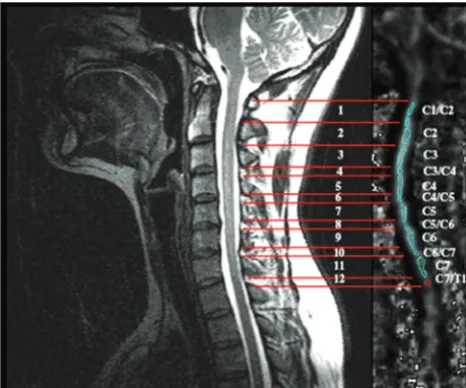
Regions of interest were selected from the FA map by using the anatomy and cord morphology from the T2WI as a reference. Twelve regions of interest were selected from a sagittal section through the midline of the cord to maximize the number of pixels analyzed within the dorsal columns on each participant, spanning C1–C7. Region-of-interest selection is demonstrated in Fig 1. We applied the whole-cord
analysis,2including both the white and gray matter as well as regions
of cord corresponding to disk levels. The separation of the gray and white matter was not attempted due to challenges of image resolution. The regions of interest were delineated to exclude approximately 2 voxels from the edge of the cord along the anterior and posterior margins. Care was taken to account for variant morphology of the cervical cord among subjects to ensure accuracy and reliability. No standardized techniques were used to normalize the cervical cords among subjects. Regions of interest were, therefore, manually delin-eated within the dorsal white matter columns to maximize the size of
evaluated cord. Average储,⬜, and FA were calculated from the
mul-tiple regions of interest along the entire cervical spinal cord. Group statistics were calculated for each of the DTI parameters and SNR. The group means and SDs were obtained by using the subject means. Quantitative statistical comparison of anisotropic parameters from the controls and patients was conducted by using normal
distribu-tions and aPvalue of .05.
Limited demographics from 8 patients with a history of CSM and SNR of T2WI
Patients Age (yr) Sex
Conventional MR Imaging:
Levels of Stenosis SNR NMG P1 61 M C4/C5, C5/C6, C6/C7 16.29 3 P2 78 F C3/C4, C4/C5, C5/C6 19.52 4 P3 43 M C5/C6, C6/C7 13.24 4 P4 64 M C3/C4, C4/C5, C5/C6, C6/C7 26.01 2 P5 48 M C4/C5, C5/C6 13.90 3 P6 64 M C4/C5, C5/C6 11.27 2 P7 48 M C3/C4, C4/C5, C5/C6, C7/T1 26.27 2 P8 70 F C4/C5, C5/C6, C6/C7 21.74 3
SPINE
ORIGINAL
Results
Figure 1 displays a sagittal T2WI of the normal cervical spinal cord demonstrating the region-of-interest selection and rela-tive spinal-level selection from the occiput through C7. The SNR in the T2WI of the 14 healthy controls with the specially constructed coil was measured as 38⫾11.9. The same value for the clinical coil from the 6 healthy controls was 20⫾8.4.
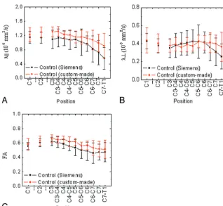
Figure 2 includes the diffusion properties of healthy con-trols obtained by using 2 different radio-frequency coils. The red line represents the results from the custom-made coil from the 14 volunteers, and the black line is the data from the 6 volunteers who were scanned a second time with the clinical coil. There are no significant differences in the obtained values between the 2 coils by using a paired-samplesttest. TheP values for each of the indices were as follows:P⫽.095 (Fig 2A), .295 (Fig 2B), and .812 (Fig 2C). This figure shows that the custom-made coil provided less variation and higher values in 储and FA compared with the clinical coil. Nearly equivalent储
and⬜values were obtained across the healthy volunteers for each cervical spinal level.
Figure 3 shows the colored scaled FA maps and RGB fiber map of 3 contiguous sections from a healthy control and a patient with CSM. FA maps from the healthy subjects gener-ally show uniform values across the entire cervical spine. In contrast, FA maps of the patients with CSM demonstrate abrupt changes in FA values at sites of pathology. The RGB map from a healthy subject in Fig 3Ashows uniform blue filling on the cord, which indicates that the direction of fiber is the same as the direction of the principal water diffusion (head-foot). We observed color degradation and decreased FA values in the patients. These results were also in concert with findings from conventional T2WIs as shown in Fig 3.
The quantitative statistical comparison of diffusivity prop-erties from the patients and controls is reported in On-line Tables 1–3. Using the mean value from each region of interest based on the clinical coil, we assumed a normal distribution. With aP⬍.05, any measured values from the patients with CSM that were outside the 95% confidence interval were deemed significantly different from those of the healthy
pa-tients on a level-by-level basis. The regions of interest from the occiput through C3 were obtained, though not included in the level-by-level analysis due to the low SNR from all subjects. On-line Tables 1–3 shows that multiple areas within the cords of the patients with CSM had significantly different values of 储,⬜, and FA compared with those of the healthy controls at
the same spinal levels based on aPvalue of .05.
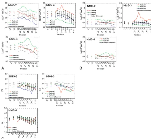
Additional quantitative comparison of DTI parameters for 8 patients and the group mean values of the healthy volunteers are presented in Fig 4, by using the NMG. There is a significant variation from the normal values at the levels where there is significant spinal stenosis. Direct-level testing of the patients’ disabilities was not available due to limitations of institutional review board approval; however, there was general agreement with the aberrancies of the diffusivity indices and their degree of clinical impairment.
Discussion
There are numerous and novel approaches to in vivo DTI spinal cord imaging as discussed previously.4,8-10However, they also have several limitations in providing robust and re-liable measurements of DTI. The greatest limitations have been related to the scanning time for DTI acquisition, distor-tion, and motion-related artifacts. Previous work has excluded cord evaluation at the level of intervertebral disks because of the significant susceptibility artifacts.2 In this report, we present the DTI results from in vivo cervical spinal cord im-aging of both healthy volunteers and patients with CSM while maintaining a short imaging time of 7 minutes. The 2D ss-IMIV-DWEPI technique allowed efficient interleaved multi-section imaging with fewer magnetic susceptibility distortion artifacts, affording an increase in the quality of images with a limited FOV.
The regions of interest were selected within the dorsal col-umns to maximize the size of the area evaluated while mini-mizing the acquisition time compared with previous axial techniques.2,10,11Although whole-cord analysis in the sagittal plane cannot avoid more partial volume effects compared with axial techniques because of the cord anatomy, a scanning time of 7 minutes with a high in-section resolution is clinically feasible for patients. Corticospinal tract evaluation within the lateral columns, while desirable, is technically challenging due to the long imaging time needed to maintain adequate SNR and FOV.
Prior studies have been conducted describing DTI of the cervical spinal cord by using imaging acquired in the sagittal plane with the section thickness ranging from 4 to 6 mm.9,12,13 These techniques had decreased in-section resolution, which caused partial volume effects. Although in-plane resolution is relatively high, partial volume effects from the gray matter may not be ignored. In our study, we minimized the section thickness to reduce the gray-white matter partial volume. We were able to obtain⬃8 clean sagittal sections of entire cord in a cross-sectional direction by using the small FOV with the IMIV technique to decrease the error related to partial volume averaging, which has been problematic in previous experiments.
Initially, the custom-made coil was developed to improve image quality, SNR, and the overall reliability of our data, in the hope that it could be used on all research subjects. Our
[image:3.594.54.287.43.237.2]dedicated coil maintained an SNR⬃2 times better than the clinical coil, but its use was limited by the body habitus of our subjects. We, therefore, used both the custom-made coil and
the clinical coil to image the healthy controls. The validity of the custom-made coil was demonstrated by comparing DTI indices with those acquired by using the clinical coil array. We
Fig 2.DTI parameters: longitudinal (A) and transversal diffusivities (B) and FA (C) of the cervical spinal cord of healthy controls by using 2 different coils (custom-made coil, 14 controls; clinical coil, 6 controls).
[image:4.594.134.452.39.332.2] [image:4.594.134.452.368.637.2]confirmed the importance of high SNR according to the Monte Carlo simulation performed by Pierpaoli and Basser,14 which showed the radical changes of储and⬜at a low SNR. In Fig 2, the clinical coil shows a large variation and, especially, a significant decrease in the indices below the C6 vertebral level due to a low SNR. In contrast, the dedicated coil provided almost constant values of indices over the length of the cervical spinal cord, in good agreement with those in the previous reports.2,10
In this report, we demonstrate the reliability of our DTI measurements by using the 2D ss-IMIV DWEPI technique for healthy controls. With the custom-made coil, the mean diffu-sivities over the entire cord were储⫽1.17⫻10⫺3(mm2/s), ⬜⫽0.40⫻10⫺3(mm2/s), and FA⫽0.59. With the clinical coil, they were储⫽0.99⫻10⫺3,⬜⫽0.38⫻10⫺3, and FA⫽ 0.54. Our results are in fair concordance with the report of Wheeler-Kingshott et al10(
储,⬜: 1.648, 0.570⫻10⫺3mm2/s
and FA: 0.61) and the report of Ellingson et al11(
1,2, and3: 1.2–.5, 0.6 – 0.7, and 0.4 – 0.5⫻10⫺3mm2/s and FA: 0.5– 0.6)
by using axial plane images. We observed a downward trend in all the diffusivities toward the lower cervical cord, as demon-strated previously. However, there is no consensus as to what values are normal. Rossi et al4reported a range of FA values from 0.69 to 0.79 in the center of the cord at the fourth cervical vertebra by using axial plane imaging, and Ries et al3reported an FA of 0.83 at the center of the spinal cord using sagittal plane imaging. The FA from our sagittal acquisition ranged from 0.46 to 0.61, which is very similar to the findings of Ell-ingson et al (0.5– 0.6).11
While maintaining an adequate SNR (20⫾8.4 for healthy controls by using the clinical coil), we established reference values and created individual quantitative maps of储,⬜, and
FA for each level of the cervical spinal cord of healthy volun-teers. These data were considered as a reference for the pa-tients with CSM (SNR⫽18.5⫾5.8). The diffusivities ob-tained from the patients with CSM were deemed abnormal on a level-by-level basis on the basis of aPvalue of .05. General trends were observed at the levels of disease across all patients
[image:5.594.50.535.39.482.2]compared with the control subjects. The储and⬜were both
increased, while FA trended low. Fifty percent of the patients had significantly lower FA values than the reference values within diseased regions of interest, as shown in Fig 4.
The FA has been used in previous experiments as a rough estimate of microstructural abnormalities,15but we suggest that these data must be used in addition to储and⬜values to
understand potential causes of abnormalities. With patient 5 as an example, the FA values were within the normal range in regions of diseased cord, while the储and⬜values were
ab-normal, likely from the low SNR that affected the entire acqui-sition. As shown in Fig 3, patient 3 has a significant change in 储and⬜, while the FA values show insignificant variation.
This is probably because the FA value is a scaled representation of储and⬜. The changes in储and⬜can cancel each other in
the calculation of FA.
Some patients with CSM had aberrantly increased储values at multiple levels, without complete agreement with findings from conventional MR imaging. For instance, patient 1 had severe stenosis at C4/C5, C5/C6, and C6/C7; however储was significantly different from the reference values only at C4/C5. This patient had elevated⬜, in accord with the measured储.
Patient 3 had elevated储at the C4 through C6 and elevated⬜ at levels C5, C6, and C7. This was in contrast to patient 4 who showed no significant changes in any parameter despite mul-tilevel disease found with conventional imaging.
The differences in DTI indices we demonstrated between patients with CSM and healthy volunteers are similar to those in the previous reports. Facon et al8found a statistically signif-icant difference in the measured FA of healthy volunteers ver-sus patients with known disease at the same level (0.67 and 0.74, respectively). Mamata et al13reported that 54% of pa-tients with spondylosis (n⫽79) showed an FA decreased from normal cords (0.66 – 0.70). Ellingson et al11reported the dif-fusion properties of the patients with chronic spinal cord in-jury based on the relative distance from a spinal cord lesion. While FA was low at the level of the lesion, they reported a gradual change in values as a function of distance from the site for the patients.
In addition to measured values, the improved quality of the DTI by using the 2D ss-IMIV-DWEPI technique allows the identification of pathology based on abnormalities identified with RGB maps. We were able to match the shape of our sub-jects’ spinal cords with FA and RGB maps and to maintain consistent region-of-interest mapping to allow direct compar-ison. Figure 3 demonstrates the high-quality DTI and a close correlation between conventional MR imaging findings and DTI results. Areas of stenosis can be clearly identified on FA and RGB maps. Altered FA values are consistent with areas of deformation of the cord, which then result in changes to the measured anisotropy. By comparing measured diffusivities between patients and controls, we found that trends in the DTI
values were consistent with conventional MR imaging find-ings and the available clinical information.
As of now, histologic correlation with abnormal 储 has been theorized to represent axonal pathology, while⬜is con-sidered to be a marker of myelin integrity.1By comparing measured diffusion properties between patients and controls, we found that trends in the DTI values were consistent with conventional MR imaging findings and the available clinical information. Additional studies are underway at our institu-tion that allow direct histologic correlainstitu-tion with both MR im-aging and DTI in the ex vivo setting.
Conclusions
The DTI data from healthy volunteers by using the 2D ss-IMIV-DWEPI technique allowed us to generate a normal ref-erence map of diffusivity values. The refref-erence DTI metrics proved useful in comparison with pathology found with con-ventional MR imaging performed on patients with CSM. By developing a normal DTI atlas, we found numerous potential clinical applications in which we may quantify the degree of microstructural impairment within the spinal cord and relate it to clinical disability and treatment effects.
References
1. Song S-K, Sun S-W, Ramsbottom MJ, et al.Dysmyelination revealed through MRI as increased radial (but unchanged axial) diffusion of water.Neuroimage
2002;17:1429 –36
2. Ellingson BM, Ulmer JL, Kurpad SN, et al.Diffusion tensor MR imaging of the
neurologically intact human spinal cord.AJNR Am J Neuroradiol2008;29:
1279 – 84
3. Ries M, Jones RA, Dousset V, et al.Diffusion tensor MRI of the spinal cord.
Magn Reson Med2000;44:884 –92
4. Rossi C, Boss A, Steidle G, et al.Water diffusion anisotropy in white and gray matter of the human spinal cord.J Magn Reson Imaging2008;27:476 – 82 5. Jeong E-K, Kim S-E, Guo J, et al.High-resolution DTI with 2D interleaved
multislice reduced FOV single-shot diffusion-weighted EPI (2D
ss-rFOV-DWEPI).Magn Reson Med2005;54:1575–79
6. Jeong E-K, Kim S-E, Kholmovski EG, et al.High-resolution DTI of a localized volume using 3D single-shot diffusion-weighted stimulated echo-planar
im-aging (3D ss-DWSTEPI).Magn Reson Med2006;56:1173– 81
7. Bihan DL, Mangin J-F, Poupon C, et al.Diffusion tensor imaging: concepts and applications.J Magn Reson Imaging2001;13:534 – 46
8. Facon D, Ozanne A, Fillard P, et al.MR diffusion tensor imaging and fiber tracking in spinal cord compression.AJNR Am J Neuroradiol2005;26:1587–94 9. Hori M, Okubo T, Aoki S, et al.Line scan diffusion tensor MRI at low magnetic field strength: feasibility study of cervical spondylotic myelopathy in an early clinical stage.J Magn Reson Imaging2006;23:183– 88
10. Wheeler-Kingshott CA, Hickman SJ, Parker GJ, et al.Investigating cervical
spinal cord structure using axial diffusion tensor imaging.Neuroimage
2002;16:93–102
11. Ellingson BM, Ulmer JL, Kurpad SN, et al.Diffusion tensor MR imaging in chronic spinal cord injury.AJNR Am J Neuroradiol2008;29:1976 – 82. Epub 2008 Aug 21
12. Clark CA, Barker GJ, Tofts PS.Magnetic resonance diffusion imaging of the human cervical spinal cord in vivo.Magn Reson Med1999;41:1269 –73 13. Mamata H, Jolesz FA, Maier SE.Apparent diffusion coefficient and fractional
anisotropy in spinal cord: age and cervical spondylosis-related changes.J Magn Reson Imaging2005;22:38 – 43
14. Pierpaoli C, Basser PJ.Toward a quantitative assessment of diffusion
anisot-ropy.Magn Reson Med1996;36:893–906